
Office Lens for Windows - image lens
The fluorophores are excited by the light in the microscope, which causes them to give off light with lower energy and of longer wavelength. It is this light that produces the magnified view, rather than the original light source. This means that fluorescent microscopy uses reflected rather than transmitted light.
For CT imaging, iodinated contrast agents are not known to pose any significant risk to the mom or baby. If you have concerns, you can speak to the radiologist to understand the potential risks and benefits of the contrast-enhanced scan.
Microbubble contrast materials can be targeted or untargeted. Untargeted contrast-enhanced ultrasound —the more common method— helps diagnose certain diseases by providing evaluation of blood flow in the heart and other organs. In targeted contrast-enhanced ultrasound, specific molecules are bound to the surface of the microbubbles. After injection, the microbubbles attach to specific targeted tissue sites, causing an increase in the ultrasound signal at the sites.
Outside links: For the convenience of our users, RadiologyInfo.org provides links to relevant websites. RadiologyInfo.org, RSNA and ACR are not responsible for the content contained on the web pages found at these links.
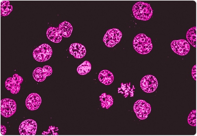
As light microscopy developed, more forms using different techniques were invented. One of the types of microscopy within the broader light microscopy group is fluorescence microscopy. Fluorescence microscopy images cells or molecules that have been tagged with a fluorescent dye. The fluorescent substances are called fluorophores, which are integrated into the sample.
"Review of the literature shows no evidence to suggest that oral ingestion by an infant of the tiny amount of gadolinium contrast medium excreted into breast milk would cause toxic effects. We believe, therefore, that the available data suggest that it is safe for the mother and infant to continue breast-feeding after receiving such an agent.
In contrast synonym
Ryding, Sara. (2023, July 21). Fluorescence Microscopy vs. Light Microscopy. News-Medical. Retrieved on November 25, 2024 from https://www.news-medical.net/life-sciences/Fluorescence-Microscopy-vs-Light-Microscopy.aspx.
It is a good idea to increase your fluid intake after an imaging exam involving a barium-based contrast material to help remove the contrast material from your body.
You should tell your doctor if these mild or moderate side effects of iodine-based contrast materials become severe or do not go away:
When the gadolinium is injected, it is normal to feel coolness at the site of injection, usually the arm for a minute or two.
Laser Diodes: 405nm, 450nm, 520nm, 635nm, 650nm, 808nm, 830nm.
For example, a commonly used label is green fluorescent protein (GFP), which is excited with blue light and emits green light with a longer wavelength. Filters around the sample can separate the fluorescent light from the surrounding radiation.
Ryding, Sara. 2023. Fluorescence Microscopy vs. Light Microscopy. News-Medical, viewed 25 November 2024, https://www.news-medical.net/life-sciences/Fluorescence-Microscopy-vs-Light-Microscopy.aspx.
Barium-sulfate contrast materials that are administered by enema (rectally) are used to enhance standard x-ray, fluoroscopy, and CT images of the lower gastrointestinal (GI) tract (colon and rectum). In some situations, iodine-based contrast materials are substituted for barium-sulfate contrast materials for rectal administration.
Contrast in art
In hospitals, quick examination of cells can be critical for doctors. In such situations, light microscopy can be used with tissues that have been frozen in carbon dioxide and sectioned using a microtome. This simpler method can be used urgently on patients who are in the operating room to guide the surgeon.
Patients with impaired kidney (renal) function should be given special consideration before receiving iodine-based contrast materials by vein or artery. While many contrast agents are safe to give in patients with kidney disease, if you have severe kidney disease and very poor kidney function you may be at increased risk of worsening kidney function when getting iodinated contrast agents. The benefits of having a contrast enhanced scan often out-weigh the risks in ensuring the radiologist can properly diagnose your medical conditions.
When iodine-based and barium-sulfate contrast materials are present in a specific area of the body, they block or limit the ability of x-rays to pass through. As a result, blood vessels, organs and other body tissue that temporarily contain the iodine-based or barium compounds change their appearance on x-ray or CT images.
Both fluorescence microscopy and light microscopy represent specific imaging techniques to visualize cells or cellular components, albeit with somewhat different capabilities and uses. At its core, fluorescence microscopy is a form of light microscopy that uses many extra features to improve its capabilities.
Anti–reflective coating (also called AR coating or anti–glare coating) improves vision, reduces eye strain and makes your eyeglasses look more ...
Contrast adjective
If the mother remains concerned about any potential ill effects, she should be given the opportunity to make an informed decision as to whether to continue or temporarily abstain from breast-feeding after receiving a gadolinium contrast medium. If the mother so desires, she may abstain from breast-feeding for 24 hours with active expression and discarding of breast milk from both breasts during that period. In anticipation of this, she may wish to use a breast pump to obtain milk before the contrast study to feed the infant during the 24-hour period following the examination."
Your questions, but not your email details will be shared with OpenAI and retained for 30 days in accordance with their privacy principles.
Thorlabs' Clear Aperture Right-Angle Brackets provide a threaded clear aperture on both mounting surfaces, allowing them to be positioned directly within a beam ...
Contrast meaning example
Barium-sulfate is the most common contrast material taken by mouth, or orally. It is also used rectally and is available in several forms, including:
You should tell your doctor if these mild side effects of barium-sulfate contrast materials become severe or do not go away:
Light microscopy does much what the name implies: visible light and magnifying lenses are used to view small objects. Light microscopes are the oldest form of higher quality imaging devices, dating back to the 1500s, and were the microscopes with which first cells were observed.
Numerical aperture (NA) refers to the cone of light that is made from a focusing lens and describes the light gathering capability of the lens (similar to ...
If you swallow the contrast material, you may find the taste mildly unpleasant; however, most patients can easily tolerate it.
Contrast materials can have a chemical structure that includes iodine, a naturally occurring chemical element. These contrast materials can be injected into veins or arteries, within the disks or the fluid spaces of the spine, and into other body cavities.
Following an imaging exam with contrast material, the material is absorbed by the body or eliminated through urine or bowel movements.
Our Half Play Spheres are solid features made from EPDM Safety Surfacing and recycled materials. Available in various sizes and 19 striking colors, ...
Ijeoma Uchegbu discusses nanomedicine's role in improving medication adherence and developing non-addictive pain relief solutions at ELRIG Drug Discovery 2024.
Mounts vary in dimensions so as to support diameters from 10 to 50mm (0.5" to 2"). Original design allows to decrease the mount's dimensions and weight.
Contrast meaning in Hindi
Fluorescence microscopy can be used in conjunction with other types of light microscopy. Due to the fact that it creates images from the reflected light (rather than the direct light), it can be used with techniques such as phase contract microscopy.
It is a good idea to increase your fluid intake after an imaging exam involving an iodine-based contrast material to help remove the contrast material from your body.
If your contrast material is given by enema, you can expect to experience a sense of abdominal fullness and an increasing need to expel the liquid. The mild discomfort will not last long.
Before arriving for your exam, you will be given specific instructions on how to prepare for the exam. Because contrast materials carry a slight risk of causing an allergic reaction or adverse reaction, you should tell your doctor about any of the following conditions. These conditions could affect the instructions you are given.
07 3810 4590 / 07 3810 4448 enrolments@sec.qld.edu.au. Quick Links. Enrol Online · Book a Tour · Campus Map · Portal · Complaints Handling · SchoolTV. © 2024 St ...
Iodine-based contrast materials injected into a vein (intravenously) are used to enhance x-ray (including fluoroscopic images) and CT images. Iodine based contrast materials are also commonly injected in the arteries during angiogram procedures. Gadolinium injected into a vein (intravenously) is used to enhance MR images. Typically, these are used to enhance:
Manufacturers of intravenous contrast provide special instructions for mothers who are breast feeding. They advise that mothers should not breast-feed their babies for 24 to 48 hours after contrast medium is given. However, both the American College of Radiology (ACR) and the European Society of Urogenital Radiology note that the available data suggest it is safe to continue breast-feeding after receiving intravenous contrast. The Manual on Contrast Media from the ACR states:
Traditional light microscopes are widely used, and often require simpler dyes to visualize contrast which is not naturally visible. This is typically a simpler technique than fluorescence microscopy. Because of this, it is used in clinical settings, such as for immediate imaging of biopsied samples in hospitals and for cervical smears.
For MR imaging, gadolinium contrast material administration is usually avoided due to unknown risk to the baby. However, it may be used when critical information can only be obtained with the use of the gadolinium-based contrast agent.
Contrast examples
Fluorescence microscopy is often applied in imaging cell structures or structural features, checking the viability of cells, imaging genetic material (both DNA and RNA), and imaging particular cells in a larger population.
Sara is a passionate life sciences writer who specializes in zoology and ornithology. She is currently completing a Ph.D. at Deakin University in Australia which focuses on how the beaks of birds change with global warming.
Copyright © 2024 Radiological Society of North America, Inc. (RSNA). To help ensure current and accurate information, we do not permit copying but encourage linking to this site.
While we only use edited and approved content for Azthena answers, it may on occasions provide incorrect responses. Please confirm any data provided with the related suppliers or authors. We do not provide medical advice, if you search for medical information you must always consult a medical professional before acting on any information provided.
In some situations, iodine-based contrast materials are substituted for barium-sulfate contrast materials for oral administration.
Contrast in English
Sep 23, 2024 — That means that objects right in front and right behind the plane of focus is already going out of focus. As you can see in this photo below, ...
When introduced into the body prior to an imaging exam, contrast materials make certain structures or tissues in the body appear different on the images than they would if no contrast material had been administered. Contrast materials help distinguish or "contrast" selected areas of the body from surrounding tissue. This helps physicians diagnose medical conditions by improving the visibility of specific organs, blood vessels, or tissues.
There is evidence that tiny traces of gadolinium may be retained in different organs of the body, including the brain, after contrast-enhanced MRI. While there are no known negative effects from this, your doctor may take gadolinium retention into account when selecting a contrast agent. There are many different gadolinium-based contrast agents available, each with its own safety profile. Decisions on which material to use may be affected by the part of the body being imaged, the cost of the material and other factors. These decisions are especially important in patients likely to undergo multiple MRI scans with gadolinium-based contrast material, such as pediatric patients, cancer patients and people with multiple sclerosis.
Nephrogenic systemic fibrosis (NSF), a thickening of the skin, organs, and other tissues, is a rare complication in patients with kidney disease that undergo an MR with contrast material. Gadolinium-based contrast material may be withheld in some patients with severe kidney disease.
A very small percentage of patients may develop a delayed reaction with a rash which can occur hours to days after an imaging exam with an iodine-based contrast material. Most are mild, but severe rashes may require medication after discussion with your physician
If a barium-sulfate contrast material (given orally or rectally) will be used during your exam, you may be asked not to eat for a few hours before your exam begins. If the contrast material will be given rectally, you may also be asked to cleanse your colon with a special diet and medication (possibly including an enema) before your exam.
Learn about the usage of process raman spectroscopy in the optimization of bioreactor monitoring and then improvement of cultivated meat production.
Ryding, Sara. "Fluorescence Microscopy vs. Light Microscopy". News-Medical. 25 November 2024. .
Ryding, Sara. "Fluorescence Microscopy vs. Light Microscopy". News-Medical. https://www.news-medical.net/life-sciences/Fluorescence-Microscopy-vs-Light-Microscopy.aspx. (accessed November 25, 2024).
The contrast material used in MRI (Magnetic Resonance Imaging) called gadolinium is less likely to produce an allergic reaction than the iodine-based materials used for x-rays and CT scanning. Very rarely, patients are allergic to gadolinium-based contrast materials and experience hives and itchy eyes. Reactions are usually mild and easily controlled by medication. Severe reactions are rare.
If you have not been sedated, no recovery period is necessary. You may resume your usual activities and normal diet immediately after the exam. Increased fluid intake will help eliminate the contrast material from your body.
Registered members can chat with Azthena, request quotations, download pdf's, brochures and subscribe to our related newsletter content.
Contrast materials are safe drugs; adverse reactions ranging from mild to severe do occur, but severe reactions are very uncommon. While serious allergic or other reactions to contrast materials are rare, radiology departments are well-equipped to deal with them.
Please type your comment or suggestion into the text box below. Note: we are unable to answer specific questions or offer individual medical advice or opinions.
A common method to visualize cells or tissue with light microscopes is to use dyes. Widely used ones might paint the main components, such as the dye combination of hematoxylin and eosin, which colors the nuclei violet and the cytoplasm pink. However, there are also more specialized dye techniques.
Fotodiox Pro Lens Adapter - Compatible with Pentax K Auto Focus Mount (PK AF) Lenses to C-Mount (1" Screw Mount) Cine & CCTV Cameras with Built-In De-Clicked ...
Contrast meaning
This website does not provide cost information. The costs for specific medical imaging tests, treatments and procedures may vary by geographic region. Discuss the fees associated with your prescribed procedure with your doctor, the medical facility staff and/or your insurance provider to get a better understanding of the possible charges you will incur.
Contrast materials are not dyes that permanently discolor internal organs. They are substances that temporarily change the way x-rays or other imaging tools interact with the body. The materials discussed in this article do not produce radiation.
When a physician needs to understand what is happening inside our bodies, they often request that a patient undergo an imaging exam. Imaging exams such as x-rays, ultrasound, computed tomography (CT), magnetic resonance (MRI), and fluoroscopy are selected based on their ability to show specific information about the structures within the body. Contrast materials, also known as contrast agents or contrast media, are used to improve the diagnostic value of those imaging exams.
Prior to any imaging exam, women should always inform their physician or technologist if there is any possibility that they are pregnant. Many imaging tests and contrast material administrations are avoided during pregnancy to minimize risk to the baby.
As the position of the pupil over the lens changes laterally, the distortion varies and becomes asymmetric. This motivates making the lens as large as possible ...
When an iodine-based contrast material is injected into your bloodstream, you may have a warm, flushed sensation and a metallic taste in your mouth that lasts for a few minutes.
Barium-sulfate contrast materials are expelled from the body with feces. You can expect bowel movements to be white for a few days. Some patients may experience changes in their normal bowel movement patterns for the first 12 to 24 hours.
The usefulness of traditional light microscopy is hampered by the fact that it uses visible light, as using visible light limits the resolution obtained from samples. On the other hand, fluorescence microscopy is not faced with this limitation, since it uses whatever light excites the fluorophores.
RadiologyInfo.org is not a medical facility. Please contact your physician with specific medical questions or for a referral to a radiologist or other physician. To locate a medical imaging or radiation oncology provider in your community, you can search the ACR-accredited facilities database.
Barium-sulfate contrast materials that are swallowed or administered by mouth (orally) are used to enhance standard x-ray, fluoroscopy, and CT images of the gastrointestinal (GI) tract, including:
Professor Nancy Ip discusses her groundbreaking neuroscience research, focusing on neurotrophic factors and innovative Alzheimer's disease treatment approaches.
As mentioned, light microscopes that are used for light microscopy employ visible light to view the samples. This light is in the 400-700 nm range, whereas fluorescence microscopy uses light with much higher intensity.
News-Medical.Net provides this medical information service in accordance with these terms and conditions. Please note that medical information found on this website is designed to support, not to replace the relationship between patient and physician/doctor and the medical advice they may provide.
Web page review process: This Web page is reviewed regularly by a physician with expertise in the medical area presented and is further reviewed by committees from the Radiological Society of North America (RSNA) and the American College of Radiology (ACR), comprising physicians with expertise in several radiologic areas.
Furthermore, there are light microscopy techniques that can image both live and fixed samples, but there can be a tradeoff between signal-to-noise ratio and sample damage during the observation process. During fluorescence microscopy, cells undergo bleaching, in which the fluorescence diminishes during extended periods of observation. To conclude, there is flexibility in both microscopy groups.
Microbubble contrast materials are tiny bubbles of an injectable gas held in a supporting shell. They are extremely small—smaller than a red blood cell—and have a high degree of "echogenicity", or ability to reflect ultrasound waves. Structures with higher echogenicity will appear brighter on ultrasound. Once the microbubbles are in the bloodstream, ultrasound technology is able capture differences in echogenicity between the gas in the microbubbles and the surrounding tissues of the body, producing an ultrasound image with increased contrast. The microbubbles dissolve, usually within 10 to 15 minutes, and the gas within them is removed from the body through exhalation. Contrast-enhanced ultrasound with microbubbles is a convenient, relatively inexpensive way to improve visualization of blood flow, and it does not use radiation. It is a useful option for patients with kidney failure or those with allergies to contrast agents used for MR and/or CT imaging.


 Ms.Cici
 8618319014500