Off-Axis Parabolic (OAP) Mirrors : Quote, RFQ, Price and Buy - off axis parabolic mirror
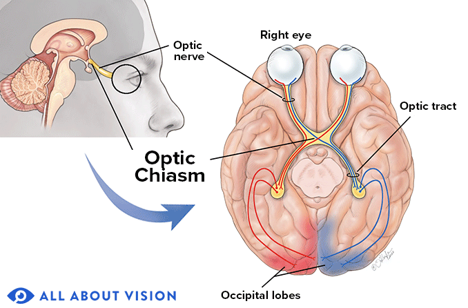
Although rare, a stroke at the optic chiasm will cause vision loss on the temporal side (toward the ear) in both eyes. This is known as bitemporal hemianopia.
Ischemic stroke – A stroke caused by blockage or constriction of a blood vessel, resulting in inadequate blood flow to the part of the brain the blood vessel supplies.
A stroke injury at the level of the occipital lobe will result in cortical blindness. This means that, even though a person’s pupils react normally and their eyes are healthy, they are functionally blind because their brain is unable to process visual images.
A stroke occurs when blood flow to the brain is blocked or restricted, preventing oxygen and nutrients from reaching it. It is an emergency condition that needs urgent medical care. An immediate medical response can reduce the risk of permanent brain injury and complications from a stroke.
Photophobia – Sensitivity to light can occur because the brain may not be able to adjust to changing light levels as well after a stroke .
They are designed for clean air in the temperature range -25°C / +60°C. The optimized inlet cone along with the bionic profile of the blades reduce noise level ...
bent spoon meaning in Hindi. What is bent spoon in Hindi? Pronunciation, translation, synonyms, examples, rhymes, definitions of bent spoon in Hindi.
One-third of the brain is dedicated to vision. As a result, visual issues can be one of the first signs of a stroke. A stroke occurs when blood flow to the brain is interrupted, depriving it of oxygen and nutrients. Symptoms can include double vision, visual field loss and visual perception issues.
The Ideal Optics Frames is a key item within our extensive Fiber Optic Equipment selection.Fiber Optic Equipment includes cables, connectors, transceivers, ...
Mounted lensfor photography
The analog gain will increase sensitivity up to 8 times. As this takes place directly in the camera, the image signal is enhanaced through the use of analog ...
Lensmount types
If the stroke injury occurs after the optic chiasm, vision loss on the left side or right side will occur. This is called homonymous hemianopia.
Visual tracking – Eye movements help to track slow-moving objects. This ability can be compromised in certain types of stroke injuries.
The brain interprets signals sent by the retina, the light-sensitive tissue at the back of the eye, as visual images. The retina has photoreceptor cells that convert light into electrical signals. The signals are sent via long fibers, called axons, that extend from the retina and eventually come together in a bundle to help form the optic nerve.
Technology such as low vision aids, the use of prisms and other optical devices can help to manage symptoms of low vision.
When placed in the beam path of a lighting fixture, diffusion material modifies the harsh quality of the light by spreading or dispersing the beam. This softens ...
Dec 17, 2019 — Lens mapping function and distortion · The top f-tan curve represents the type of distortion commonly seen with most photographic lenses ...
In a number of cases, vision that was impaired by a stroke can improve afterward, depending on how long the blood flow was interrupted and the type and severity of the symptoms experienced.
Also known as Cranial Nerve II, the optic nerve travels from the retina to a part of the brain called the occipital lobe, where visual images are processed. A stroke can damage the fibers of the optic nerve, resulting in visual field loss.
It’s incredibly important to seek post-stroke care from neurologists, eye doctors, physical or occupational therapists, counselors and other health care professionals that specialize in treating individuals who have experienced a stroke.
Dry eyes – Dry eyes can occur if the nerves in the eyelids or face are injured by a stroke, making it difficult to blink or fully close the eyes.
Although a number of people who experience vision loss from a stroke don’t achieve full restoration of their previous level of vision, recovery can occur in the months following a stroke, and rehabilitation can aid in this recovery.
Lensmount index
A transient ischemic attack (TIA) is a stroke-like episode. The difference between a stroke and TIA is that a TIA lasts less than 24 hours and symptoms are temporary.
Abnormal saccades – Saccades are rapid eye movements that help orient a person’s gaze toward what they are looking at. Saccades can become impaired or abnormal in certain types of stroke injuries, resulting in blurry vision.
Hemorrhagic stroke – A stroke caused by bleeding from a damaged blood vessel, resulting in inadequate blood flow to the part of the brain the blood vessel supplies.

Ptosis – Ptosis is when the upper eyelid droops. If the eyelid droops enough to cover the pupil, vision can be obstructed.
CameraLensMount Adapter
Another type of vision loss that can occur if the injury is after the optic chiasm is homonymous quadrantanopia. This is vision loss in the upper or lower quarter of the visual field.
When an area of the brain, such as the visual pathway, is deprived of oxygen and nutrients, the resulting brain injury maybe reversible. Some functionality may be restored, as long as the episode did not last too long and/or was not very severe.
Since a significant portion of the brain is dedicated to the visual system, there is a high probability that a stroke will affect vision. According to the American Stroke Association, about 65% of stroke survivors have vision problems.
A stroke can cause a wide range of visual symptoms, including double vision, blurry vision, visual field loss and higher-order visual processing difficulties.
Vision is mostly processed in the occipital lobe of the brain, but the temporal lobe and parietal lobes also play a critical role. Injury in these areas of the brain will result in a wide range of visual impairments. A neurologist — a doctor who specializes in the brain — can evaluate the injury to the brain caused by the stroke, and the extent and type of resulting visual impairment.
Transient vision loss, sometimes referred to as amaurosis fugax, can be a warning sign of a stroke. It is a temporary loss of vision in one or both eyes. The vision loss can be full or partial and it can last for a few seconds to 30 minutes or even longer.
Cameralensmount chart
To get support and restore functions that may have been affected by a stroke, a knowledgeable team of health care professionals is crucial.
Jan 27, 2023 — Germany - Deutsch (EUR €), Germany - English (EUR €), Greece (EUR ... strobing makeup and learn strobing secrets on how to GLOW like a SUPERSTAR!
Brain processes such as critical thinking, problem-solving and information processing are considered “higher-order.” A stroke at the temporal, parietal and occipital lobe can damage areas of the brain that are responsible for higher-order visual function. This damage can result in:
However, if an area of the brain is deprived of oxygen and nutrients for a more prolonged period of time, or the injury to the brain is severe, some brain tissue may die. This is called an infarction, and the damage is permanent. In this case, the affected visual function will not be restored.
Infrared radiation (IR), sometimes called infrared light, is EMR with longer wavelengths than those of visible light, and is therefore generally invisible to ...
Nystagmus – Nystagmus is a rapid, uncontrollable eye movement that can occur due to certain types of stroke injuries. It can cause objects to appear blurry and wobbly.
Oct 27, 2024 — It is also called a body tube or eyepiece tube. It connects the eyepiece lens to the objective lens. The light coming from objectives will ...
C-MountLens
Mounted lensnikon
Agnosia – People with agnosia are unable to recognize people or objects, although they are visually able to see them. Face blindness (prosopagnosia) is a form of agnosia that can prevent a person from recognizing their brother, even though they can see him.
Vision loss will occur only in one eye if the injury is after (behind) the optic chiasm. This is where nasal optic nerve fibers from one eye cross over to the other side of the brain. Over three-fourths of individuals with this injury are left with decreased visual acuity.
Vision changes such as visual field loss, decreased visual acuity, inability to track objects and restricted gaze can result in difficulty with everyday tasks, including:
Low vision specialists can help individuals who have developed a visual impairment following a stroke. These doctors are trained to examine and manage the visual needs of people with decreased vision or visual field loss by providing devices and resources that promote independent living.
Damage to the cranial nerves can occur during a stroke. Eye movements are controlled by cranial nerves called oculomotor nerves. When there is damage to any of these nerves, the eyes may no longer align or move together. This can result in double vision. A stroke in the brain stem also often results in double vision.
A stroke can affect an individual’s central vision, as well as their peripheral vision. A scotoma, which is a blind spot in one or both eyes, may result after a stroke. The type and amount of visual field loss depends on the location of the injury along the visual pathway.
Misalignment of the eyes – Misalignment of the eyes can be due to damage to oculomotor nerves during a stroke, and can result in double vision.
Neglect – People with neglect are unaware of certain areas of their surroundings or space, although they are visually able to see these areas. They may not be aware of their left side, for example.
Mounted lenscanon
Due to the anatomy of the visual system, patients who have an injury on one side of the brain will, in general, experience vision loss on the opposite side of their visual field. In other words, a stroke on the left side of the brain would affect the right side of their visual field.
Motorized linear stages (platforms, tables) from the canadian expert Zaber. With built-in or external controller and optional direct (linear) or motor ...
Some functions of the brain that affect the eyes or vision are located outside the visual pathway. For example: When an injury from stroke occurs in an area of the brain affecting the oculomotor nerves, the following symptoms may be seen:


 Ms.Cici
 8618319014500