
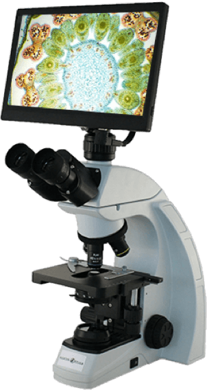
Nikon Microscope - nikon microscope
Application imagingpdf
Through specialized methods like MRI oximetry, we study neurobiochemistry and stroke risk. We also develop methods to evaluate the damage caused by stroke. Our researchers use novel radiotracers and develop sophisticated radioligands for PET/MR imaging of the brain to investigate the role of cell receptors in amyotrophic lateral sclerosis (ALS), also known as Lou Gehrig’s disease, and examine the mechanism and effects of Tau protein buildup, a phenomenon that accompanies many neurodegenerative conditions.
Our XD-GRASP technique solves these problems and indexes the acquired MR signal in relation to the periodic beating of the heart and inflations of the lungs. The resulting image data can be sorted in reconstruction to “freeze” either kind of motion. The method allows scientists to study cardiac physiology and the relationship between breathing and heartbeat with unprecedented detail, especially in conditions with challenging cardiac dynamics like arrhythmia. Our scientists are combining this technique with sodium MRI to better understand how the heart handles one of its fundamental metabolites. The technology also applies to the imaging of other organs in which contrast-enhanced MRI can indicate pathology by tracing vasculature.
MedicalImagingScience salary
Sep 15, 2018 — Rotate the collimator until the laser dot is at the lowest point of the circle traced by the laser. If you tighten the screw that is now top- ...
We are currently developing sodium MRI methods to investigate the metabolism of leg muscles. In particular, we use sodium imaging to study the relationship between exercise and microvascular dilation, oxidative stress, and biomolecules that promote the health of nerve cells. We do so to determine whether supervised exercise can reverse diabetic neuropathy. Our researchers also investigate how circulation in small vessels affects the brain in a condition known as small vessel disease.
The ultimate goal of researchers at NYU Langone’s Department of Radiology is the improvement of human health. In clinical practice, doctors rely on imaging to accurately diagnose diseases and effectively guide therapies. In medical research, scientists depend on imaging to study basic phenomena and to evaluate the safety and efficacy of new treatments. Our researchers develop novel imaging technologies to advance medical science and clinical care in several areas.
Anti glare glasses, or lenses treated with Anti glare coatings, also known as anti reflection, reflection free, AR or MAR coatings, were developed to reduce ...
Oct 30, 2012 — Line pairs per picture height is a real world comparable measurement for evaluation of the impact in an image, just because it is not related to ...
Our department is currently developing a variety of sophisticated methods, such as diffusion MRI and MR fingerprinting, to obtain precise quantitative maps and biophysical measurements of articular cartilage. The technology has the potential to help guide hip and knee replacements, enable earlier diagnosis of degenerative joint disease, and facilitate the search for effective treatment of osteoarthritis.
Medicalimagingapplications
Division of Breast Imaging, Division of Abdominal Imaging, Division of Nuclear Medicine, Division of Interventional Neuroradiology
We develop new methods for imaging of the heart and the vasculature of muscles and organs to study cardiovascular diseases and advance treatment. Our team has invented an MRI technique called XD-GRASP that produces high-quality heart images from a continuous scan. Conventional cardiac imaging protocols rely on a battery of carefully calibrated acquisitions—an error-prone and time-consuming approach that is labor intensive for the technologists who are tasked with management of complex settings and burdensome for the patients who are repeatedly required to hold their breath.
What are the 4 types of medicalimaging
Both refracted light beams are polarized - one in a direction parallel to the surface and the other in a direction perpendicular to the surface. Since these two ...
Diagnosticimaging
Our team developed a method to obtain high-quality MRI of the liver in the presence of respiratory motion, eliminating the need to have patients hold their breath during imaging. The technique, called GRASP, makes small lesions easier to identify and is now clinically available worldwide. We are expanding this technology and adapting it to applications in abdominal and breast MRI.
In this chapter we therefore examine the resonant frequencies and spatial distributions of the electromagnetic fields which form the laser cavity modes. We ...
Other areas of research include imaging methods to assess bone quality and predict fracture risk and patient responses to osteoporosis therapy. We also develop advanced MRI techniques, like diffusion tensor imaging, to investigate the biokinetics and physiology of muscle tissue with the aim of advancing knowledge and treatment of conditions such as dermatomyositis and diabetic neuropathy.
LabDex is a global manufacturer of innovative Lab Equipment & Analytical Instruments for numerous applications in R&D, Quality control & Testing Laboratory.
We also apply sophisticated imaging methods like diffusion MRI to the study, diagnosis, and evaluation of prostate cancer, and create new, highly accurate ways of imaging white matter nerve bundles to provide precise information about where and how neuronal tracts adjoin brain tumors.
We develop leading-edge imaging methods to study cancer and guide oncologic therapies. Our researchers create AI algorithms that improve the accuracy of breast cancer detection from mammograms and design neural networks that learn from a combination of mammograms and clinical data in order to lower recall rates for mammography screening. We tailor specialized imaging methods, such as gradient–echo spectroscopic MRI, to aid in the search for new biomarkers of breast cancer. Our research teams also apply vanguard imaging technologies, such as metabolic sodium MRI, to assess early response of breast cancer to neoadjuvant chemotherapy.
Application imagingexamples
Browse through the largest collection of home design ideas for every room in your home. With millions of inspiring photos from design professionals, you'll find ...
Imagingtechnologies
Application imagingrequirements
Symbiont Service Corp is a Facilities Services, Commercial & Residential Construction, and Geothermal Pool Heating company located in Englewood, ...
by CY Han · 2021 · Cited by 5 — where k = 2πn/λ is the propagation number, n is the refractive index of the environmental medium, and f is the focal length of the test sample. For converging ...
In preclinical research, we develop and use leading-edge diffusion MRI in combination with MR microscopy and computational neuroanatomy to investigate how genetic mutations and early brain injuries affect neural development. We are also making theoretical and technical advances to obtain cellular-level information about the brain’s micro-neuroanatomy in vivo through diffusion MRI.
We create new imaging methods and tools for the diagnosis, treatment, and study of muscle and joint conditions. Our scientists pioneered the application of artificial intelligence (AI) to MR image reconstruction, significantly accelerating MRI exams of the knee. We invented ultraflexible low-impedance MRI coils that open the door to noninvasive study of the biodynamics of the hand. We’ve also trained deep learning algorithms to predict which patients with early symptoms of osteoarthritis are likely to need total knee replacement within a decade of diagnosis.
We use cookies and similar tools to give you the best website experience. By using our site, you accept our websites privacy policy . . Opens in a new tab
Best headshot aiming codes · 1. Green oval fitting the enemies' heads · 2. Two dots · 3. Small purple crosshair · 4. Yellow circle with a line 0;P;c;8;u;FCF003FF;h; ...
We create and adapt novel imaging technologies to learn more about the brain and the neurological diseases that affect it. Investigators in our department develop advanced MR spectroscopy methods to study the earliest manifestations of Alzheimer’s disease, detect early-stage neurodegeneration in multiple sclerosis, and investigate the role of demyelination in a range of cognitive disorders. Our researchers combine MR spectroscopy with sodium MRI to better understand the metabolic damage that accompanies traumatic brain injury.


 Ms.Cici
 8618319014500