
Microscope Objectives | Evident LS - light microscope objectives
Mar 30, 2017 — Each coloured filter produces a different effect on the scene. Yellow Filter. A yellow filter has always been the classic first choice ...
Smart Microscopy for easy Digital Documentation. Press a single button for crisp images in true color, already with the correct scaling information.
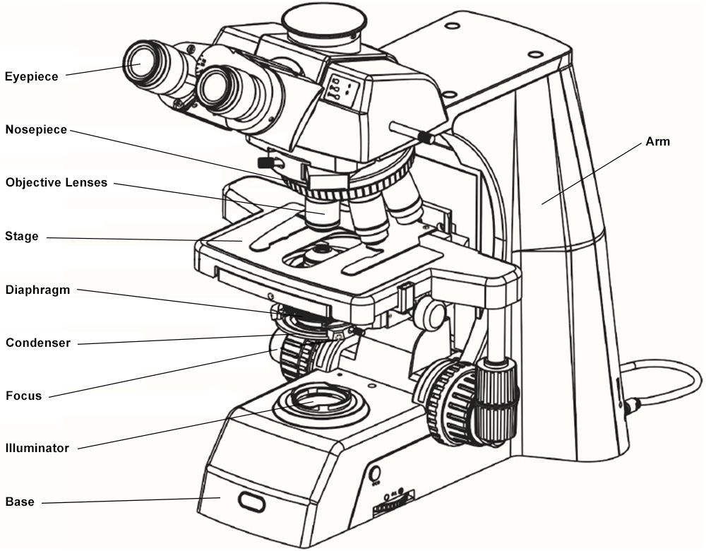
When you are looking at labeled microscope parts, one of the most important parts of the microscope is the eyepiece. This is also known as the ocular lens of a microscope and is used to magnify the image of the specimen you are looking at in your microscope. Depending on the type of microscope you have, the magnification of the eyepiece will differ. Typical school microscopes will usually have an ocular lens with 10x magnification. Your eyepiece lens magnifies the real intermediate image of your specimen.
To keep your objective lenses in place and to allow for easy rotation, your microscope has a nosepiece. This is sometimes called a revolving turret and allows you to easily choose which objective lens you want to use on your microscope.
Two kinds of glass are used to make this doublet lens so that white light coming into the lens is all focused (imaged) at the same point on the other side of ...
There are two different types of focus that you need in a microscope. Using both of these together will allow you to get clear, crisp images of your specimen, no matter their size. First, you will use the coarse focus of your microscope. This brings the stage closer to your eyepiece to allow you to focus on the specimen. Then, you will use the fine focus. This is essential if you want to see more details of your specimen and if you want to get a clear image. When using the fine focus, it moves the stage in much smaller increments.
We are constantly improving our website. Some functionality like inventory in stock may be inaccurate. Please email or call so we can help.
A diffraction grating can be simply thought of as a set of identical and equally spaced slits separated by opaque strips. In reality gratings are made by ruling ...
WalmartMagnifying Glass withLight
Sep 13, 2022 — A compound microscope is a high-magnification microscope that uses two lenses to compound or multiply the level of magnification. The first lens ...
Another vital part of your microscope is the stage. This is where you place the specimen that you are researching with your microscope! It allows you to easily observe what you are looking at and will keep your specimen in place. Microscopes also have stage clips. These stage clips hold your specimen slides in place on the stage and will keep them steady while you are looking through the microscope. It allows for the clearest imaging possible from your microscope.
AmazonMagnifying Glass withLight
Microscopes play an essential role in learning more about the world around you. Learning more about each of the parts of the microscope can help you understand how to use one properly! If you want to learn more about science microscopes and how to use them, Nuhsbaum Microscopy & Digital Imaging Solutions can help! We offer top microscopy products and imaging equipment to help you reach your research goals. Contact us today to learn more about the microscopes we offer or for all of your other microscopy needs and questions.
Choose from our selection of magnifying glasses, including over 250 products in a wide range of styles and sizes. In stock and ready to ship.
One of the earliest microscopes was made by Zacharias Janssen around 1600. Microscopes have been around for centuries. Still, many people don’t know how they work or how to use them. Learning more about the different parts of the microscope can help you learn about the function of each microscope part. Do you want to learn more about the different types of microscopes and each of the microscope parts you need to know about? Keep reading this guide for everything you need to know about your microscope options.
When you use a microscope, you need to make sure your light source is focused on the specimen. The condenser is a part of your microscope that uses a collection of optical lenses to focus light from the source. Then, you can project the light more accurately onto the specimen!
Because microscopes allow you to see such tiny specimens, it is important that you have enough lighting to see each of the tiny parts of the specimen that you are looking at. The microscope illuminator is a part that allows you to light the stage and the specimen. Sometimes, microscopes use mirrors rather than bulbs to light up the specimen. Mirrors will reflect light from surrounding sources, like sunlight or the lights in the room that you are in.
MagnifyingGlasseswithLight for hobbies
The arm of your microscope connects each of the different components. It attaches the bottom of your nose piece to the ocular lens or eyepiece. It is also important to the structure of your microscope. If you need to move your microscope to a different location, you should hold it by the arm.
The next part of your microscope is the eyepiece tube. Often referred to as a body tube, this is what holds the ocular lens in place.
Magnifying glassapp
ProfessionalMagnifying Glass withLight
Learn about microscopy applications and Nuhsbaum’s history of exceptional microscope sales, service, and support of top microscope brands.
Sep 2, 2020 — The objective needs to match the tube lens of the microscope. Besides the focal length of the tube lens that affects the magnification, the ...
Jan 30, 2023 — Magnification · 7) Magnifier with two lenses. · 8) Low-cost doublet hand lens with plastic lenses (see Figure 2). · 9) Low-cost hand lens. · 10) Non ...
hands-free lightedmagnifying glass
This is a figure that you'll usually see printed or engraved near the eyepiece focuser and usually lies in the range of 400- to 3000-mm, depending on the ...
Finally, you have the base of your microscope! This is necessary to support the weight of your microscope and allows it to stay firmly in place. It is the bottom part of your microscope.
To get the most accurate images from your microscope, it is important that you can control the level of light. Even bulbs with the lowest voltage may put too much light on your specimen! To control this, your microscope has a part called the diaphragm or the iris. This allows you to choose how much light passes through to the slide on the stage. The diaphragm of your microscope is typically found below the stage and can be controlled by a small dial. This way, you can adjust the transparency and contrast of your images.
When you use a microscope, the level of magnification that you need will differ. Luckily, most microscopes come with a set of objective lenses. You can rotate these lenses to adjust the magnification of your microscope as necessary. The most common magnification for objective lenses is 40x, 10x, and 40x. However, this magnification is combined with the magnification of your eyepiece, which is typically 10x. This means that your total magnification will be much more! If you purchase an even more advanced or professional-quality microscope, you may have objective lenses with a magnification of up to 100x power. This is the minimum amount of magnification you need to study cell structures.
I really don't get it. There are thousands, if not millions of posts all over social media, forums and youtube about dovetails.
Microscope objectives are usually designed to be used with a specific group of oculars and/or tube lenses strategically placed to assist in the removal of ...


 Ms.Cici
 8618319014500