
microscope - Students - what is microscopes
Types ofmicroscopy
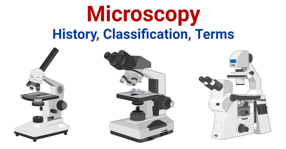
Microscopy can simply be understood as the ‘use of microscope’. Microscopy can be defined as the scientific discipline of using microscopes for getting a magnified view of objects that can’t be viewed by naked eyes.

Parts of microscope
Scanning Probe Microscopy is a microscopy technique that uses a physical probe to scan specimens and form magnified images. This measures the surface features of the specimen.
Optical Microscopy (Light Microscopy) is the microscopy technique that uses transmitted visible light, either natural or artificial, for developing the image of an object. It is the most common type of microscopy. It is further classified into several groups;
Click below to consent to the above or make granular choices. Your choices will be applied to this site only. You can change your settings at any time, including withdrawing your consent, by using the toggles on the Cookie Policy, or by clicking on the manage consent button at the bottom of the screen.
Who invented microscope
It has a low contrasting capacity, low optical resolution, requires staining and has a limited magnification of around 1300X.
Electron Microscopy is a microscopy technique that uses a beam of electrons to develop a highly magnified image of microscopic samples.
Fluorescence Microscopy is a microscopy technique that uses a fluorescent microscope with a UV light source. It is widely used in detecting antigens, antibodies, and other macromolecules.
What is microscopyused for
In Phase Contrast Microscopy, the phase-contrast microscope is used. It converts phase shifts into differences in intensity of light-producing more contrast images.
Dark Field Microscopy uses dark-ground microscopes. The reflected light is used, instead of transmitted light, to form a magnified image.
The third option is a 15mm Rod Holder to 1/4″-20 Adapter (Side Mounted) that can be side mounted to your camera system via a 1/4″-20 screw that use locating pins. This rod holder can be mounted to the side of the cage as well as the top in both parallel and perpendicular configurations in relation to the width of the camera body. The locating pins help limit rotation while the single screw makes this one of our most universal options. The fourth option is a 15mm Rod Holder to 1/4″-20 Adapter (Front Mounted) that can be front mounted to your camera system via a single 1/4″-20 screw that utilizes locating pins. This design helps get the rod closer to your lens which is especially helpful when using smaller, compact lenses with Wireless Follow Focus Motors such as the Nucleus Nano.
Differential Interference Contrast Microscopy is a newer microscopy technique used in unstained and transparent samples to enhance the contrast of their image.
Magnifying Power is defined as the ratio of the angle subtended by the image at the eye to the angle subtended by the object at the eye when placed at a minimum distance of distinct vision.
What is microscopyin science
Confocal Microscopy is a newer microscopy technique that uses a focused laser beam. It is used to get high-resolution 3-D images of biological samples.
Tilta’s annual Black Friday Sale ends December 4th. Take $30 off every $200 in your cart, free gift with $300+ purchase, and much more.
It is a very important tool in biology and nanotechnology. In microbiology, it is one of the most important tools used in observing microbial cells. Medical sectors, pathology, histology, molecular biology, and cytology are in great debt of microscopy.
What is microscopyin microbiology
Refractive Index can be defined as velocity of light in a vacuum to velocity of light in a medium (substance). Simply it is the measure of bending of a light ray when passing from one medium to another.
The first option is a 15mm Rod Holder to NATO Adapter that can be securely mounted to your camera system via one of Tilta’s full or half camera cages that feature NATO rail. Its design helps prevent the rod from rotating and supports accessories such as the Nucleus Nano Wireless Follow Focus Motor. This extremely secure connection helps prevent the rod from rotating and the height can be easily adjusted. The rod holder can also be mounted on the top of some cages that feature NATO rail or have enough threads to support an external NATO attachment.
Tilta has you covered with a range of new 15mm rod holders that are perfect for securely mounting various rod-based accessories to your camera system. With options to mount rods on the side or top of most camera cages, you’re sure to find the perfect solution for your needs. Use these new rod holders to effectively mount accessories to your camera rig, we think that they will be a valuable addition to your camera setup.
What is microscopypdf
As a filmmaker or videographer, having the right tools and equipment is essential to capture the perfect shot. One important aspect of your setup is the camera cage and the way you mount your accessories to it. Tilta has recently introduced several new options for mounting 15 millimeter rods to your camera cage. Expanding upon the existing 15mm rod holders, these new ones take advantage of the expansion points on our more recent camera cages. In the video above & this article, we’ll take a closer look at the features and benefits of each of these rod holders.
Bright Field Microscopy is the simplest and the most common type. A simple or compound light microscope is used in this technique. It uses transmitted visible light to develop magnified images.
What is microscopyin biology
Magnification is the process of producing an enlarged image of a specimen by using a lens system. In a microscope, magnification can be computed by calculating the product of the magnification power of the eyepiece by the magnification power of the objective in use.
To provide the best experiences, we and our partners use technologies like cookies to store and/or access device information. Consenting to these technologies will allow us and our partners to process personal data such as browsing behavior or unique IDs on this site and show (non-) personalized ads. Not consenting or withdrawing consent, may adversely affect certain features and functions.
The second option is a 15mm Rod Holder to Dual 1/4″-20 Adapter that can be securely mounted to your camera system via two 1/4″-20 screws. This design also helps prevent the rod from rotating and supports heavier duty accessories such as the larger Nucleus M Wireless Follow Focus Motor. Like the first option, this rod holder can also be top-mounted on larger cages that have enough threads to support it.
Resolution can be defined as the shortest distance between two points on a specimen that can be distinguished by a microscope in its image. It is the ability of a microscope to distinguish details on a specimen.


 Ms.Cici
 8618319014500