
Mach-Zehnder-Interferometer - mach zehnder interferometer
Types ofobjectivelenses
In our vacuum focused geometry, the Rayleigh range of the initial beam is only 8.5 cm. Therefore, the observed filamentation can only be associated with a true “light bullet", in contrast to the “moving focus" (a macroscopic beam self-focused at a distance decreasing with increasing intensity as observed in solids in the 70's) or nonlinear axicon focusing [A. Dubietis, E. Gaisauskas, G. Tamosauskas, and P. Di Trapani. Light _laments without self-channeling. Phys. Rev. Lett., 92:253903-1—253903-5, (2004)]. Furthermore, it has been shown that the nonlinear reshaping takes place before Fraunhofer diffraction. Beams with super Gaussian profile create for this reason a ring of multiple filaments when focused in air. If focused in vacuum, the focus profile is a Fourier transform of a square profile, thus with a parabolic profile in the middle, which, when launched in air, will create a single filament. New research efforts are aimed at extending the range of filaments by creating nested filaments of different wavelengths, and high repetition rate filaments to establish a stationary waveguide in air. Sponsors: Walgreens Weekly Ad and CVS weekly ad.
Your objective lens isn't just for increasing the size of your subject; it can also provide better resolution. For example, achromatic lenses contain two smaller lenses (convex and concave) that are used to limit the refracting light of your subject, and phase-contrast lenses use phase plates to pick up miniscule changes in wavelength amplitude, making moving subjects easier to observe. Lenses like these help reduce ghost images so that the real image is projected to your eyepiece.
Oil immersionobjectivemicroscopefunction
Figuring out the total magnification power of your microscope is easy: just multiply the power of your objective lens by your ocular lens. For instance, if your eyepiece has 10x magnification and you're using a low-power lens (10x), you have 100x magnification in total. Switch to your scanning lens (4x), and magnification becomes 40x. It's important to keep in mind that the ocular lens and objective lens total magnification is ultimately what you're viewing. If you were viewing your subject through a single lens, then that lens would have to be extremely powerful to match what you can easily get with both. Therefore, one lens isn't nearly as effective without the other.
Stage microscopefunction
The objective and ocular lens are found on different parts of the microscope. The ocular lens is part of the eyepiece and therefore closer to your eye as you look into the microscope. The location of the eyepiece always indicates the correct observing position at or near the top of the microscope.
Often, your microscope will have at least three objective lenses on a rotating disc, each with a different magnification level. If you find your current lens lacking, it's easy to switch to one of the others. Objective lenses with higher magnification have shorter focal lengths, or less space between the lens and the surface of the subject. Since depth of field decreases as magnification increases, those wanting a broader field of view should stick to shorter lenses. For example, if your current objective lens has 100x magnification but you need a wider field of view, you'll want to switch to a lens with lower magnification, such as 40x.
Objectivelens magnification
Oil immersionobjective
We have pioneered the field of filamentation in air, starting in 1995 when we reported the first observation of UV filaments with femtosecond pulses [Xin Miao Zhao, Patrick Rambo, and Jean-Claude Diels. Filamentation of femtosecond UV pulses in air. In QELS, 1995, volume 16, page 178 (QThD2), Baltimore, MA, 1995. Optical Society of America.]. Both UV and IR filaments are created in our laboratory out of a beam waist, prepared in vacuum and launched into the atmosphere through an aerodynamic window. This unique initial condition contrasts with all other studies, where the filaments are let to evolve from a macroscopic beam (several cm in diameter).
There are many other kinds of objective lenses out there, so you have no shortage of options. Do some research and find out which lens best suits your needs and goals.
While it may initially seem redundant to have two separate lenses in your microscope, they do far more together than they ever could on their own.
In contrast, your microscope's eyepiece will usually have only one ocular lens, though you can usually swap the eyepiece as well. The standard magnification level of the ocular lens is 10x, but there are stronger ones available. When selecting an eyepiece, you should think about eye relief, or the required distance between your eyes and the lens. Eyepieces with large eye relief give you some space, while those with small eye relief require you to be up close.
This is why a microscope is such a good investment for anyone interested in science. If you want to understand and examine the world around you, there's no better tool. AmScope's selection is built to last, and we carry all kinds of objective lenses as well, so a microscope from us will serve you well for many years.
Scanningobjectivelens
High power objective functionmicroscope
The microscope is one of the most iconic and commonly used tools in many scientific fields. We rely on these devices to observe things that are so small that they are otherwise invisible to the naked eye. To do this, the microscope makes use of both an ocular and an objective lens. If you don't know the difference, don't worry; this article will tell you everything you need to know about these two lens types and how they function together to make microscopes work.
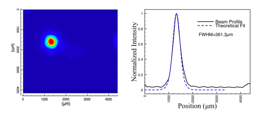
Figure 1. Left: Direct image of filament at a position of 3m away from the geometric focus point. At lower power, the beam is linearly diffracted to a FWHM of around 15mm. At high power (above the threshold for filamentation) the beam collapses into a filament. We measure a FWHM of 360 μm. Right: the solid line is the measured beam profile. The dotted line is a theoretical beam profile based on eigenfunction solution of the modified nonlinear Schrodinger equation.
Mediumpower objectivemicroscopefunction
AmScope exclusive ALL-IN-ONE 3D DIGITAL INSPECTION MICROSCOPE. View different angles and perspectives of objects with ease.
The objective lens, on the other hand, looms over your subject, typically near the middle of the microscope. This is because the objective lens is responsible for gathering light reflections from your subject. It then shoots a beam of light into the microscope, which becomes an image that you observe from the eyepiece containing the ocular lens.
Everyone knows that microscopes are a crucial tool in science, but few realize how versatile and adaptable they can be. Thanks to the variance in lenses, microscopes can serve all kinds of purposes for all kinds of people, from the doctor identifying cancer cells to the child wanting to get a closer look at their favorite bug. Once you know how all of the optical elements work together, like the ocular lens vs objective lens, it's easy to maximize the efficiency of your microscope.
In figure 1, a typical direct image of filament is shown on the left. On the right its intensity profile is compared to a theoretical fit obtained by solving the modified nonlinear Schrodinger equation. The beam is first gently focused inside a vacuum tube to prevent any nonlinear effect, then launched via an aerodynamic window into the air. We observe the filament at 2 to 4 m away from the aerodynamic window. Using a similar technique as for the IR filaments, we directly image the filament using a CCD camera after linear attenuating the high intensity filament by a grazing incident mirror.We are also interested in studying the effect on the photon ionized plasma by adding a 750nm source of 1J in 1μs in order to heat up the electron and sustain the plasma channel. With the combination of UV filament and the 750nm "plasma heater", we expect to be able to trigger a discharge over a longer gap between electrodes, and be a step closer to trigger lightning! Figure 2 shows the experimental setup of high voltage electrodes for studying the discharge. Sponsored links; ALDI Ad, Meijer Ad, Safeway Ad, and Walmart Ad. Reference:1. O. Chalus, A. Sukinin, A. Aceves, and J.-C Diels, “Propagation of non-diffracting intense ultra-violet beams” , Optics Communication 281, 3356-3360 (2008)
We were the first group to experimentally demonstrate filamentation in air in the ultra-violet regime (248 and 266nm), with 3 orders of magnitude more energy (>200mJ) than the near-infrared filament.UV filament is a balance among diffraction, self-focusing, and plasma defocusing during pulse propagation through nonlinear medium (air in our case). UV is interesting because, first, it requires much lower intensity due to a lower critical power for self-focusing, and less intensity to ionize major molecules in air (3-photon process versus 8-photon process for oxygen). Hence it involves nonlinear effects of a lower order, less nonlinear loss, making the modeling simpler. Second, UV filamenting pulses can be much longer, carrying more energy. Because of the shorter optical period of the UV light, the heating (inverse Bremstrahlung) of the electron plasma by the pulse is much less efficient, and it takes a longer time (180 ns at 266 nm) to reach avalanche breakdown. The UV pulses to make filament can be as long as 180ns, without avalanche breakdown of the air, trapping energy as high as 180J. We demonstrated single UV filaments of 0.2ns, 0.2J in air [1]. Simulation of UV filament propagation in air shows that with selected initial condition, single filaments can be sustained for longer than 600m, making it promising for filament-triggered lightning study.
There are four main types of objective lenses, each with a different diameter of field of view, and therefore a different magnification level:


 Ms.Cici
 8618319014500