
Mach-Zehnder Interferometer - mach interferometer
Coarse and Fine Focus knobs are used to focus the microscope. Increasingly, they are coaxial knobs - that is to say they are built on the same axis with the fine focus knob on the outside. Coaxial focus knobs are more convenient since the viewer does not have to grope for a different knob.
Function ofbasein microscope
May 28, 2010 — Magnifying glasses work by using a convex lens to bend and focus light rays, making objects appear larger and clearer. 2. What is the difference ...
TECHSPEC C Series Fixed Focal Length Lenses are optically designed with requirements for factory automation and inspection. ✓ Shop now with Edmund Optics!
Here, we break down the anatomy of an objective lens into easy-to-understand terms and discuss the common parts that make up an objective.
Function ofarmin microscope
Stage Clips are used when there is no mechanical stage. The viewer is required to move the slide manually to view different sections of the specimen.
Quick Reference. 1 A deviation from what is normal, usual, or right. See also chromosomal aberration. 2 A temporary lapse of behaviour or mental function.
Knowledge is power when it comes to objective lenses. Many objective designs require you to make a tradeoff in one area of performance when you improve another. However, advances in objective technology enable our latest optical designs to overcome this common limitation.
Eyepiece Tube holds the eyepieces in place above the objective lens. Binocular microscope heads typically incorporate a diopter adjustment ring that allows for the possible inconsistencies of our eyesight in one or both eyes. The monocular (single eye usage) microscope does not need a diopter. Binocular microscopes also swivel (Interpupillary Adjustment) to allow for different distances between the eyes of different individuals.
Objective Lenses are the primary optical lenses on a microscope. They range from 4x-100x and typically, include, three, four or five on lens on most microscopes. Objectives can be forward or rear-facing.
Rebecca holds a bachelor's degree in journalism from Endicott College and writes about trends and technologies in science and industry. She works closely with Evident engineers and scientists to write pieces about the latest laser scanning, super-resolution, multiphoton, upright, stereo, and inverted microscope systems, as well as leading-edge optics, cameras, and software. Follow her work to learn about Evident's latest for numerous applications, including cytology, pathology, education, and more.
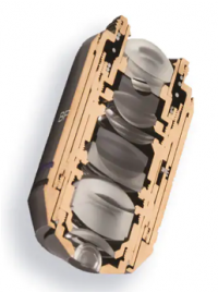
Keep in mind, objectives with more optical corrections for aberrations and flatness typically contain many lenses. For instance, sophisticated plan-apochromatic objectives have about 15 lens elements—while common achromatic objectives contain significantly fewer lenses.
Function ofbody tubein microscope
Aspheric lenses excel in focusing light to a precise point, resulting in minimal blur and enhanced image quality.
Illuminator is the light source for a microscope, typically located in the base of the microscope. Most light microscopes use low voltage, halogen bulbs with continuous variable lighting control located within the base.
Function ofnosepiecein microscope
Explore the universe with precision-crafted optics from William Optics. Discover an unparalleled range of Refractor telescopes, binoculars, digital camera ...
Oct 1, 2024 — The Fresnel lens is used particularly in lighthouses and searchlights to concentrate the light into a relatively narrow beam. It would be almost ...
What iseyepiece in microscope
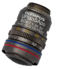
The objective lens is one of the most important parts of a microscope, since it determines its basic performance and function. Yet, these precision pieces of optical equipment are often not well understood.
For instance, Olympus X Line objectives are packed with many ultra-thin convex and concave lenses to offer exceptional flatness, aberration correction, and numerical aperture in one lens system. The result? Bright, high-quality images throughout the field of view.
Microscopeparts and functions
Eyepiece or Ocular is what you look through at the top of the microscope. Typically, standard eyepieces have a magnifying power of 10x. Optional eyepieces of varying powers are available, typically from 5x-30x.
Stage is where the specimen to be viewed is placed. A mechanical stage is used when working at higher magnifications where delicate movements of the specimen slide are required.
In addition to these core components, some objectives include a spring-loaded retractable assembly to protect the front lens elements and specimen from collision damage.
While objective designs vary based on factors like their intended purpose, microscopy method, aberration correction, and manufacturer, all microscope objectives share some similar characteristics. Here are four common components to know:
Nosepiece houses the objectives. The objectives are exposed and are mounted on a rotating turret so that different objectives can be conveniently selected. Standard objectives include 4x, 10x, 40x and 100x although different power objectives are available.
Structure andfunction of eyepiece in microscope

Condenser is used to collect and focus the light from the illuminator on to the specimen. It is located under the stage often in conjunction with an iris diaphragm.
Function ofstagein microscope
JavaScript seems to be disabled in your browser. For the best experience on our site, be sure to turn on Javascript in your browser.
Features · Molded Glass Aspheric Lenses Designed for Infinite Magnification · Focus or Collimate Light Without Introducing Spherical Aberration · Available ...
Eyepiece: It is also referred to as the ocular. This area is used to view objects through the microscope. It is located at the tip of the microscope. Eyepiece ...
The compound microscope has two systems of lenses for greater magnification, 1) the ocular, or eyepiece lens that one looks into and 2) the objective lens, or ...
MTF Services Ltd MTB413B B4 2/3" to 1/3" Bayonet Adaptor · For B4 Mounted Lens on 1/3" Chip Cameras · Offers a Magnification Factor of 1.833x · Compatible with ...
A high power or compound microscope achieves higher levels of magnification than a stereo or low power microscope. It is used to view smaller specimens such as cell structures which cannot be seen at lower levels of magnification. Essentially, a compound microscope consists of structural and optical components. However, within these two basic systems, there are some essential components that every microscopist should know and understand. These key microscope parts are illustrated and explained below.
Iris Diaphragm controls the amount of light reaching the specimen. It is located above the condenser and below the stage. Most high quality microscopes include an Abbe condenser with an iris diaphragm. Combined, they control both the focus and quantity of light applied to the specimen.


 Ms.Cici
 8618319014500