
Light absorption by the plant. - light absorbtion
An important trait of fluorescence is the Stokes shift. It describes the differences in the energy level of an exciting and an emitted photon. The photon emitted by a fluorochrome is of greater wavelength than an exciting photon. This is due to the release of energy to the surroundings after the fluorochrome is excited but before it emits the photon. The resulting shift in wavelength makes it possible to distinguish between the exciting and the emission light. The absorption and emission of energy can be seen as specific characteristics of a molecule species.
Fluorescence and phosphorescence

Fluorescence microscopy
Not all products or services are approved or offered in every market, and approved labelling and instructions may vary between countries. Please contact your local representative for further information.
As a result of technical advancements, one can also find more efficient microscopes like scanning probe microscopes and scanning acoustic microscopes.
Fluorescence in diamonds
Microscope is a tool that produces enlarged images of small objects, allowing the observer to have an exceedingly close view of minute structures in a slide. It is primarily used for examination and analysis. Here, let us learn more about different types of microscopes and also their parts and functions.
The stage is where the specimen to be viewed is placed. A mechanical stage is often used when working on a specimen at a higher magnification. This is when delicate movement of the specimen is required. Stage clips are operated to hold the slide in place. To see different areas of the specimen, the observer must physically move the slide. A separate knob is present to move the slide in the mechanical stage. The aperture is a tiny hole in the stage via which the transmitted light enters the stage.
The primary function of a microscope is to study biological specimens. A microscope solely functions on two concepts – magnification and resolution. Magnification is simply the ability of the microscope to enlarge the image. Whereas the ability to analyse minute details depends on the resolution.
Most fluorescence microscopes are epi-fluorescence microscopes. The illuminator and objective lens are positioned on the same side of the specimen and the light does not pass through the specimen. Besides the excitation and the emission filter, a dichroic mirror is needed for this kind of fluorescence microscope. A dichroic mirror allows light of a certain wavelength to pass through, while light of other wavelengths is reflected. The filters and the dichroic mirror are often plugged in together in a filter cube.
Fluorescence vs phosphorescence
Fluorescence microscopy is widely used and offers great specificity. Various techniques make it possible to address different problems and even to circumvent the diffraction limit that was described by Ernst Abbe.
Different types of filters are used in fluorescence microscopy. Band pass, long pass and short pass filters can be distinguished. Band pass filters transmit a band of wavelengths, whereas light with a greater or smaller wavelength will be blocked. Long pass and short pass filters are edge filters. Long pass filters transmit light of long wavelengths. Light with a wavelength above a certain cutoff value will not be able to pass through. In contrast, short pass filters transmit short wavelengths and block long ones.
Fluorescence spectroscopy
A compound microscope is a high-power microscope that has higher magnification levels than a low-power or dissection microscope. It is used to examine tiny specimens like cell structures that cannot be viewed at lower magnification levels. A compound microscope is made up of both structural and optical components. The 3 basic structural components are – the head, arm and base.
The ocular or eyepiece is what an observer looks through and is present in the upper portion of the microscope. The eyepiecetube clasps the eyepieces which are positioned above the objective lens. The objective lenses are the main optical lenses. They range in various magnifications from 4x to 100x and generally include 3 to 5 lenses on a single microscope. Nosepiece houses the objective lenses.
A fluorescence microscope (upright or inverted) is similar to an ordinary light microscope, except that the illumination is provided by a laser as monochromatic light or a bright and powerful light source like a mercury-vapor or a xenon arc lamp. In addition it contains an excitation filter and an emission filter. The excitation filter transmits only light that is able to excite the specimen with its particular dye. The light emitted by the specimen has to pass through the emission filter before it reaches the detector. The emission filter is only translucent for light with a distinct wavelength, like the light emitted by the specimen.
A simple microscope is a basic light microscope that has only one lens. The condenser part is absent in simple microscopes. They work on natural light and there is less usage of hooks and knobs for adjustments. On the other hand, compound microscopes have 2 adjustment knobs – fine and coarse. Their magnification is also higher than the simple microscope.
Fluorescence in Chemistry
Instead of light, these microscopes use beams of electrons to generate images. The two well-known electron microscopes are:
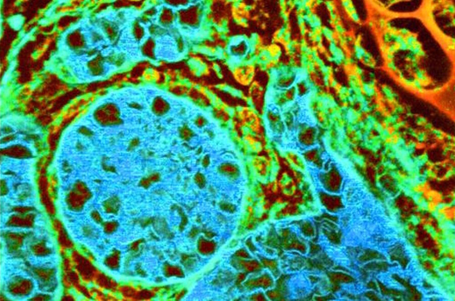
The localization of a molecule species can be determined with a co-staining of organelles, e.g. the cytoskeleton or membranes. Confocal laser scanning microscopy (CLSM) makes it possible to observe areas in the specimen without signals from the outside of the focal plane and allows optical sectioning. Total internal reflection (TIRF) microscopy is a technique that allows the observation of a thin region close to the cell surface. An evanescent field excites the fluorochromes in this area.
In recent years, LEDs (light emitting diodes) have also been used as light source for fluorescence microscopes. The wavelength of the light emitted by LEDs depends on the material used for production. However, an excitation filter is needed in most cases, as LEDs often emit light in a rather broad wavelength range.
The basic objective of a microscope is to magnify small objects. More than magnification, the most important function of a microscope is to provide resolution. It should render high-quality details of the desired specimen in order to proceed with the experiment and analysis. Simple and compound are some of the earliest known microscopes that have been recently replaced by electron and fluorescent microscopes. The different types of microscopes are as follows:
These are few applications associated with each microscope. Keep exploring BYJU’S Biology to learn more such exciting topics.
A technique for observing the dynamics of a molecule species is fluorescence recovery after photobleaching (FRAP). Fluorochromes in a restricted area are photobleached and the diffusion of unbleached molecules into this area can be measured. Interaction studies can be performed with fluorescence energy transfer (FRET) microscopy. An excited donor chromophore can transfer energy to an acceptor fluorochrome and excite it. This is only possible if both are brought together very closely. If these dyes are coupled to different proteins, they will only be able to transfer energy and fluoresce if the proteins interact with each other.
Fluorescence pronunciation
An illuminator acts as the light source and is typically located at the microscope’s base. Most light microscopes operate on halogen bulbs with low voltage and also have variable and continuous lighting control within the base. A condenser is typically used to gather and focus the illuminator’s light onto the specimen. It is found beneath the stage and is often observed in conjunction with a diaphragm or iris. Iris or Diaphragm regulates the amount of light that reaches the specimen. It is situated above the condenser but beneath the stage.
Both simple and compound microscopes can be used for microbiological studies. Specimens like fungi and algae can be viewed under these microscopes. Microscopes can also be used to study soil particles.
Compound and dissection microscopes are the two types of microscopes that are mostly used in schools for educational purposes.
Fluorescence examples
The fine and coarse focus knobs are the adjustment knobs that are often used to focus the microscope. They are coaxial knobs. This means the focusing system of both fine and coarse focus are mounted on the same axis. There is also a condenser focus knob which moves the condenser up or down to control the lighting
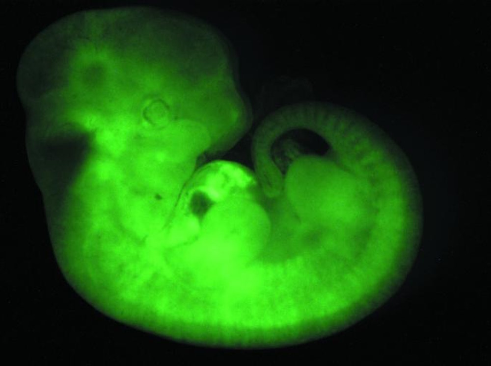
Fluorescence microscopy is a special form of light microscopy. It uses the ability of fluorochromes to emit light after being excited with light of a certain wavelength. Proteins of interest can be marked with such fluorochromes via antibody staining or tagging with fluorescent proteins. It allows the determination of the distribution of a single molecule species, its amount and its localization inside a cell. Furthermore, colocalization and interaction studies can be performed, ion concentrations observed using reversibly binding dyes, e.g. Ca2+ and fura-2, and cellular processes like endocytosis and exocytosis observed. Today it is even possible to image sub-resolution particles with the help of fluorescence microscopy.
The excitation light passes through the excitation filter and is directed to the dichroic mirror. This reflects the light through the objective towards the specimen. Fluorochromes in the specimen are excited and emit photons. This emission light passes back through the objective to the dichroic mirror. The emitted light has an appropriate wavelength and is able to pass. Excitation light that is reflected by the specimen is not able to pass through the dichroic mirror and will be blocked. If excitation light is able to pass through the dichroic mirror it will be blocked when it reaches the emission filter. Light passing through the emission filter can be measured with a detector.
These are basic microscopes that use light to magnify objects. The lenses in these microscopes refract the light for the objects beneath them to appear closer. The different types of light or optical microscopes are:
Techniques that allow sub-resolution images are stimulated emission depletion (STED) microscopy, ground-state depletion (GSD) microscopy, single molecule and ground state depletion microscopy followed by individual molecule return (GSDIM), and photoactivated localization microscopy (PALM) and stochastic optical reconstruction microscopy (STORM). The sub-resolution microscopes used for these techniques are also referred to as nanoscopes, as they resolve images at the nanometer scale.


 Ms.Cici
 8618319014500