
LEXAN ULG1003 Sheet. High Optical Clarity Polycarbonate - optical polycarbonate
Diopter adjustmentfunction
Coarse and Fine Focus knobs are used to focus the microscope. Increasingly, they are coaxial knobs - that is to say they are built on the same axis with the fine focus knob on the outside. Coaxial focus knobs are more convenient since the viewer does not have to grope for a different knob.

In this image a medium depth of field allows the viewer to focus on multiple subjects without creating confusion for your eyes Photo by Sebastian J. Sciotti Jr. In this image a medium depth of field allows the viewer to focus on multiple subjects without creating confusion for your eyes Download Image Share Image: X Facebook Email Photo by: Sebastian J. Sciotti Jr. VIRIN: 170525-D-SS007-019C
Eyepiecelensfunction
Eyepiece or Ocular is what you look through at the top of the microscope. Typically, standard eyepieces have a magnifying power of 10x. Optional eyepieces of varying powers are available, typically from 5x-30x.
What iseyepiecein microscope
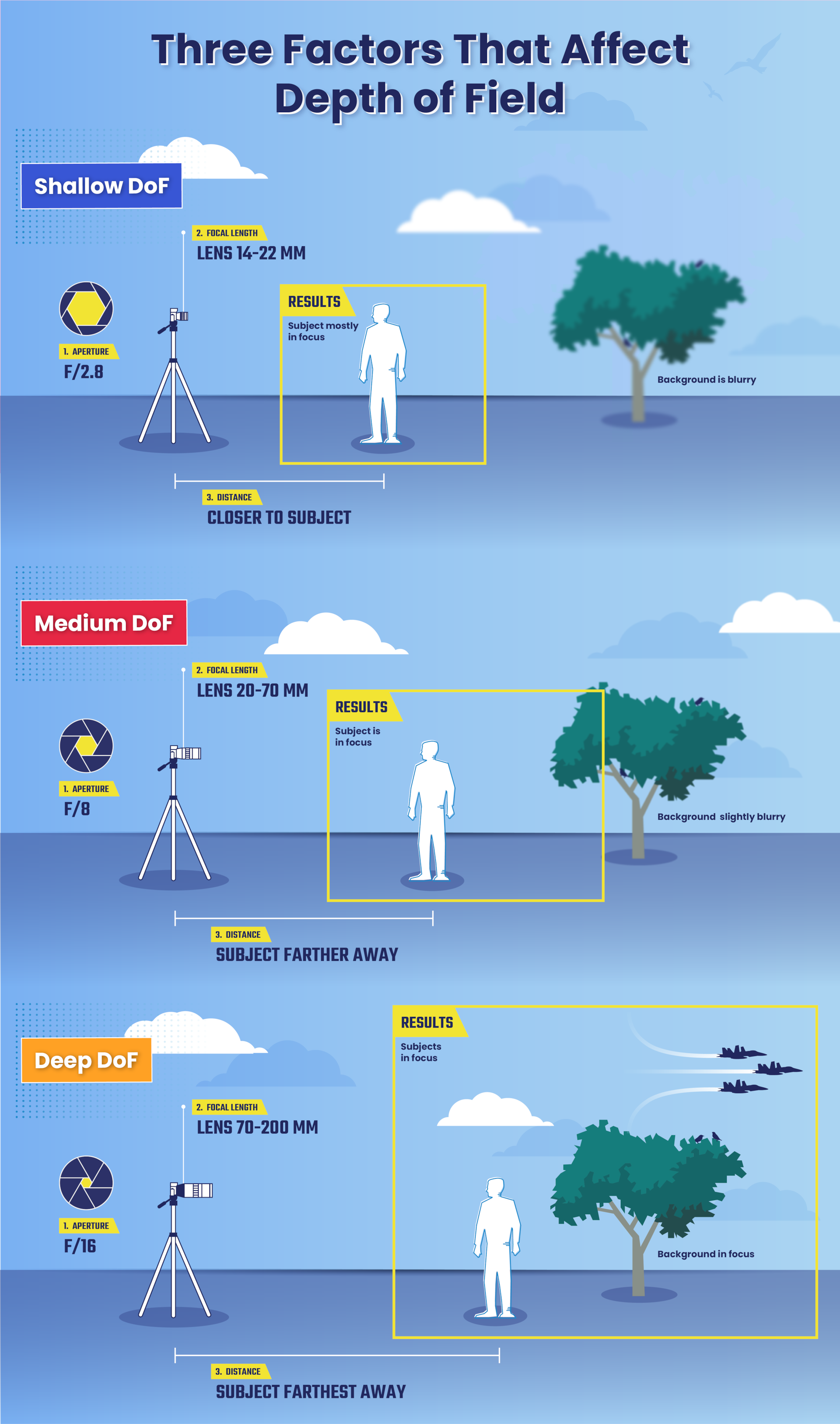
You can affect the depth of field by changing the following factors: aperture, the focal length and the distance from the subject.
Eyepiecediagram
Nosepiece houses the objectives. The objectives are exposed and are mounted on a rotating turret so that different objectives can be conveniently selected. Standard objectives include 4x, 10x, 40x and 100x although different power objectives are available.
Condenser is used to collect and focus the light from the illuminator on to the specimen. It is located under the stage often in conjunction with an iris diaphragm.
Distance to subject refers to the length between the camera and the focus of the image. The closer the camera is to the subject it is focusing on, the narrower the depth of field will be. Inversely, the farther away the subject is from the camera, the wider the depth of field will be.
JavaScript seems to be disabled in your browser. For the best experience on our site, be sure to turn on Javascript in your browser.
Depth of field (DoF) is the area between the nearest and farthest points from the camera that are acceptably sharp in an image. A deep DoF means all or most of your photo will be in focus, including the foreground, subject and background. Use a deep DoF in group photos, landscape shots and when elements in the background or foreground add to the message the photo is attempting to communicate. A shallow DoF means more narrow range will be acceptably sharp in the image. Shallow DoF is good to use when you want to isolate your subject from their surroundings, such as in a portrait or when elements in the background or foreground may be distracting.
Function of eyepiecein microscope
Infographic illustrates how changing the aperture, the focal length and the distance from the subject affect the depth of field. Download Image Share Image: X Facebook Email Photo by: DINFOS PAVILION Team VIRIN: 200907-D-PA656-0002
Arm microscopefunction
Objective Lenses are the primary optical lenses on a microscope. They range from 4x-100x and typically, include, three, four or five on lens on most microscopes. Objectives can be forward or rear-facing.
In this image a deep depth of field allows the viewer to take in many subjects, including an artillery shell mid-flight. Photo by Staff Sgt. Steven Schneider In this image a deep depth of field allows the viewer to take in many subjects, including an artillery shell mid-flight. Download Image Share Image: X Facebook Email Photo by: Staff Sgt. Steven Schneider VIRIN: 170918-O-N0132-7230C
Function ofbody tube in microscope
Stage Clips are used when there is no mechanical stage. The viewer is required to move the slide manually to view different sections of the specimen.
Describe thefunction ofthe mirror
Iris Diaphragm controls the amount of light reaching the specimen. It is located above the condenser and below the stage. Most high quality microscopes include an Abbe condenser with an iris diaphragm. Combined, they control both the focus and quantity of light applied to the specimen.

Stage is where the specimen to be viewed is placed. A mechanical stage is used when working at higher magnifications where delicate movements of the specimen slide are required.
The following graphic illustrates how changing these factors: aperture, focal length and the distance from the subject affect the depth of field.
In this image you can see how a shallow depth of field keeps the focus on the action. Photo by Samuel King In this image you can see how a shallow depth of field keeps the focus on the action. Download Image Share Image: X Facebook Email Photo by: Samuel King VIRIN: 170908-F-OC707-0517C
The focal length of the lens determines the image magnification. The wider the lens, the shorter the focal length. This allows you to capture a wider depth of field. The longer or more zoomed in the camera lens, the less depth of field you capture.
Eyepiece Tube holds the eyepieces in place above the objective lens. Binocular microscope heads typically incorporate a diopter adjustment ring that allows for the possible inconsistencies of our eyesight in one or both eyes. The monocular (single eye usage) microscope does not need a diopter. Binocular microscopes also swivel (Interpupillary Adjustment) to allow for different distances between the eyes of different individuals.
The aperture is the opening created by a set of overlapping metal blades, known as the diaphragm, inside a photographic lens. This opening controls the amount of light coming through the lens. The wider the aperture, the less depth of field you capture. The smaller the aperture, the deeper the depth of field.
A high power or compound microscope achieves higher levels of magnification than a stereo or low power microscope. It is used to view smaller specimens such as cell structures which cannot be seen at lower levels of magnification. Essentially, a compound microscope consists of structural and optical components. However, within these two basic systems, there are some essential components that every microscopist should know and understand. These key microscope parts are illustrated and explained below.
Illuminator is the light source for a microscope, typically located in the base of the microscope. Most light microscopes use low voltage, halogen bulbs with continuous variable lighting control located within the base.


 Ms.Cici
 8618319014500