
Lenticular lenses | PPT | Free Download - lenticular lenses glasses
CollimatedLED
Overall, the eyepiece on a microscope plays a crucial role in magnifying and enhancing the image of the specimen, as well as providing a comfortable and effective viewing experience for the user.
Eyepiece design and construction have evolved over time to improve the quality and comfort of the viewing experience. Modern eyepieces are typically designed with multiple lens elements to minimize aberrations and distortions, resulting in a clearer and more accurate image. Some eyepieces also incorporate advanced coatings to reduce glare and improve contrast.
The eyepiece on a microscope, also known as the ocular lens, is the lens at the top of the microscope through which the viewer looks. It is the lens closest to the eye when using the microscope. The primary function of the eyepiece is to magnify the image produced by the objective lens, which is the lens closest to the object being observed. This magnification allows the viewer to see a larger and more detailed image of the specimen.
TPLbacklight
For more information on the CCS America MFU Series Collimated Backlight, please contact R.J. Wilson, Inc. at 781-335-5500 or [email protected].
Shandong Yuying provides the large size solar concentrated Fresnel lens ,including CPV lens which is made of SOG,Linear lens and Fresnel lens.
Their spectrometers are known for their advanced technology and exceptional performance. One of the popular spectrometers offered by AVANTES is the AvaSpec 3648 ...
In addition to magnification, the eyepiece also helps to focus the light rays coming from the objective lens and to direct them into the viewer's eye. This helps to create a clear and sharp image of the specimen under observation. The eyepiece also often contains a reticle or a graticule, which is a grid or scale that can be used to measure the size or dimensions of the specimen.
Looking for allen hex keys? BC Fasteners has metric an standard sizes available.
Jan 17, 2022 — Retroreflectors in vehicle lighting often use advanced materials such as glass or special plastics. These materials are developed to reflect ...
An eyepiece on a microscope is a lens that is positioned at the top of the microscope and is used to view the magnified image of the specimen. It is also known as an ocular lens and is an essential component of the microscope's optical system. The eyepiece typically contains a set of lenses that further magnify the image produced by the objective lens, allowing the viewer to see a highly detailed and enlarged image of the specimen.
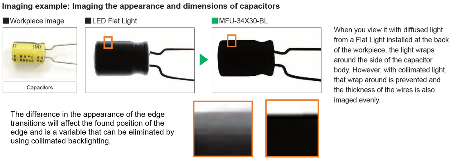
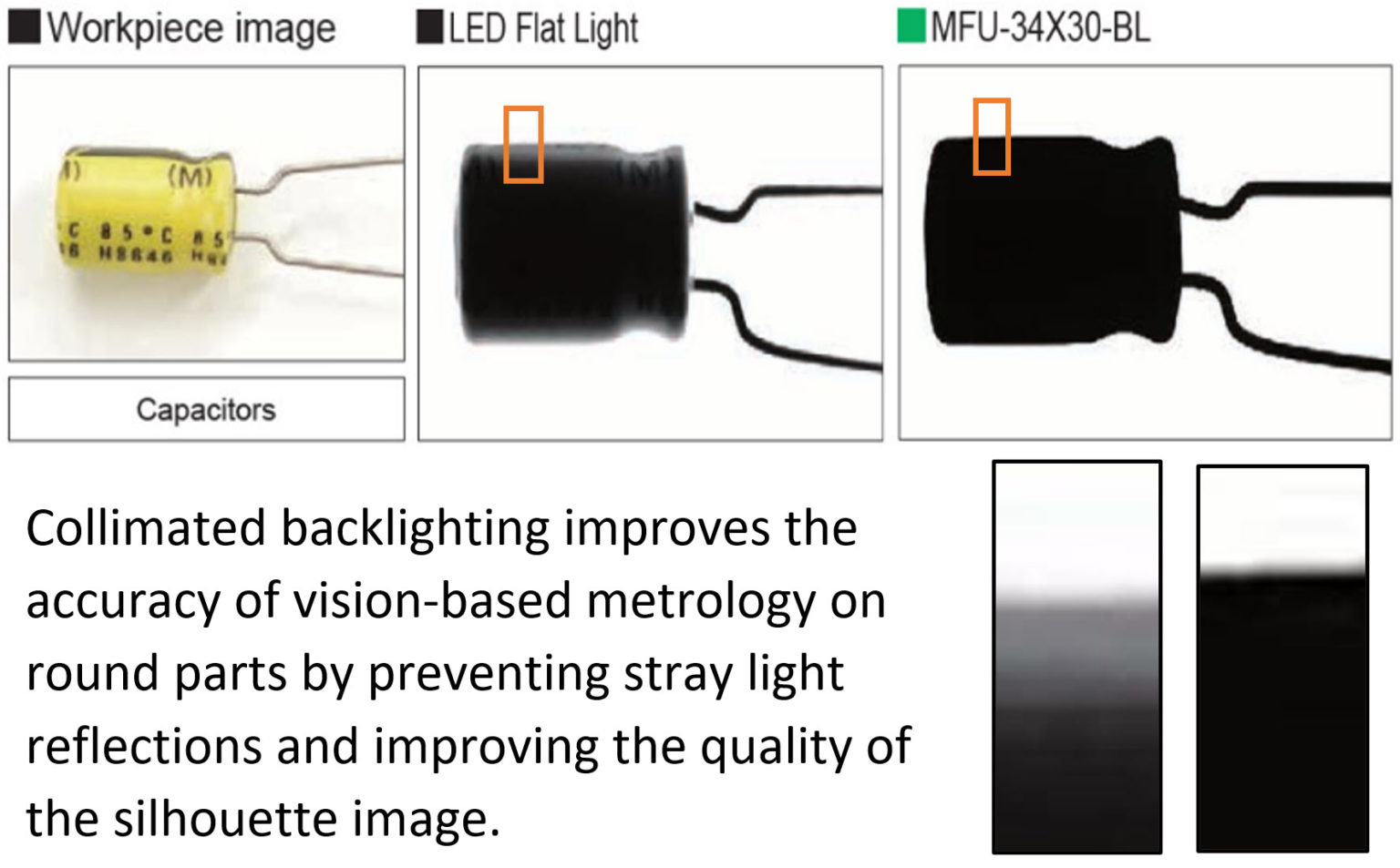
The more consistent the appearance of an edge is to the vision software, the more reliable and accurate its measurements will typically be.
The magnification power of the eyepiece is a measure of how much the image is enlarged when viewed through the microscope. This is usually expressed as a number followed by an "x" (e.g., 10x, 20x), which indicates the number of times the image is magnified. For example, if the eyepiece has a magnification power of 10x and the objective lens has a magnification power of 40x, the total magnification of the microscope would be 400x (10x multiplied by 40x).
The Fresnel lighthouse lens used a large lamp at the focal plane as its light source. It also contained a central panel of magnifying glasses surrounded above ...
In summary, the eyepiece on a microscope is a crucial component that contributes to the overall quality of the viewing experience. Its design and construction have evolved to prioritize optical performance, user comfort, and versatility, making it an essential part of modern microscopy.
Collimated backlighting will enhance the performance of vision-based metrology systems when inspecting round parts because the light won’t “wrap around” rounded or curved edges, presenting a higher-quality silhouette image of the feature to the edge detection function of the vision software. The silhouette quality is improved whether a standard or telecentric lens is being used on the camera.
The eyepiece, also known as the ocular lens, is the lens at the top of the microscope that you look through to view the specimen. It typically contains a magnifying lens that further enlarges the image produced by the objective lens. The eyepiece is usually removable and interchangeable, allowing for different magnifications to be achieved depending on the specific needs of the user.
An eyepiece on a microscope, also known as an ocular lens, is the lens at the top of the microscope that you look through to view the specimen. It is the part of the microscope that is closest to your eye and is responsible for magnifying the image of the specimen. The eyepiece typically contains a set of lenses that work together to magnify the image produced by the objective lens, which is the lens closest to the specimen.
From the latest point of view, advancements in microscope technology have led to the development of eyepieces with variable magnification power, allowing users to adjust the level of magnification based on their specific needs. Additionally, some modern microscopes are equipped with digital eyepieces that can capture and display images on a computer screen, enabling users to easily share and analyze the microscopic images. These digital eyepieces often come with software that allows for further image enhancement and analysis, expanding the capabilities of traditional eyepieces.
3. Wide-field eyepiece: This type of eyepiece is designed to provide a larger and more comfortable viewing area, allowing the viewer to see more of the specimen at once. It is particularly useful for applications that require prolonged observation.
We provide selected LED and fiber optic lighting, lenses & filters, cameras, sensors, and accessories from the top partners in the industry. Call us at 781-335-5500 to learn more.
From a modern perspective, the eyepiece on a microscope may also be designed to reduce eye strain and provide a comfortable viewing experience. Some eyepieces are equipped with adjustable diopter settings to accommodate individual differences in vision, and others may incorporate anti-glare or anti-reflection coatings to improve image clarity.
Metaphasebacklight
Clarity refers to the ability of the lens to provide a clear and sharp image. A high-quality lens with excellent clarity ensures that the magnified image is ...
1. Huygenian eyepiece: This is a simple eyepiece design that consists of two plano-convex lenses with the convex sides facing each other. It provides a relatively narrow field of view and is commonly used in older microscopes.
With a highly collimated light, the light rays are nearly perfectly parallel and provide a high-contrast silhouette image that supports improved metrology. Coupled with a telecentric lens, using highly collimated backlighting will provide the best possible vision-based metrology performance on rounded features and edges.
Machine visionBacklight
For example, an Edge tool’s consistent performance and its ability to detect the correct edge position may be affected by variations in the appearance of an edge, by the parameters set in the vision tool itself, by variations in the part’s surface finish that may reflect more or less light, and by changes in the radius of the measured part that affects how wide the greyscale transition is between the foreground and the background.
The latest point of view on eyepiece design emphasizes the importance of ergonomic design to reduce eye strain and improve user comfort during extended periods of use. This includes features such as adjustable eye relief and eyecups to accommodate different users and provide a more comfortable viewing experience. Additionally, advancements in materials and manufacturing techniques have allowed for the production of lightweight yet durable eyepieces that are well-suited for various applications.
The eyepiece on a microscope, also known as an ocular lens, is the part of the microscope that is looked through to view the magnified specimen. It is located at the top of the microscope and is the lens closest to the eye of the observer. The eyepiece is designed to magnify the image produced by the objective lens, which is the lens closest to the specimen being observed.
Verschiebungen im Markt und disruptive Veränderungen des Technologieeinsatzes kündigen sich an · Machine Vision als Basis und künstliche Intelligenz befördert ...
An IR lens captures infrared light that is virtually impossible to see with the naked eye. While a normal camera lens captures images of objects that radiate ...
An application video on lens scratch detection follows and illustrates the performance difference, in this application, between standard diffused backlights and collimated backlights.
Collimated backlightvs led
Collimation film is often used on standard backlights to make them more directional. This film behaves the same as privacy film used on laptop screens, where you can see your screen, but your neighbor can’t. The collimating film is providing about +/- 30° of directional control, or collimation, of the light rays. This can help minimize light diffusion on rounded edges in the appropriate applications, but collimating film will not provide highly collimated light, such as that provided by a telecentric backlight or the MFU described above.
In recent years, there has been a growing interest in digital eyepieces, which incorporate digital imaging technology to capture and display the magnified image on a computer or other digital device. This allows for easier sharing of images and facilitates analysis and documentation of the specimens. Additionally, there has been a focus on ergonomic designs to improve user comfort and reduce eye strain during prolonged use. These advancements aim to enhance the overall microscopy experience and make it more accessible to a wider range of users.
Laser cutting is a technology that uses a laser to vaporize materials, resulting in a cut edge. While typically used for industrial manufacturing ...
When using a standard backlight, the light rays exit the diffuse surface at all random angles, which can direct light to the sides of rounded features. Because this light is visible in the acquired image, the acquired image may contain multiple grey values within the vision tool’s region-of-interest.
Jan 6, 2019 — Regular gloves have lots of lint so it is better to use surgical gloves, or the types that apraisers use to handle delicate items on antiques ...
2. Ramsden eyepiece: This design features two plano-convex lenses with the convex sides facing away from each other. It offers a wider field of view compared to the Huygenian eyepiece and is commonly used in modern microscopes.


 Ms.Cici
 8618319014500