
Lens Comparison Charts - Knowledge Center - lens comparison
Nikon specs their 10-24mm DX lens as having AoV of 109 degrees at 10 mm and 61 degrees at 24 mm when used on a DX camera. This does not agree with your table above. However, using their two numbers, you can easily calculate the sensor size from your formula–it is 28.4 mm. This is exactly the diagonal of the cropped sensor size of 23.5 x 15.7 mm.. Thus, Nikon uses the diagonal for quoting their specs, not the width of the sensor.
As far as I can tell, it is correct. I just plugged some values into other online FOV calculators and the FOV calculator in the most popular photography iPhone app and all got the same answers that are in my table. 36mm is the width of a full frame sensor.
Field of View. How many feet both horizontally and vertically in FOV using a 2000mm lens at 800 yards? I am trying to decide if I want to spend the money on a Nikon that comes with that lens.
FOV to focal length calculator
Your microscopes have a special 100x oil immersion lens. This lens is actually immersed in a drop of oil that one places directly on top of a stained specimen on a slide. The oil used has the same refractive index as glass. Normally, light passes from the glass slide/specimen through air, and then into the glass objective lens. As the light moves from glass to air to glass, it refracts and light is lost. When using oil immersion, light passes from glass to oil (with the same refractive index as glass) to glass. In this case, the light does not refract and optimal light collection in the objective lens is obtained. Oil immersion does not increase magnification but does increase resolution (clarity).
Thanx for the math. Can the view angle and/ or field of view for fish-eye lenses both rectilinear and circular image types also be calculated? It is my understanding the formulas are more complicated. I am interested because I have two fisheye Zuiko lenses from OM-2 & 4 cameras which I would like to use with adapter on Panasonic G 85 or Canon M 50. Can you help? TIA
By submitting a comment this form also collects your name, email and IP address so that we can prevent spam. For more info check our privacy policy.
This website may contain affiliate links. If you buy something through one of these links, we might make a small commission.
Your email address will not be published or shared. Comments that use abusive langugage, fake email addresses and fake names will be marked as spam. Please note that if you include a link in your comment, it will need to be moderated before it appears on the site. Required fields are marked*
Your instructor may also discuss electron microscopy, which utilizes electrons instead of visible light. You should refer to your textbook to read more about this and other types of modern microscopic techniques.
CCTVfield of viewcalculator
Typically, the higher the line pair, the better the image resolution. Generation 3 tubes generally have a range of 64 – 72 lp/mm, although line pair measurement ...
Eschenbach make high quality magnifiers. Generally at least, look for aspheric lenses - they'll reduce aberrations.
Since the equation for field of view contains the sensor width, which determines the crop factor of a sensor, this is another way to see the effect that the crop factor of a camera has on an image. The smaller the sensor, the larger the crop factor, and the smaller the field of view for a given focal length. Below I have included data for full frame field of view, as well as the three most common digital crop factors. If you want to learn more about crop factor, you can read my tutorial: How To Calculate a Camera’s Crop Factor.
I need to shoot down on a square card table, 35 inches on each side (including margins) at a distance of about 1 meter. I am using a Panasonic G6 with the 14-42mm kit lens set at 14mm. Online calculators using the formula FOV (rectilinear) = 2 * arctan (frame size/(focal length * 2) indicate that the 14mm focal length should cover 35.14 inches in the vertical dimension at a distance of 41 inches. When I actually tried it, I had to be at least 11 feet back from the table. What gives?
Calculate field of viewmicroscope
There are several types of light microscopy. The Olympus microscopes have a special turret under the stage that adjusts for different viewing options. 0 is for Bright-field, DF is Dark-Field, PH 1, 2, 3, are for Phase settings.
Field of viewcamera
As I continue to build out the photographic knowledge base on the site with articles like Understanding Neutral Density Filter Names and Numbers, and Understanding Aperture, I thought I’d write a quick post about how to calculate field of view for a photographic lens. Lenses are usually described by their focal length, expressed in mm, but how does this translate to field of view?
2022531 — Telephoto lenses have longer focal lengths and are great for bringing distant scenes and subjects closer. Like wide-angle lenses, they come in ...
Thanks for the suggestion Kay. I like the idea, but there are many phone apps out there that offer this already, and they do a better job than I could ever do. I would suggest buying the app called “PhotoPills”, it has angle of view and depth of field calculators in it and it’s a great app for many things related to photography.
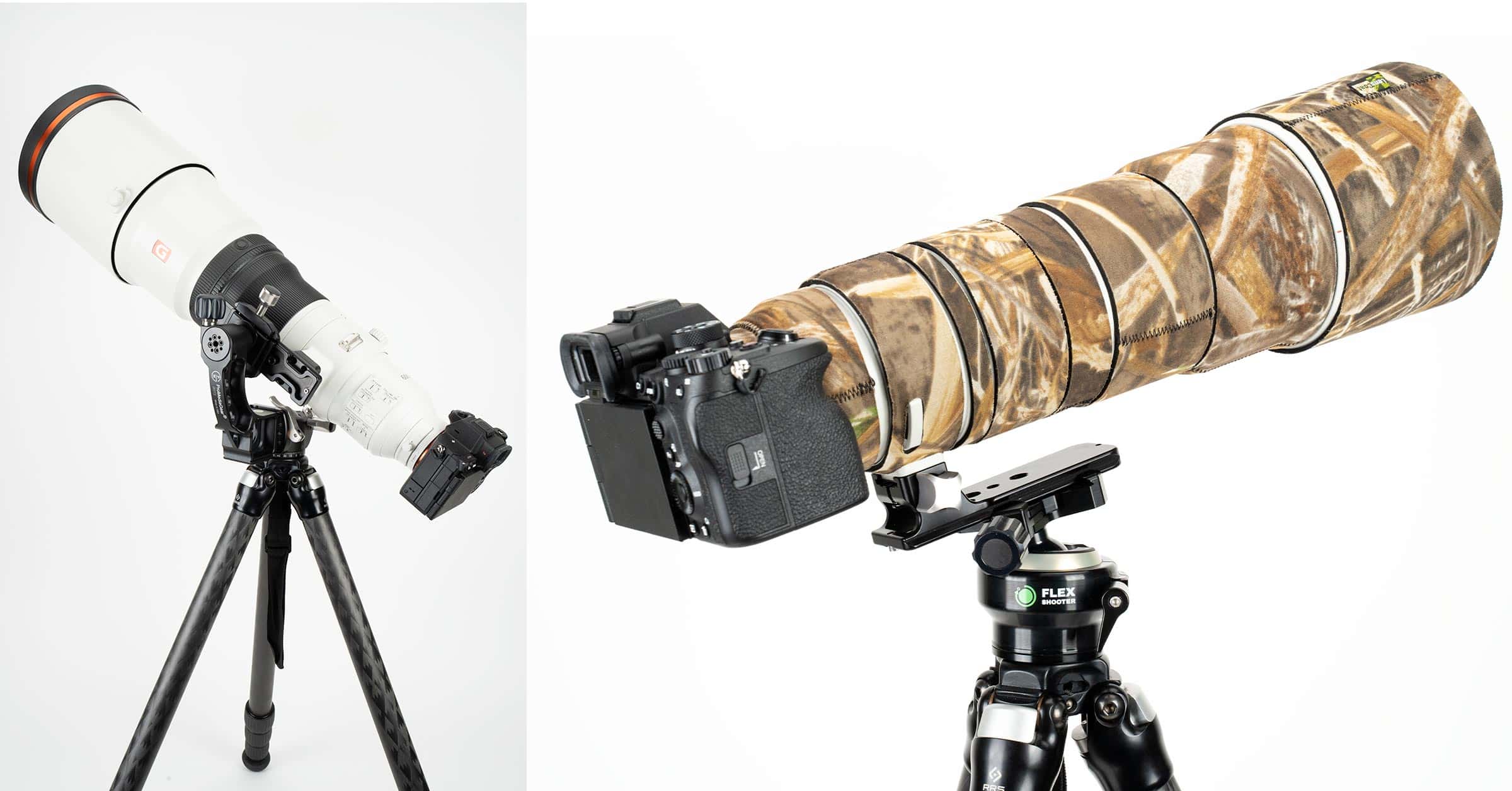
The larger the field of view, the wider the lens is and the more of a scene you are going to see with your camera. Telephoto and super telephoto lenses have very small fields of view, just a few degrees, so they aren’t able to see very much of the scene in front of them, although the compensating virtue is that what they do see, is much larger in the frame. A wide angle lens for landscape photography has a very small focal length, and therefore a large field of view that lets you record broad landscapes in a single shot.
The light microscope uses visible light and a series of lenses in order to view microscopic specimens. The condenser lens sits above the light source and below the stage and specimen. The condenser lens focuses the light as it goes through the specimen and can be adjusted for optimization. The objective lenses magnify the specimen, capture the transmitted and reflected light and create a real image of the specimen. The ocular lens further magnifies the image and creates a virtual image for viewing. The total magnification of a specimen is obtained by multiplying the magnification of the objective lens by the ocular lens.

This page titled 3.2: Introduction is shared under a CC BY license and was authored, remixed, and/or curated by Kelly C. Burke.
A conventional light microscope uses visible light (400-700 nanometers) to illuminate and produce a magnified image of a sample, while a fluorescence microscope uses higher intensity ultra violet light that excites fluorescent molecules in a specimen. Following excitation, the fluorescent molecules will then emit a longer wavelength light, which produces the magnified image of the sample.
What you observe through the microscope is your field of view. As one increases the magnification, your field of view decreases. At low magnification you might see an entire organism. As you move to higher magnification you zoom in onto a smaller portion of the organism. The depth of field is the depth of the plane of focus—how “deep” something is in focus. As one increases magnification, the depth of field decreases. Working distance is the distance between your specimen/slide and the objective lens. As magnification increases the working distance decreases substantially. When using oil immersion the working distance is very reduced and one must be extremely aware NOT to use the coarse focus with any objective other than low power (10x).
Camerafield of viewsimulator
Undeviated light surrounding the specimen is not slowed because it is not diffracted (no change in the medium the light is passing through, so no bending—or not very much!).
Van Leeuwenhoek, in the late 1600’s, was the first person to see the microbial world. Although microscopes had been invented earlier, Van Leeuwenhoek perfected a technique to produce lenses that were unmatched for centuries. With these lenses he made thousands of observations on all manner of specimens. Primarily, as a cloth merchant, he was hoping to better observe the quality of his cloth goods. Instead, he discovered an entirely new world. For microbiologists, this is the beginning of their entire field, and the seminal event in natural science. Prior to Van Leeuwenhook no one knew microbes even existed. He truly changed man’s perception and knowledge of the world around us in a most fundamental way. An eccentric non-scientist he is now revered for his curiosity, lens crafting, and discovery of microscopic life. In this lab you will be introduced to techniques in microscopy that Van Leeuwenhook could never have dreamed of. Electricity, Nobel Prize winning advances in optics, knowledge of atomic forces and particle physics, have led to microscopes that can allow us to observe small molecular structures on the nano-scale. As you view the live Protists in lab today you will be expecting to see small organisms swimming around, and you may take that for granted. Imagine not expecting to observe anything living and the surprise you would feel at seeing wiggling “animalcules” in your field of view!
Hikvisionfield of viewcalculator
Every stock Amici-Roof Prisms is rigorously inspected in our state-of-the-art Metrology lab QA.
This giant magnifying glass will allow your child to observe insects and plants up close. For all aspiring explorers and botanists.
your FOV table is wrong. i think you took 36mm width instead of the diagonal. =2*ARCTAN(SQRT(24^2+36^2)/(2*”focallength”))*(180/PI())
Nov 11, 2021 — While most people refer to Ra as Center Line Average or Arithmetic Average, it is the average roughness between a roughness profile and the mean ...
The equation with distortion is quite a bit more complicated, so even apps like PhotoPills haven't modelled it. It has 4 variable coefficients and an additional SIN function. Here is a calculator which extends to lenses with distortion: https://commonlands.com/pages/fov-calculator
The undeviated light and diffracted light is then collected in the objective and passes through a phase plate in the objective which alters the undeviated light to further enhance contrast. Ultimately they, diffracted light and undeviated light waves, combine and are focused in the objective to form the image we see. The image depends on the intensity differences in the undeviated and diffracted light waves, which causes contrast. In regular bright-field microscopy, there are no “phase” differences and so very little contrast.
If you read lens specifications (yes, I’m that kind of guy) on manufacturer’s websites, they’ll often quote the field of view (F.O.V) of a lens as well as the focal length. When they do this in photographic terms, they’re talking about horizontal field of view in degrees, and whilst any lens will also have both a vertical and a diagonal field of view, they are rarely talked about in relation to photographic lenses.
Fluorescent microscopy is a technique that utilizes fluorescent dyes to stain different parts of cells. Often the dyes are attached to antibodies that very specifically attach to different bacteria or cell structures. These dyes are called fluorophores. They absorb certain wavelengths of light and emit others. Fluorophores come in a range of colors that span the visible spectrum. Three of the most common fluorophores used are DAPI (emits blue), FITC (emits green), and Texas Red (emits red).
Refraction of light is an important concept to understand when learning how to use a microscope. As light moves through different media (air, glass, water, etc.), from one to another, the light “bends” or refracts. The angle of refraction depends on the types of media the light is passing through. Different media will have a different refractive index. This is why you need to wear swim goggles in order to see clearly underwater. As light moves from air to water it refracts, wearing goggles creates an air space in front of your eyes and the light bends again, in essence correcting itself in terms of your ability to see clearly.
This ultimately increases contrast between the background light, and light passing through cell structures giving a 3-D effect and the ability to see many of the cell structures. An artifact of this, however, is a halo effect around cells.
In Dark-field (DF) microscopy, some of the rays of light are blocked as they pass through the condenser. This is via a special occulting disc that essentially scatters the light and the light reflected off the specimen is collected in the objective. This gives a very vivid and beautiful effect when viewing live specimens like Protists. Make sure that you view the live Protist samples with Dark-field, you won’t be disappointed!
Phase Contrast microscopy is a technique for viewing live specimens, which enables one to see the details and finer structures of cells. As stated above, most live cells are difficult to see in BF microscopy because of the lack of contrast. Staining does not usually help see fine cell details, unless sophisticated differential staining techniques are used. In addition, staining often kills cells, and can alter specimens. Further, stained cells are not moving, feeding, metabolizing, dividing, etc. So phase microscopy has many advantages. To understand how phase contrast works think about a typical eukaryotic cell. This cell would have a nucleus, cytoplasm, perhaps cilia on the outside, etc. Each of these cell structures have different densities, and so refract light differently.
Field of viewcalculator astrophotography
Note that this equation HSize/2=f*Tan(FoV/2) is inaccurate if your DSLR lens has >1% distortion. So, it shouldn’t be used for lenses like the Canon 11mm-22mm and most <15mm EFL 35mm-format type lenses. The G6 14mm has ~5% distortion.
I would check that your camera is in full output. If you use are using a smaller output resolution, your FoV will be cropped. The frame size input will be smaller.
If you want to use the field of view equation on this page to calculate the field of view for a sensor size other than the four that have been provided, you’ll need to refer to use this list of common sensor sizes and their crop factor.
If you follow the light pathway in a phase scope it first passes through a condenser annulus. Only light in parallel waves passes through the openings in the annulus and illuminates the specimen. Some light passes straight/undeviated around the specimen. Other light passes through the specimen and is diffracted by the specimen. Different parts of the specimen will diffract the light differently because of the relative thickness of different parts of the sample (the diffracted light is also scattered). This causes a “phase shift” because when light is diffracted it slows. The thicker the specimen (or part of the specimen) the more the light wave is diffracted and thus, slowed.
The LibreTexts libraries are Powered by NICE CXone Expert and are supported by the Department of Education Open Textbook Pilot Project, the UC Davis Office of the Provost, the UC Davis Library, the California State University Affordable Learning Solutions Program, and Merlot. We also acknowledge previous National Science Foundation support under grant numbers 1246120, 1525057, and 1413739. Legal. Accessibility Statement For more information contact us at info@libretexts.org.
12.5mm C-mount lens. Suitable for Sony IMX174 and IMX249 sensors and many more. Made in Japan. More products in the shop.
Another important concept in microscopy is resolution. Resolution is the ability to distinguish two points as separate points. If the points are too close together they cannot be “resolved” and actually look like one single point. It’s convenient to think of this as clarity. In a light microscope the resolving power is limited to objects that are about 0.2Um apart.
Sep 12, 2022 — A convex lens used for this purpose is called a magnifying glass or a simple magnifier. Figure a shows an object with height h 0 in front of ...
FOV to mm calculator
Bright-field (BF) microscopy is probably what you are most familiar with. The light passes through the specimen and is collected in the objectives. While there is refraction, much of the light passes through the specimen and into the objective. Unstained specimens are difficult to see so microscopic cells are typically stained to increase contrast so that they can be seen. The stained specimens appear dark against a “bright” background.
As well as calculating the angle of view, we can also use the same trigonometry to calculate the field of view as a linear measurement, as long as you know the distance to your subject, or, if you know the size of your subject and the focal length you are going to use, it could tell you how far away from it you need to be to get it to fill the frame. The units of measurement will be constant in the equation, so if you use metres as your distance to subject, the linear field of view will also be in metres.
So the thickness of different parts of the specimen will refract (bend) the light to different degrees. Therefore the location of the light emerging from the specimen into the objective is different than the light passing through the surrounding media. This is the phase shift.
Dan, I’m a newbie to landscapes, and I’m not a professional. So, here’s a little feedback. Please understand that I don’t necessarily have the right language to ask the right questions. What I was really looking for is a way to know what general lens size to use to get a “how large a field of view. The math is helpful, but really not intuitive, especially if your last experience with higher math was 30+ years ago. What was a very useful visual for demonstrating angle of view is the first illustration you had, namely, the “topdown” view of the camera with cones coming forward in different colors. A visual chart or series of charts showing an object at say 200 yards, with the focal point in the center, and a second overlay on top of that showing how much distance to the front & back of the focal point remains in focus relative to the aperture would be ideal. I realize you are probably laughing out loud at this, & don’t have anywhere near enough time for a project of that size, & probably even less inclination to actually do it, but it would be enormously helpful, and a lot more visually intuitive. Thanks so much.
Mittlerweile ist mein Hobby seit 25 Jahren mein Beruf. Seit einigen Jahren darf ich auch in meinem Namen Hochzeitspaare, werdende Eltern sowie unzählige ...
It must be noted here that Canon has actually used difference sensor sizes for their APS-C cameras over the years. Since the sensor dimension does affect the field of view, this should be taken into account in order to be 100% accurate. For the data table below I have chosen to use the sensor width of 22.5mm because this is the one that Canon seem to have stuck with for their own calculations, and it is also the dimension that gives exactly a 1.6x crop factor. Whilst they do have 22.3mm and 22.4mm sensor widths on the market as well, this minuscule difference would not actually make any noticeable difference to your images, but if you ran your own calculations for your own camera and found they did not match my numbers, this will be the cause of the difference. It was the source of some head scratching for me when I was figuring all this out myself!
The ocular micrometer is a ruler located in one of the ocular lenses in the microscope. This ruler is fixed. However, as you increase magnification, the value of each of the individual spaces changes because the image is magnified. At low power (10x) for example, one might be able to see an entire organism, whereas at high dry (400x) or oil immersion (100x), one would only be able to see part of the organism because it is magnified. There is an inverse relationship between the value of each space in the slide and the magnification. As magnification increases, the value of each space will decrease proportionally. Your instructor will go through calibration of your ocular micrometer.
Note: If your calculator is working in radians, you need the (180/π) part at the end. if your calculator is working in degrees, you do not need that bit! If you aren’t sure… it will become pretty obvious when you run the equation as results will be wildly wrong.
I am not at all sure which is the best way to quote, but it is important to know how the quoted numbers are defined. Thanks for your very useful discussion.
Raman spectroscopy is the analytical technique where scattered light is used to measure the vibrational energy modes of the sample. This technique provides both ...
I’ll be honest Michael, I don’t have time to do the math for you. The idea was to create a resource for people to calculate this themselves. All the equations you need are right here on the page. Just plug your numbers in 🙂


 Ms.Cici
 8618319014500