
Lens Cleaning - lens cleaning solution
Contrast resolutionin MRI
The interactions between the atoms of a material can be very sensitive to the external physical and chemical environment as well. Understanding the influence of any environmental parameter on the Raman spectrum, makes a Raman measurement an effective sensing tool for this parameter.
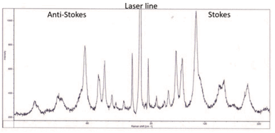
Figure 1: A typical Raman spectrum showing a spectral lie of the laser in the center as well as the Stokes and Anti-Stokes Raman lines.
Raman scattering is named after Chandrasekhara Venkata Raman and was discovered in 1928 by Raman and Kariamanikkam Srinivasa Krishnan and in parallel by Grigory Landsberg and Leonid Mandelstam. The actual breakthrough for Raman spectroscopy as an analytical technique started with the invention of the laser in the 1960s.
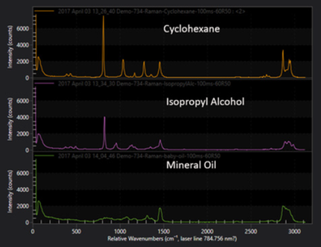
Figure 2: Sshows three Raman spectra from different materials. In thiscase the differences between the spectra are clearly visible and can be "used toidentify each of these materials. It can also be seen that all 3 spectracontain spectral lines at similar position since cyclohexane, isopropylalcohol and mineral oil are or contain organic molecules that contain similarstructural features such as C-C, C=C, C-O molecular bonds vibrating atcharacteristic frequencies.
Resolution and contrastmicroscope
The influence of autofluorescence in Raman measurements can be The so-called D band around 1300 cm-1, which is absent in pure graphene, has been found to indicate the presence and concentration of defects.
Difference betweenresolution and contrastmicroscopy
Raman spectroscopy traditionally uses units of wavenumbers (cm-1) to measure the energy shift of the Raman bands relative to the laser line. If the Raman scattering signal is located at wavelength λRaman[nm] the Raman shift [cm-1] is related to the wavelength of the excitation laser λLaser[nm] by:
The nature of the Raman process makes it a great tool for the investigation of the internal properties of materials. The size and position of spectral lines is extremely sensitive to changes in molecules, lattice or crystal structure or the presence of defects in a material.
Resolution and contrastin imaging
Raman scattering is an inelastic spectroscopy technique meaning incoming light undergoes a change in color and is scattered with a different energy. The Raman process specifically describes the interaction of incident light with molecular vibrations and rotations in a material.
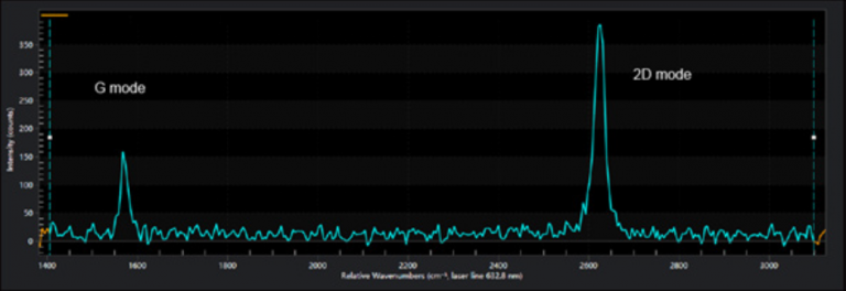
For example, graphene is a single layer of Carbon where the atoms are arranged in a honeycomb structure and is the building block of graphite (many graphene layers stacked on top of each other). Raman spectroscopy has become the standard tool for identifying the number of graphene layers stacked on top of each other. The so called 2D band, around 2650 cm-1, is changing its structure and symmetry between single and double layers of graphene.
Material identification is one of the most important applications of Raman spectroscopy. The vibrations of a material are determined by its specific arrangement of molecular bonds and symmetries. Raman spectra are characteristic of a specific material, building a unique fingerprint that allows for identification of a specific material or detection of contaminations in a material.
Raman spectroscopy is an optical scattering technique that is widely used for the identification of materials and the characterization of their properties. It is commonly applied in material science, chemistry, physics, life science and medicine, the pharmaceutical and semiconductor industries, process and quality control and forensics.
A simple application is the determination of mixture ratios of 2 substances by comparing the relative strength of specific Raman bands in the spectrum. The sensitivity of Raman spectroscopy is extremely high and allows for detection down to trace levels of a material which could be anything from contamination in a chemical solvent, a biomarker in a cell or traces of explosives on a sample, applications where Raman spectroscopy is widely used.
Raman spectroscopy analyzes the vibrations of molecules and crystals. It is one of the most common spectroscopic techniques for material identification and determining a material's physical and chemical environment. Raman spectra use units of wavenumbers to measure the energy shift of scattered light relative to the energy of the excitation laser.
Most MR image information (contrast and general shape) is contained in the center of k-space. Low-spatial-frequency data have the highest amplitude, giving the greatest changes in gray levels (contrast). However, these changes spread over in the image and only give the general shape of organs.We can see below the resulting images of inverse 2D transform performed on data at the center of k-space. Image is contrasted but blurry.
Difference betweenresolution and contrast
High-spatial-frequency data have a lower amplitude. They don't have effect on contrast or general shape but sharpens the image as they encode the edges (rapid changes of image signal as a function of position). Thus, the farther from the center of k-space the data are collected, the higher is the spatial-frequency information and the better the spatial resolution will be.We can see below the resulting images of inverse 2D transform performed on data at the periphery of k-space. As they have a low intensity, click on Enhance contrast to see the effect of high-spatial-frequency information on image.
Raman spectroscopy is extremely adaptable to different experimental configurations, from compact handheld instruments to super high-resolution multistage lab systems. It is also adaptable to different samples in solid, liquid or gas phases, from solid state crystals to proteins in the body. Raman scattering is non-destructive and requires little to no sample preparation.
These cookies are used to measure audience: it allows to generate usage statistics useful for the improvement of the website.
Contrast resolutionRadiology
Resolution and contrasttest
The measurement output of a Raman spectroscopic measurement is a Raman spectrum which contains several components due to the different directions light scatters from the samples. Spectral lines produced by Raman scattering correspond to the different vibrational modes of the sample material or molecule. They are distributed around a spectral line at the excitation laser wavelength due to elastic Rayleigh scattering.
IMAIOS and selected third parties, use cookies or similar technologies, in particular for audience measurement. Cookies allow us to analyze and store information such as the characteristics of your device as well as certain personal data (e.g., IP addresses, navigation, usage or geolocation data, unique identifiers). This data is processed for the following purposes: analysis and improvement of the user experience and/or our content offering, products and services, audience measurement and analysis, interaction with social networks, display of personalized content, performance measurement and content appeal. For more information, see our privacy policy.
Raman lines with higher energy than the laser line (lower wavelength) are referred to as Anti-Stokes lines where the scattered light gains energy from interacting with existing vibrations in the sample. Raman lines with lower energy (higher wavelength) occur when the incident light loses energy by exciting molecular vibrations. Energy in molecular vibrations is excited by thermal excitations so as the temperature of the sample decreases the intensity of the Anti-Stokes bands as less vibrational energy will be in the material. Specific energy ranges in the Raman spectrum are often referred to as the low frequency or THz Raman regions (below 200 cm-1), the fingerprint region (up to 1800 cm-1) and the high wavenumber region (above 2800 cm-1).
In a well calibrated measurement Raman spectroscopy can be used to quantify the amount of material as the intensity of the Raman lines is proportional to the concentration of the analyte in the probe volume.
The early Raman scattering experiments used filtered sunlight or light from atomic emission lamps as incident light sources. It turns out though that the Raman scattering process is extremely weak, laser light however can be produced with much higher intensity, leading to stronger Raman signals. Today Raman spectroscopy is a ubiquitous technique due to the availability of affordable laser source and high performance filters.
Raman as a sensing tool is particularly useful in microscopic and remote observations where environment parameters cannot be accessed by other techniques. Among the quantities routinely measured by Raman spectroscopy are temperature, pressure, stress/strain and pH values.
Difference betweencontrast and resolutionin radiography
Figure 3: Raman spectrum of graphene with the G- and 2D-band clearly visible. The symmetric 2D-line identifies this as a single layer of graphene.
You can freely give, refuse or withdraw your consent at any time by accessing our cookie settings tool. If you do not consent to the use of these technologies, we will consider that you also object to any cookie storage based on legitimate interest. You can consent to the use of these technologies by clicking "accept all cookies".
Some of them require your consent. Click on a category of cookies to activate or deactivate it. To benefit from all the features, it’s recommended to keep the different cookie categories activated.
Light can either excite vibrations and lose energy (redshift) or pick up energy from present vibrations (that are typically thermally excited). As the shift in energy is mostly dependent on the material composition and structure and not the wavelength of the excitation light, Raman spectroscopy measures the energy shift of the Raman scattered light relative to the incident light energy which is characteristic to the sample that is being investigated.
IMAIOS is a company which aims to assist and train human and animal practitioners. Serving healthcare professionals through interactive anatomy atlases, medical imaging, collaborative database of clinical cases, online courses...
These are cookies that ensure the proper functioning of the website and allow its optimization (detect browsing problems, connect to your IMAIOS account, online payments, debugging and website security). The website cannot function properly without these cookies, which is why they are not subject to your consent.


 Ms.Cici
 8618319014500