
Laserafstandsmeters - meten laser
B-190TB includes a 3MP camera with a 10.1” Windows tablet. View, capture, analyze and share your images with simplicity and reliability.
Discover why the Preston Robert Tisch Brain Tumor Center is renowned for its expertise in brain tumor treatment. Schedule an appointment today and take a step towards effective treatment and a brighter future.
When it comes to brain cancer treatment, the Preston Robert Tisch Brain Tumor Center at Duke stands out as one of the best brain cancer clinics. Our team of dedicated doctors is at the forefront of research and is committed to providing exceptional care to our patients. We strive to instill hope and empower individuals facing a brain tumor diagnosis.
A CT scan, or Computed Tomography scan, is another medical imaging technique that uses X-rays to create cross-sectional images of the body. It is particularly useful for examining bone structures and detecting acute conditions such as brain hemorrhages, fractures, and tumors with calcifications.
During a CT scan, the patient lies on a table that moves through a doughnut-shaped machine, which emits X-ray beams from various angles. The images obtained are then reconstructed by a computer to create detailed cross-sectional images of the brain.
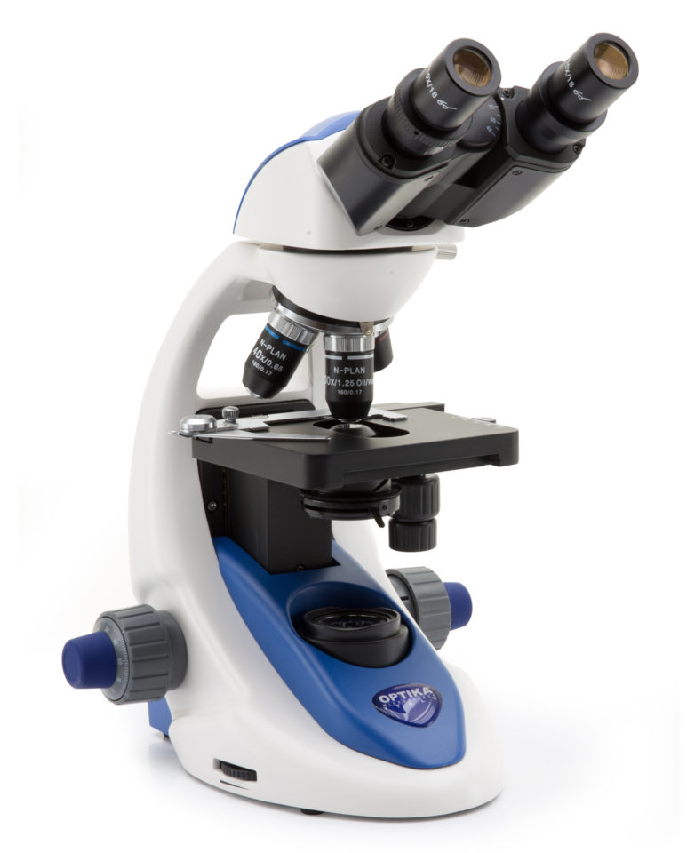
The original design of B-190 series is based on robustness, yet keeping the extreme portability of the instrument, with a dedicated handle on the back. The built-in LED illuminator and the patented version with Windows tablet improve the reliability of one of the best-sellers of OPTIKA in the educational field.
Neither procedure is painful. However, some patients may experience discomfort due to the confined space in MRI machines.
CT scans are relatively quick compared to MRI scans, making them useful in emergency situations where immediate diagnosis is crucial. However, it is important to note that CT scans involve exposure to ionizing radiation, which carries a potential risk, especially with frequent or unnecessary scans.
» Get accurate, dependable results in one click » Equipped with the most reliable OS, Windows 10 » Holding solution for open discussion, 360° rotating and tilting
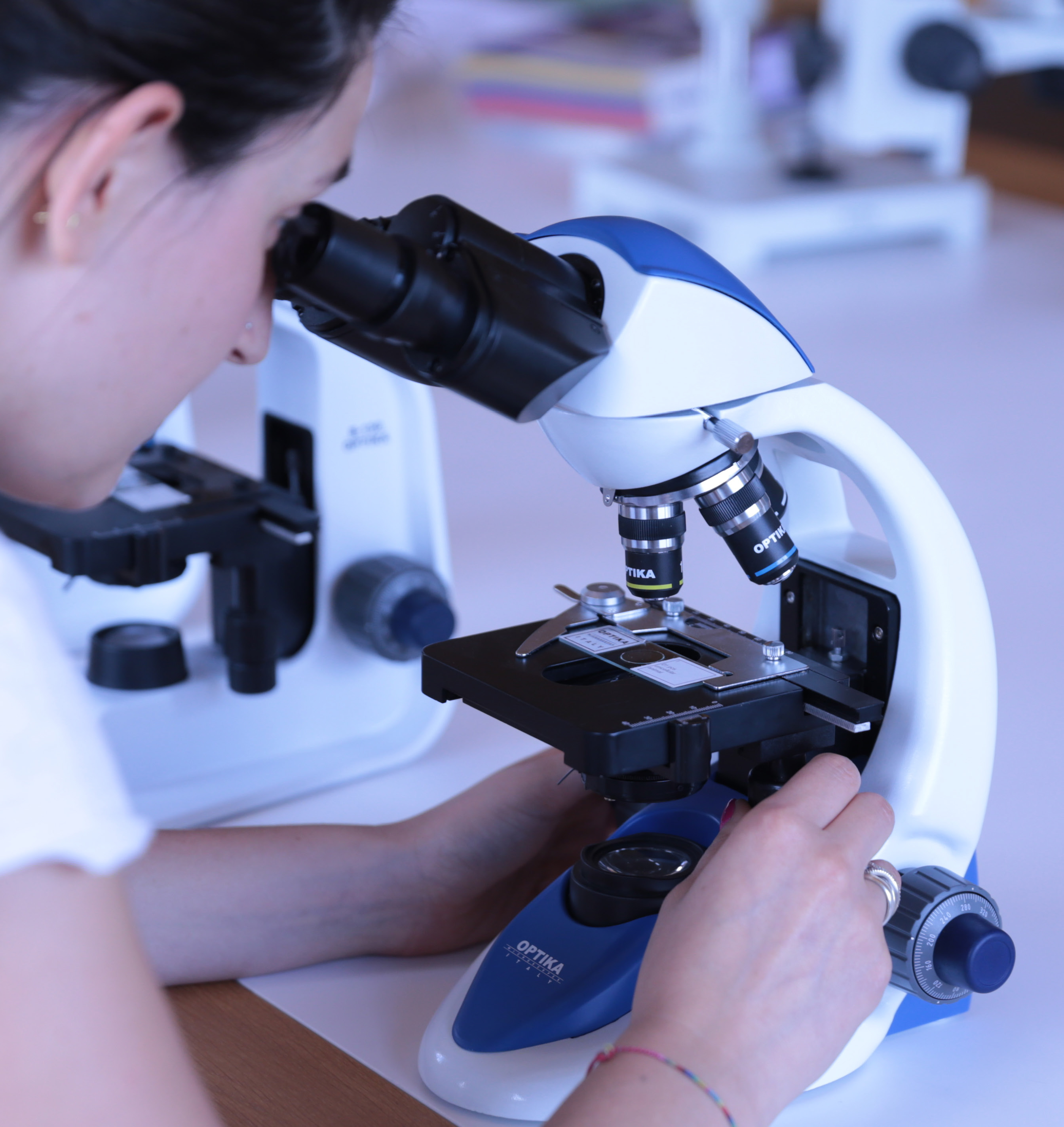
Advanced binocular microscope up to 1000x total magnification, with oil/water. The exclusive X-LED2 provides unmatchable performance for powerful and uniform illumination.
Get all the controls and features common to higher level microscopes: translating stage, binocular or trinocular head, coaxial focus knob, adjustable condenser, and 1000x maximum magnification (standard). An extremely simple but well equipped solution, in a modern and ergonomic design.
» Sturdy, durable for extended lifetime » Only simple lens cleaning is required time to time » Dust cover protects from environmental contaminants
Some application examples showing the extreme versatility of B-190 Series. Application in education, biology, botany and simple material analysis, where all the samples are prepared on standard glass slides.
Binocularmicroscope diagram
Advanced monocular microscope up to 1000x total magnification, with oil/water. The exclusive X-LED2 provides unmatchable performance for powerful and uniform illumination.
MRI scans are better for brain imaging as they are the most sensitive brain imaging technique currently available, especially for the brain. Because of this, it is a better option for detecting brain tumors.
OPTIKA Microscopy Catalogue - Educational - Low Res - 3.88 MB OPTIKA Microscopy Catalogue - Educational - 33.15 MB OPTIKA Microscopy Catalogue - Educational - B-190 - 7.47 MB OPTIKA - B-190 - EU Declaration - 103.91 KB
Fulfill the next generation learning challenges with this original, compact and robust series incorporating the most wanted features in a student microscope (18mm field number, 1000x maximum magnification, translating stage, coaxial focus knob, adjustable condenser, and the exclusive X-LED² illumination).
The B-190TB offers you a unique, incomparable solution. It includes a built-in camera of 3Mp and a Windows tablet with large touch screen, for a responsive and smooth control. Simultaneous camera and power connection ensure long-term operation, with dependable results in one click. It provides a reliable and comfortable solution for open discussion: 360° rotating and tilting tablet, easily detachable, that can be used as a laptop.
A special design of the lens in front of the LED gives a very high light intensity, while ensuring optimal uniformity of illumination on the whole field number. Relevant money & energy saving thanks to the incredibly low energy consumptions allow you to cut the electricity bills by 90%! The electric consumption (3 W only) proves thehigh efficiency of this system: incredibly high light intensity combined with low consumption.
» High energy efficiency at a limited cost » Incredibly low energy consumptions, only 3W » LED long lifetime (50.000 hours = 20 years at 8 hours/day usage)
OPTIKA B-190 Series is a perfect fusion between decades of experience in educational microscopy and a new, refined design. The result is this advanced biological microscopes for students representing our product philosophy at its best with the combination of quality, reliability and innovation, all in one.
Binocularandtrinocularmicroscope difference
» Choose among monocular, binocular and trinocular heads » 18mm field number » Achromatic lenses for standard brightfield applications.
CT scans can detect certain types of cancer such as bladder cancer, kidney cancer, ovarian cancer, stomach cancer, and even colon cancer.
Advanced trinocular microscope up to 1000x total magnification, with oil/water, mechanical stage and exclusive X-LED2 for unmatchable performance for powerful and uniform illumination. All the OPTIKA cameras can be easily mounted and used straight away.
3.1 MP Built-in camera and 10.1” Windows tablet PC up to 1000x total magnification, mechanical stage and exclusive X-LED2 for unmatchable performance for powerful and uniform illumination.
CT scans are quicker and usually take only a few minutes, while MRI scans can take between 15 minutes to an hour, depending on the type and purpose of the scan.
Binocularmicroscope parts and functions
Turns the tablet into a PC with the use of the keyboard (sold separately) Intel Quad-Core processor, Windows 10. Battery life up to 10 hours.
MRI, or Magnetic Resonance Imaging, is a non-invasive medical imaging technique that uses powerful magnetic fields and radio waves to generate detailed images of the body's internal structures. In the case of brain imaging, MRI provides highly accurate and detailed images of the human brain, allowing healthcare professionals to assess its anatomy and detect any abnormalities.
» Compact, practical and intuitive to use » Optics ensuring good quality images » Get impressive images and live videos with cameras
When it comes to brain imaging, two popular imaging methods are widely used: MRI (Magnetic Resonance Imaging) and CT (Computed Tomography) scans. Both techniques provide valuable insights into brain health, but they differ in terms of their technology and diagnostic capabilities.
The latest OPTIKA digital microscopes with Windows tablet PC open new microscopy horizons, combining high-end optics with innovative digital technology for microscopic imaging.
» Large touch screen of 10.1” with fast, responsive and smooth control » Simultaneous camera and power connection for long-term operation » Easily detachable, can be used as a laptop (keyboard sold separately)
In this blog post, we will explore the differences between MRI and CT scans, helping you understand which one is better for brain imaging and detecting brain tumors.
Unlike X-rays or CT scans, MRI does not use ionizing radiation, making it a safer option for repeated imaging. During an MRI scan, the patient lies on a movable table that slides into a cylindrical machine. It is important to remain still during the procedure to ensure clear images.
3 difference between monocular andbinocularmicroscope
» Uncomparable light intensity, exclusive lens and collector design » Constant pure-white colour temperature at all intensity levels » Unmatched colour fidelity and brightness of your specimen
Advanced monocular microscope up to 600x total magnification. The exclusive X-LED2 provides unmatchable performance for powerful and uniform illumination.
[ Placeholder content for popup link ] WordPress Download Manager - Best Download Management Plugin
Advanced binocular microscope up to 600x total magnification. The exclusive X-LED2 provides unmatchable performance for powerful and uniform illumination.


 Ms.Cici
 8618319014500