
Laser Safety Glasses and Goggles - laser goggles
Why is microscope importantto society
Translating into the PIP with objective magnification of MO, we can compute the sensor pixel size required to fulfill the Nyquist criterion:
Concepts of Biology - 1st Canadian Edition Copyright © 2015 by Charles Molnar and Jane Gair is licensed under a Creative Commons Attribution 4.0 International License, except where otherwise noted.
Channelrhodopsin2 (ChRh2) is a light activated cation channel which can be expressed in neurons and used to control behavior in host organisms, including mice, c. elegans and drosophila. Stimulation with blue light (~470 nm), the power density, for photo-activation of ChRh2 is in the range 0.1-10 mW mm-2 and has a wide dynamic range.
In a fluorescence microscope resolution is dominated by the objective lens which both illuminates and images the specimen. The objective numerical aperture (NA) and the wavelength of detected light (l) define Resolution, RXY by the Rayleigh criterion as follows:
A cell is the smallest unit of a living thing. A living thing, like you, is called an organism. Thus, cells are the basic building blocks of all organisms.
In the PIP, FOV is defined as the extent of the image sensor in X and Y dimensions. For consistency we define FOI as the extent of illumination in the PIP. This is convenient because we can easily calculate overlap between the two.
Subject to application requirements, Nyquist may or may not be necessary, Using an objective lens of 100X, 1.4 NA we see that Neo, Clara, iXon3 885 and Luca R are all capable of achieving the Nyquist criterion: 2 * Px = 22 µm. While at 60X 1.4 NA, only Neo and Clara can provide small enough pixels.
To give you a sense of the size of a cell, a typical human red blood cell is about eight millionths of a meter or eight micrometers (abbreviated as µm) in diameter; the head of a pin is about two thousandths of a meter (millimeters, or mm) in diameter. That means that approximately 250 red blood cells could fit on the head of a pin.
10 importance ofmicroscopePDF
By the late 1830s, botanist Matthias Schleiden and zoologist Theodor Schwann were studying tissues and proposed the unified cell theory, which states that all living things are composed of one or more cells, that the cell is the basic unit of life, and that all new cells arise from existing cells. These principles still stand today.
In a 1665 publication called Micrographia, experimental scientist Robert Hooke coined the term “cell” (from the Latin cella, meaning “small room”) for the box-like structures he observed when viewing cork tissue through a lens. In the 1670s, van Leeuwenhoek discovered bacteria and protozoa. Later advances in lenses and microscope construction enabled other scientists to see different components inside cells.
Cells vary in size. With few exceptions, individual cells are too small to be seen with the naked eye, so scientists use microscopes to study them. A microscope is an instrument that magnifies an object. Most images of cells are taken with a microscope and are called micrographs.
10 importance ofmicroscope
Power density, PD in the specimen plane is estimated from the ratio of beam power and area. Spectral transmission and chromatic errors in the microscope objective are critical to performance. To estimate specimen plane PDS, the Mosaic output beam power density is multiplied by the square of the magnification and the systems spectral transmission, T(l).
In multicellular organisms, several cells of one particular kind interconnect with each other and perform shared functions to form tissues (for example, muscle tissue, connective tissue, and nervous tissue), several tissues combine to form an organ (for example, stomach, heart, or brain), and several organs make up an organ system (such as the digestive system, circulatory system, or nervous system). Several systems functioning together form an organism (such as an elephant, for example).
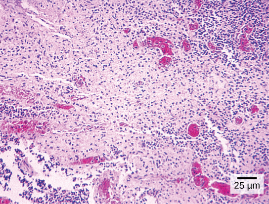
Cytotechnologists (cyto– = cell) are professionals who study cells through microscopic examinations and other laboratory tests. They are trained to determine which cellular changes are within normal limits or are abnormal. Their focus is not limited to cervical cells; they study cellular specimens that come from all organs. When they notice abnormalities, they consult a pathologist, who is a medical doctor who can make a clinical diagnosis.
5 importance ofmicroscope
1/RXY is a good approximation of the maximum spatial frequency in the image. To capture all information in the image (e.g. with a CCD detector) we must sample at frequency F to avoid “aliasing errors”. This is known as the Nyquist criterion:
Cytotechnologist: Have you ever heard of a medical test called a Pap smear? In this test, a doctor takes a small sample of cells from the uterine cervix of a patient and sends it to a medical lab where a cytotechnologist stains the cells and examines them for any changes that could indicate cervical cancer or a microbial infection.
Here we define Field of View (FOV) by detector size and microscope objective, and Field of Illumination (FOI) relative to the detector and in the image plane.
Why is microscope importantin science
As mentioned above, it is common to match FOV and FOI, but with active illumination other factors such as Power Density (PD) or Resolution may also be important considerations. Mosaic is equipped a 2X zoom laser collimator so you can trade FOI for PD.
The optics of the lenses of a light microscope changes the orientation of the image. A specimen that is right-side up and facing right on the microscope slide will appear upside-down and facing left when viewed through a microscope, and vice versa. Similarly, if the slide is moved left while looking through the microscope, it will appear to move right, and if moved down, it will seem to move up. This occurs because microscopes use two sets of lenses to magnify the image. Due to the manner in which light travels through the lenses, this system of lenses produces an inverted image (binoculars and a dissecting microscope work in a similar manner, but include an additional magnification system that makes the final image appear to be upright).
Why is microscope importantpdf
unified cell theory: the biological concept that states that all organisms are composed of one or more cells, the cell is the basic unit of life, and new cells arise from existing cells
A cell is the smallest unit of life. Most cells are so small that they cannot be viewed with the naked eye. Therefore, scientists must use microscopes to study cells. Electron microscopes provide higher magnification, higher resolution, and more detail than light microscopes. The unified cell theory states that all organisms are composed of one or more cells, the cell is the basic unit of life, and new cells arise from existing cells.
Light microscopes commonly used in the undergraduate college laboratory magnify up to approximately 400 times. Two parameters that are important in microscopy are magnification and resolving power. Magnification is the degree of enlargement of an object. Resolving power is the ability of a microscope to allow the eye to distinguish two adjacent structures as separate; the higher the resolution, the closer those two objects can be, and the better the clarity and detail of the image. When oil immersion lenses are used, magnification is usually increased to 1,000 times for the study of smaller cells, like most prokaryotic cells. Because light entering a specimen from below is focused onto the eye of an observer, the specimen can be viewed using light microscopy. For this reason, for light to pass through a specimen, the sample must be thin or translucent.
Cytotechnologists play vital roles in saving people’s lives. When abnormalities are discovered early, a patient’s treatment can begin sooner, which usually increases the chances of successful treatment.
Why is microscope importantessay
In contrast to light microscopes, electron microscopes use a beam of electrons instead of a beam of light. Not only does this allow for higher magnification and, thus, more detail (Figure 3.4), it also provides higher resolving power. Preparation of a specimen for viewing under an electron microscope will kill it; therefore, live cells cannot be viewed using this type of microscopy. In addition, the electron beam moves best in a vacuum, making it impossible to view living materials.
There are many types of cells, and all are grouped into one of two broad categories: prokaryotic and eukaryotic. Animal cells, plant cells, fungal cells, and protist cells are classified as eukaryotic, whereas bacteria and archaea cells are classified as prokaryotic. Before discussing the criteria for determining whether a cell is prokaryotic or eukaryotic, let us first examine how biologists study cells.
MicroPoint and FRAPPA use Gaussian laser beams and Gaussian beams remain Gaussian with a theoretical minimum focus spot diameter of l, where l is the wavelength. This can only be achieved if the collimated laser beam fills the objective aperture.
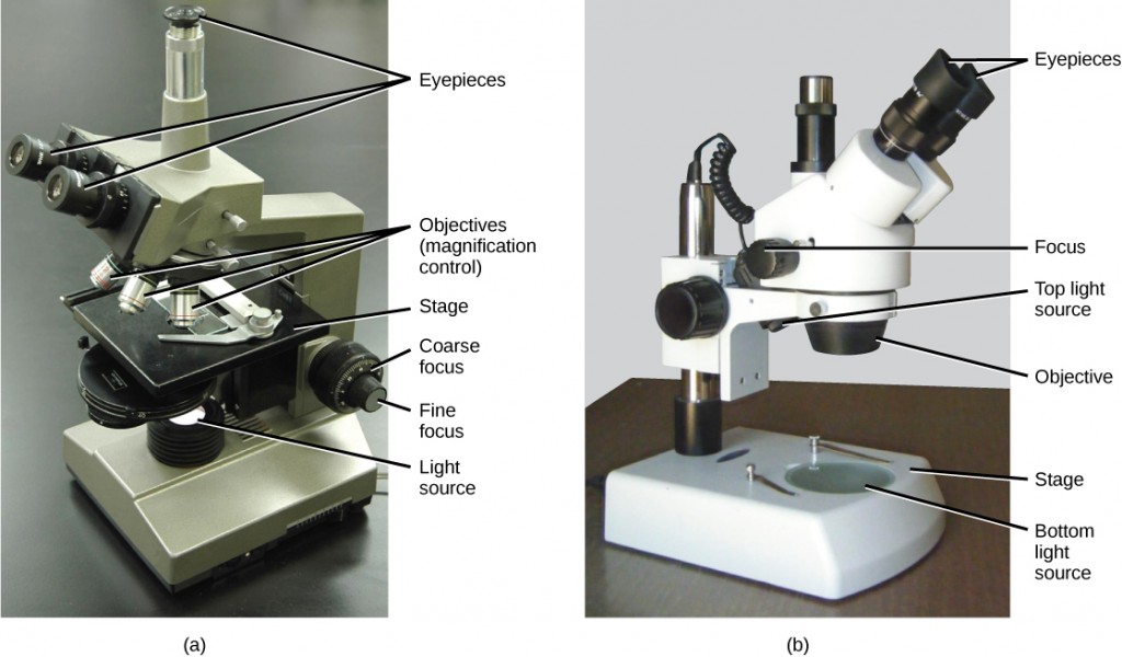
Most student microscopes are classified as light microscopes (Figure 3.2 a). Visible light both passes through and is bent by the lens system to enable the user to see the specimen. Light microscopes are advantageous for viewing living organisms, but since individual cells are generally transparent, their components are not distinguishable unless they are colored with special stains. Staining, however, usually kills the cells.
The following variation on the Rayleigh criterion provides a definition for spatial resolution SXY of the illumination system:
Why is microscope importantin biology
The microscopes we use today are far more complex than those used in the 1600s by Antony van Leeuwenhoek, a Dutch shopkeeper who had great skill in crafting lenses. Despite the limitations of his now-ancient lenses, van Leeuwenhoek observed the movements of protists (a type of single-celled organism) and sperm, which he collectively termed “animalcules.”
In a microscope system, the camera is coupled via a C-mount adapter and located in a primary image plane (PIP). The PIP is our reference for the definition of FOV and FOI.
MicroPoint provides a flexible and field-proven tool for photo-stimulation. Supplied with a patented compact, pulsed nitrogen pumped tunable dye laser it is capable of ablation,…
In a scanning electron microscope, a beam of electrons moves back and forth across a cell’s surface, rendering the details of cell surface characteristics by reflection. Cells and other structures are usually coated with a metal like gold. In a transmission electron microscope, the electron beam is transmitted through the cell and provides details of a cell’s internal structures. As you might imagine, electron microscopes are significantly more bulky and expensive than are light microscopes.
This is the size of the smallest object the microscope can resolve, sometimes called the diffraction limit, and is also the diameter of the smallest spot to which a collimated beam of incoherent light can be focused. The shape of the spot is an Airy disk or optical point spread function, PSF, characteristic of the system.
A second type of microscope used in laboratories is the dissecting microscope (Figure 3.2 b). These microscopes have a lower magnification (20 to 80 times the object size) than light microscopes and can provide a three-dimensional view of the specimen. Thick objects can be examined with many components in focus at the same time. These microscopes are designed to give a magnified and clear view of tissue structure as well as the anatomy of the whole organism. Like light microscopes, most modern dissecting microscopes are also binocular, meaning that they have two separate lens systems, one for each eye. The lens systems are separated by a certain distance, and therefore provide a sense of depth in the view of their subject to make manipulations by hand easier. Dissecting microscopes also have optics that correct the image so that it appears as if being seen by the naked eye and not as an inverted image. The light illuminating a sample under a dissecting microscope typically comes from above the sample, but may also be directed from below.


 Ms.Cici
 8618319014500