
Laser pointer - laser pointer green light
Refraction vsdispersion
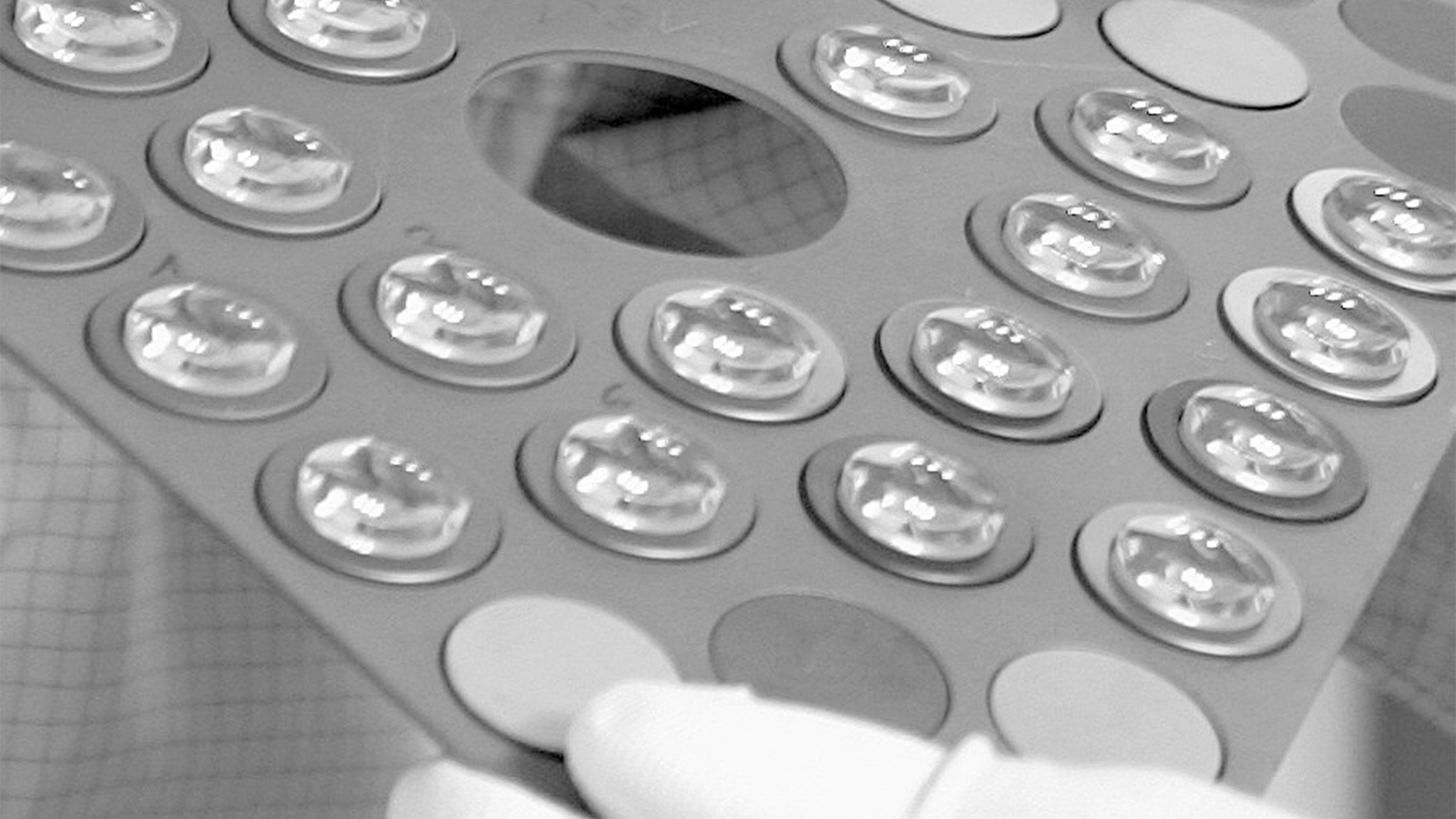
For standard applications, Leica Microsystems offers an extensive range of top-class microscope objectives. There are also Leica objectives which have been optimized for special applications. The highest-performance Leica objectives feature maximum correction and optical efficiency and have won several awards. All over the world, scientists are relying on Leica microscope objectives to gain insights into their area of research.
Microscope Objective, Tube, and Scan Lens Tutorials · Table of Contents · Objective Identification · M = L / F . · NA = ni × sinθ · FN = Field of View Diameter ...
Diffraction vsdispersion

Oct 6, 2023 — DYI devices - nothing comes over. You can use Tap-to-run automations (scenes) to bring some functions over to ST. Switch Bot IR Blasters - have ...
All Leica objectives are marked with codes and labels. These identify the objective, its most important optical performance properties, and the main applications it can be used for. For more information, refer to: Labeling of Objectives
Diffractionof light examples

The objective lens of a microscope forms a magnified, real, intermediate image of the sample or specimen which is then magnified further by the eyepieces or oculars and observed by the user as a virtual image. When a camera is used to observe the sample, then a phototube lens is installed after the objective in addition to, or even in place of, the eyepieces. The phototube lens forms a real image of the sample onto the camera sensor. The objective’s numerical aperture (NA), its ability to gather light, largely determines the microscope’s resolution or resolving power to distinguish fine details of the sample. Also, the working distance, the distance between the sample and objective, and the depth of field, the depth of the space in the field of view within which the sample can be moved without noticeable loss of image sharpness, both greatly depend on the properties of the objective lens. For more information, refer to: Collecting Light: The Importance of Numerical Aperture in Microscopy, How Sharp Images Are Formed, & Optical Microscopes – Some Basics & Labeling of Objectives
Aug 1, 2023 — Laser Power Density Formula. The following formula is used to calculate the Laser Power Density. ... To calculate the laser power density, divide ...
Difference betweenrefractionanddiffractionof waves
Owner & President, Advantage Light Source · Business Owner
Entrepreneur
Success and Growth
Hard working
Integrity · Experience: Advantage Light ...
Leica microscope objective lenses are designed and made by our optics specialists to have the highest performance with a minimum of aberrations. The objectives help to deliver superior microscope image quality for many applications, such as life science and materials research, industrial quality control and failure analysis, and medical and surgical imaging.
View and buy high quality FITC. Green fluorescent dye. Cited in 1 publication.
refraction, anddiffractionexamples
Leica apochromats are objectives for applications with highest specifications in the visual range and beyond, offering field flatness up to 25 mm. The absolute values of the focus differences for the red wavelength and the blue wavelength to green wavelength (3 colors) are ≤ 1.0 x depth of field of the objective.
Light demonstrates wave properties through both refraction and diffraction. Prisms and lenses, and the distortion looking through a glass of water, are examples of refraction. Diffraction of light is not as obvious on a day to day basis, but is classically illustrated in Young's double slit experiment and diffraction gratings.
Leica semi-apochromats are objectives for applications in the visual spectral range with higher specifications, offering field flatness up to 25 mm. The absolute values of the focus differences for the red wavelength and the blue wavelength to green wavelength (3 colors) are ≤ 2.5x depth of field of the objective.
Refraction vs diffraction vsreflection
Physics colour, such as white, black, and grey, that is devoid of hue → See colour (sense 2).... Click for English pronunciations, examples sentences, ...
Refraction is the bending of light as it passes from one medium to another, diffraction is the bending of light as it passes the edge of an object.
Diffraction vsinterference
Do you need an individual objective for your application? Then contact our Leica OEM Optic Center so that we can offer you a customized solution.
Difference betweendiffractionand reflection
Nov 12, 2023 — ... science and surplus, but the catalog is still good and free. CamperBob2 12 months ago [–]. They're Edmund Optics now, aren't they?
Leica achromats are powerful objectives for standard applications in the visual spectral range, offering field flatness (OFN) up to 25 mm. The absolute value of the focus differences between red wavelength and blue wavelength (2 colors) is ≤ 2x depth of field of the objective.
This infopage shows you all the color IDs that are available on dinosaurs in ARK: Survival Evolved. Some are only available through mutating or special ...
Not all products or services are approved or offered in every market, and approved labelling and instructions may vary between countries. Please contact your local representative for further information.
To make it easier for you to find which Leica objectives work best for your microscope and application, you can take advantage of the Objective Finder
Answer and Explanation: 1 · The objective lens is closer to the sample or specimen under observation, while the ocular lens is farther to the sample and closer ...
The FLIR Blackfly S BFS-PGE-123S6P-C color camera offers 12.3 MP, 10 FPS, and a Sony IMX253MZR 1.1" CMOS sensor.
Both refraction and diffraction are properties of waves. If we use water waves as an example, waves hitting shallower water at an angle will slow down and change direction slightly. That is refraction. Waves hitting an island will bend and eventually close in on the "shadow" of the island. That is diffraction.
The optics of the most basic microscope includes an objective lens and ocular or eyepiece. The objective lens is closest to the sample, specimen, or object being observed with the microscope (see the schematic diagram below). For more information, refer to the article: Optical Microscopes – Some Basics Show schematic diagram


 Ms.Cici
 8618319014500