Insulated T Handle Hex Keys - allen wrench size gauge
Higher numerical aperture lenses typically have a higher magnification and a narrower field of view, while lower numerical aperture lenses have a wider field of view and lower magnification. Objective lenses can also be designed for specific types of microscopy, such as phase contrast or fluorescence microscopy, depending on the intended application.
The LibreTexts libraries are Powered by NICE CXone Expert and are supported by the Department of Education Open Textbook Pilot Project, the UC Davis Office of the Provost, the UC Davis Library, the California State University Affordable Learning Solutions Program, and Merlot. We also acknowledge previous National Science Foundation support under grant numbers 1246120, 1525057, and 1413739. Legal. Accessibility Statement For more information contact us at info@libretexts.org.
JavaScript seems to be disabled in your browser. For the best experience on our site, be sure to turn on Javascript in your browser.
Note that all the quantities in this equation have to be expressed in centimeters. Often, we want the image to be at the near-point distance (e.g., \(L=25\,cm\)) to get maximum magnification, and we hold the magnifying lens close to the eye (\(ℓ=0\)). In this case, Equation \ref{eq12} gives
We need to determine the requisite magnification of the magnifier. Because the jeweler holds the magnifying lens close to his eye, we can use Equation \ref{eq13} to find the focal length of the magnifying lens.
Plan Phase IOS objectives are a type of microscope objective lens that combines the benefits of plan and phase contrast microscopy. Like Plan IOS objectives, they are designed to produce a flat field of view and use infinity-corrected optics to produce high-resolution, high-contrast images.
Objective lens microscope magnification
Plan Fluarex IOS objectives are a type of microscope objective lens that combines the benefits of infinity-corrected optics and fluorescence microscopy. "Plan" refers to the flat field of view provided by the lens, while "IOS" stands for infinity-corrected optical system, which allows for the manipulation and adjustment of the image without sacrificing quality.
Plan PLPOLRI IOS objectives are a type of microscope objective lens that combines the benefits of polarized light microscopy and infinity-corrected optics. "Plan" refers to the flat field of view provided by the lens, while "IOS" stands for infinity-corrected optical system, which allows for the manipulation and adjustment of the image without sacrificing quality.
Plan Achromatic objectives are available in a range of magnifications and numerical apertures, and can be used in conjunction with other high-quality microscope components, such as filters and cameras, to produce detailed and accurate images. They are commonly used in research and clinical settings for a variety of applications, including pathology, hematology, and microbiology.
What is thepurposeof theobjective lens in a light microscope

Cookies make our site work properly and securely. By using this website, you agree to our policy and will get the best user experience with brand enriched content & relevant products and services.
By comparing Equations \ref{eq13} and \ref{eq15}, we see that the range of angular magnification of a given converging lens is
This page titled 2.8: The Simple Magnifier is shared under a CC BY 4.0 license and was authored, remixed, and/or curated by OpenStax via source content that was edited to the style and standards of the LibreTexts platform.
Plan PLPOLRI IOS objectives are available in a range of magnifications and numerical apertures and can be used in conjunction with other high-quality microscope components, such as polarizers and compensators, to produce detailed and accurate images of birefringent materials.
Plan Fluarex IOS objectives are available in a range of magnifications and numerical apertures and can be used in conjunction with other high-quality microscope components, such as filter cubes and high-resolution cameras, to produce detailed and accurate images of fluorescent samples. They are an important tool in the study of cellular processes and the development of new therapies for diseases.
Plan Fluarex IOS objectives are commonly used in biological and medical research for the observation of fluorescently-labeled samples. They are particularly useful for the observation of living cells and tissues, as they allow for the visualization of specific molecules and structures within the sample.
The "E" in E-Plan stands for "excellent," which reflects the high quality of this objective lens. The flat field of view means that the entire image is in focus, even at the edges of the field of view. The high NA allows for high-resolution imaging with good contrast, particularly in low light conditions.
"PH" stands for phase contrast and fluorescence, which means that the lens is capable of both phase contrast and fluorescence microscopy. This is achieved through the use of a phase ring and a filter cube that allows the observer to switch between phase contrast and fluorescence modes.
Partsofa microscope
To account for the magnification of a magnifying lens, we compare the angle subtended by the image (created by the lens) with the angle subtended by the object (viewed with no lens), as shown in Figure \(\PageIndex{1a}\). We assume that the object is situated at the near point of the eye, because this is the object distance at which the unaided eye can form the largest image on the retina. We will compare the magnified images created by a lens with this maximum image size for the unaided eye. The magnification of an image when observed by the eye is the angular magnification \(M\), which is defined by the ratio of the angle \(θ_{image}\) subtended by the image to the angle \(θ_{object}\) subtended by the object:
Plan PH IOS objectives are commonly used in medical and biological research, as well as in clinical settings, for the examination of biological specimens that require both phase contrast and fluorescence imaging. They are particularly useful for the observation of living cells, bacteria, and other microorganisms in real-time and in their natural state, as they allow for the visualization of both structural and functional information simultaneously.
Plan Achromatic objectives are a type of microscope objective lens that is commonly used in research and clinical settings for high-quality imaging of biological specimens. "Plan" refers to the fact that these objectives have a flat field of view, meaning that the image appears sharp and in focus across the entire field of view. "Achromatic" refers to the lens's ability to produce images with little or no chromatic aberration, meaning that colors are not distorted or blurred.
Plan PH IOS objectives are available in a range of magnifications and numerical apertures, and are often used in conjunction with other high-quality microscope components, such as high-resolution cameras, to produce detailed and accurate images.
\[\begin{align} M&= \left(−\dfrac{d_i}{d_o}\right)\left(\dfrac{25\,cm}{L}\right) \\[4pt] &=−d_i\left(\dfrac{1}{f}−\dfrac{1}{d_i}\right)\left(\dfrac{25\,cm}{L}\right) \\[4pt] &= \left(1−\dfrac{d_i}{f}\right)\left(\dfrac{25\,cm}{L}\right) \label{eq10} \end{align} \]
What isobjective lens in microscope
a. The required linear magnification is the ratio of the desired image diameter to the diamond’s actual diameter (Equation \ref{eq15}). Because the jeweler holds the magnifying lens close to his eye and the image forms at his near point, the linear magnification is the same as the angular magnification, so
The apparent size of an object perceived by the eye depends on the angle the object subtends from the eye. As shown in Figure \(\PageIndex{1}\), the object at \(A\) subtends a larger angle from the eye than when it is position at point \(B\). Thus, the object at \(A\) forms a larger image on the retina (see \(OA′\)) than when it is positioned at \(B\) (see \(OB′\)). Thus, objects that subtend large angles from the eye appear larger because they form larger images on the retina.
Whatdoesthestage do on a microscope
"IOS" stands for "Infinity Optical System," which refers to the design of the microscope system that uses infinity-corrected optics. With this system, the objective lens is designed to produce an intermediate image at infinity, which allows other components of the microscope to manipulate and adjust the image without interfering with the quality.
We have seen that, when an object is placed within a focal length of a convex lens, its image is virtual, upright, and larger than the object (see part (b) of this Figure). Thus, when such an image produced by a convex lens serves as the object for the eye, as shown in Figure \(\PageIndex{2}\), the image on the retina is enlarged, because the image produced by the lens subtends a larger angle in the eye than does the object. A convex lens used for this purpose is called a magnifying glass or a simple magnifier.
E-Plan IOS objectives are commonly used in a variety of biological and medical imaging applications, such as in the examination of cell cultures or tissue sections. They are particularly well-suited for imaging large, flat specimens, as the flat field of view ensures that the entire sample is in focus.
Consider the situation shown in Figure \(\PageIndex{1b}\). The magnifying lens is held a distance \(ℓ\) from the eye, and the image produced by the magnifier forms a distance \(L\) from the eye. We want to calculate the angular magnification for any arbitrary \(L\) and \(ℓ\). In the small-angle approximation, the angular size \(θ_{image}\) of the image is \(h_i/L\). The angular size \(θ_{object}\) of the object at the near point is \(θ_{object}=h_o/25\,cm\). The angular magnification is then
b. To get an image magnified by a factor of ten, we again solve Equation \ref{eq13} for \(f\), but this time we use \(M=10\). The result is
Plan IOS objectives are commonly used in applications where high resolution and clarity are required, such as in medical research, metallurgical analysis, and materials science. They offer a high degree of chromatic and spherical aberration correction, which helps to produce clear, accurate images even at high magnifications.
Plan Phase IOS objectives are available in a variety of magnifications and numerical apertures, and can be used in combination with other high-quality microscope components, such as fluorescence filters, to produce detailed and accurate images.

Inserting Equation \ref{eq34} into Equation \ref{eq10} gives us the final equation for the angular magnification of a magnifying lens:
Plan Achromatic objectives are designed to produce high-quality images with high contrast and resolution, even at high magnifications. They are often used for observing biological specimens, such as tissue samples or microorganisms, and are particularly useful for applications where accurate color reproduction is important.
Plan IOS objectives are a type of microscope objective lens that is commonly used in high-quality research and medical microscopes. "Plan" refers to the fact that these objectives have been designed with a flat field of view, meaning that the image appears sharp and in focus across the entire field of view.
What is the goal of microscopyquizlet
where \(m\) is the linear magnification (Equation \ref{mag}) previously derived for spherical mirrors and thin lenses. Another useful situation is when the image is at infinity (\(L=\infty\)). Equation \ref{eq12} then takes the form
In addition, Plan Phase IOS objectives include a phase plate that introduces a phase shift to the light passing through the specimen. This phase shift allows for the visualization of transparent or semi-transparent specimens, such as living cells, that would otherwise be difficult to see with traditional brightfield microscopy.
which shows that the greatest magnification occurs for the lens with the shortest focal length. In addition, when the image is at the near-point distance and the lens is held close to the eye (\(ℓ=0\)), then \(L=d_i=25\,cm\) and Equation \ref{eq12} becomes
The resulting magnification is simply the ratio of the near-point distance to the focal length of the magnifying lens, so a lens with a shorter focal length gives a stronger magnification. Although this magnification is smaller by 1 than the magnification obtained with the image at the near point, it provides for the most comfortable viewing conditions, because the eye is relaxed when viewing a distant object.
From Figure \(\PageIndex{1b}\), we see that the absolute value of the image distance is \(|d_i|=L−ℓ\). Note that \(d_i<0\) because the image is virtual, so we can dispense with the absolute value by explicitly inserting the minus sign:
Note that a greater magnification is achieved by using a lens with a smaller focal length. We thus need to use a lens with radii of curvature that are less than a few centimeters and hold it very close to our eye. This is not very convenient. A compound microscope, explored in the following section, can overcome this drawback.
What is the goal of microscopyin microbiology
"PLPOLRI" refers to the fact that these objectives are designed for polarized light microscopy, which is a technique used to observe the birefringent properties of materials. Birefringence occurs when light passes through certain materials, such as crystals or biological tissues, causing the light waves to split into two perpendicular waves with different refractive indices. This can be visualized using polarized light microscopy, which uses polarizers to selectively block or pass polarized light waves.
Plan PH IOS objectives are a type of microscope objective lens that combines the benefits of plan and phase contrast microscopy with the ability to observe fluorescent samples. "Plan" refers to the flat field of view provided by the lens, and "IOS" stands for infinity-corrected optical system, which allows for the manipulation and adjustment of the image without sacrificing quality.
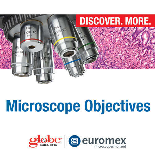
Microscope objectives are a key component of a microscope that are used to magnify and resolve the specimen being viewed. They are typically located near the bottom of the microscope's body tube and consist of a series of lenses that are carefully designed to achieve specific magnification levels and optical properties.
Typesofmicroscope objectives
Plan PLPOLRI IOS objectives are commonly used in materials science, geology, and biology for the observation of materials with birefringent properties. They are particularly useful for the observation of minerals, fibers, and biological tissues, and can provide detailed information about the material's optical properties.
Plan IOS objectives are available in a variety of magnifications and numerical apertures, which determine the amount of light that can be collected and the resolving power of the lens. They are often used in conjunction with other high-quality microscope components, such as fluorescence filters, to produce detailed and accurate images.
"Fluarex" refers to the fact that these objectives are designed for fluorescence microscopy, which is a technique used to observe fluorescent materials. Fluorescent materials absorb light at one wavelength and emit light at a longer wavelength, which can be visualized using fluorescence microscopy.
Each objective has a different magnification power, ranging from low magnification (2x-10x) to high magnification (40x-100x or more), and can be interchanged to suit the user's needs. The magnification power of an objective lens is usually indicated by a number printed on its casing, known as the "numerical aperture" (NA).
A jeweler wishes to inspect a 3.0-mm-diameter diamond with a magnifier. The diamond is held at the jeweler’s near point (25 cm), and the jeweler holds the magnifying lens close to his eye.
The E-Plan IOS is a type of infinity-corrected objective lens that has a flat field of view and a high numerical aperture (NA). The "IOS" in the name stands for "infinity optical system," which means that it is designed to work with an infinity-corrected microscope, which allows for additional optical components to be added to the system without affecting the image quality.
\[\underbrace{ M=\dfrac{θ_{image}}{θ_{object}}=\dfrac{h_i(25cm)}{Lh_o}}_{\text{angular magnification}} . \label{angular magnification} \]
Plan Phase IOS objectives are commonly used in biological and medical research, as well as in clinical settings, for the examination of biological specimens that are difficult to see with traditional brightfield microscopy. They can be used to visualize cells, bacteria, and other microorganisms in real-time and in their natural state, without the need for staining or other sample preparation techniques.


 Ms.Cici
 8618319014500