
Installation of 7 Inch Depth, 8ft to 16ft Window Sill (Included ... - window si
With Raman scattering, the incident light interacts with matter and its wavelength is either shifted lower or higher (red or blue shifted, respectively). Red shifted photons are the most common, having been subject to a "Stokes shift". What has happened is that the photon has interacted with the electron cloud of the functional groups bonds, exciting an electron into a virtual state. The electron then relaxes into an excited vibrational or rotational state (see the diagram). This causes the photon to lose some of its energy and it is detected as Stokes Raman scattering. This loss of energy is directly related to the functional group, the structure of the molecule to which it is attached, the types of atoms in that molecule and the molecule's environment.
Convexconcavefunction
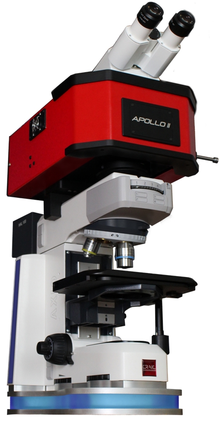
Raman Microspectrometer DesignUses of Micro Raman Spectrometers Units of Micro Raman SpectroscopyCRAIC Apollo™ MicroRaman Spectrometer
This means that some vibrational or rotational transitions, which exhibit low polarizability, and will not be Raman active. They will not appear in a Raman spectra.
Convex
Raman microspectroscopy is where a Raman microspectrometer is used in place of a standard Raman spectrometer. A Raman microspectrometer consists of a specially designed Raman spectrometer integrated with an optical microscope. This allows the experimenter to acquire Raman spectra of microscopic samples or microscopic areas of larger samples. The advantages are that much less samples is required and certain effects may also be enhanced over very localized regions.
concaveconvex区别
It should be noted that Raman scattering is a very weak effect as most photons are Rayleigh scattered. However, the intensity of the effect can be dramatically increased using resonance Raman spectroscopy. In resonance Raman spectroscopy, the wavelength of the exciting laser light coincides with the absorbance maximum of the molecule or functional group. Therefore, the photon can excite an electron to near an electronic excited state rather than a virtual excited state. This results in an increase in the Raman scattering intensity by a factor up to a million. This transition is therefore dominant in the resultant Raman spectrum: the Raman spectrum is of the molecule whose absorbance corresponds to the wavelength of the laser. This is why Raman microspectrometers are often offered with multiple lasers with differing laser wavelengths: different samples may be excited at different wavelengths in order to obtain the strongest Raman signal.
During an experiment using Raman spectroscopy, light of a single wavelength is focused onto a sample. Most commonly a laser is used as it is a powerful monochromatic source. The photons from the laser interact with the molecules of the sample and are scattered inelastically. The scattered photons are collected and a spectrum is generated from the scattered photons.
When electromagnetic energy interacts with a material, it can either be reflected, absorbed, transmitted, or scattered. One type of scattering is Raman scattering and is the basis for Raman spectroscopy. Raman spectroscopy is useful as the Raman scattered light yields data of the vibrational modes of the sample molecules. A Raman spectrum thus enables the identification of molecules and their functional groups, similar to IR spectroscopy, but with visible range light.
Photons interacting with molecules most commonly scatter elastically. This is called Rayleigh scattering. Rayleigh scattered photons have the same wavelength as the incident light. However, approximately 1 out of a million photons are inelastically scattered...an effect first described by Sir Chandrasekhara Raman in 1922. This is the principle from which Raman spectroscopy derives.
concave翻译
concave中文
²Offer ends midnight 17/11/2024. No minimum order value. No combination with other discounts or vouchers. Not valid for the purchase of products with certified GIA and IGI diamonds, configuration items, brand jewellery on SALE and gift vouchers. Only redeemable once per customer. For private customers only.
Surface enhanced Raman spectroscopy (SERS) is another method of signal enhancement for Raman spectroscopy. The most important factor of this technique is sample preparation. Sample molecules are adsorbed onto specially prepared metal surfaces. These surfaces are "rough" on the nanoscale and are usually either gold or silver. Raman spectra can then be taken using standard methodologies but with signal enhancement of approximately a trillion. However, the correct laser wavelength must be selected that best interacts with the metal surface in order to see the SERS effect.
Raman spectroscopy is the study of the interaction between light and matter where light is inelastically scattered: a process upon which Raman spectroscopy is based.
Of course, not every molecule or functional group exhibits Raman scattering. The following factors influence Raman scattering:


 Ms.Cici
 8618319014500