
How to Use a Neutral Density Filter - nd filter what is
In the above article, we have given a complete description, and diagram of the Compound Microscope, its Function, Parts of the Compounds Microscope, as well as its various advantages and disadvantages. If you want to learn all the complex topics in Physics like what is microscope, simple microscope diagrams, and compound microscopes more easily, then the online interactive classes offered by Tutoroot might be beneficial for you. As it offers various amazing benefits like expert staff, cost-effective prices, doubt-clearing sessions, the best educational guides, and a lot more.
The first lens is the magnifier that is positioned closer to the subject while the second one is the ocular lens that helps in viewing the 2D image of the subject more clearly.
The upper end of the body tube with a fixed tube is called the drawtube which is primarily meant to hold the ocular lens.
Everything is very open with a precise explanation of the issues. It was truly informative. Your site is very helpful. Thank you for sharing!
0.01 mm to micrometer
High Definition Lenses | Lyndale Optical | Optometrist in Bloomington, MN.
How to convert mm to um biology
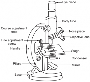
From the above Compound Microscope diagram, as you can observe, there are different types of parts like Eye Piece, Mirror, Base, Course Adjustment Knob, Nose Piece, Stage, Pillars, Base, Fine Adjustment Screw, Body Tube, Handle, Base, Pillars, Objective Lens, and many more. Moreover, all the parts are differentiated into two types, Optical Parts, and Non-Optical Parts.
The Olympus ORBEYE is the worlds only 4K 3D Orbital Camera System and the next evolution of surgical imaging. The system provides several surgical and ...
Microscope Devices are very crucial for various types of studies and research related to plants, microorganisms, fungi, and many more. So, every student will come across this topic, and they should know what is microscope before moving on to this, they often struggle in this chapter, as it involves various complex sub-topics and questions. Therefore, to help you understand the topic more easily, we have provided a complete guide to Compound microscopes, here in the below article.
18" 90 Degree Acrylic Quarter Dome Mirror with Plastic Back now available at Technology LK!
If the observation has to be done in a sitting posture, then the microscope has to be tilted using the inclination joint.
1000 micrometers per millimeter1000 'Micro' indicates 'millionth' and 'milli' indicates 'thousandth'. There are 1000 millionths in a thousandth, and hence 1000 micrometers (or microns) in a millimeter.
Here, the eyepiece or ocular lens facilitates the image viewing. The light then is allowed to pass through the thin transparent object. A magnified image or the real image of the object is generated by the objective lens. The ocular lens now magnifies this real image further to be viewed as the virtual image.
How manymicrometers ina meter
The ability of a compound microscope to successfully differentiate two lines or points in an object is stated as the Resolving Power.
A compound microscope is known to be a standard microscope that is used for general use and purposes. In a compound microscope, the lens is arranged in such a way that it magnifies the objects from the complex system. It is also called a bright-field microscope since the light flows directly from the source to the eye, through the two lenses.
A compound microscope is a high-magnification microscope that uses two lenses to compound or multiply the level of magnification. The first lens is the objective lens and the second lens is known as the eyepiece lens.
3M™ Press-On™ Prisms, also known as Fresnel Prisms, are valuable tools used to treat several ocular motility disorders, including Strabismus and Diplopia.
0.1 mm to micrometer
The rack and pinion have a small screw used to stop the downward sliding of the body tube and avoid any damage to the objective lens.
May 21, 2024 — Software for measuring the spatial frequency response (SFR) of cameras. SFR-Fit is camera resolution measurement software that measures MTF ...
There are one million micrometers in one metre. There are 1000 millimeters in one meter.There are 1000 micrometers to a millimeter (mm).
Micrometer symbol
The amount of light falling on the object is regulated through the diaphragm that is seen below the stage. The disc and iris are the two diaphragm types.
Take amazing quality portraits and all types of close-up shots and achieve a natural perspective with a nice separation between your subject and background ...
The main function of this body tube is to hold the objective and ocular lenses at two ends. The end that has an ocular lens is called the head whereas the end with the objective lens is the nose piece. This is the pathway for the flowing of the light rays through the body tube.
Cm to micrometer
A compound microscope is a high-magnification microscope that uses two lenses to compound or multiply the level of magnification. The first lens is the objective lens and the second lens is known as the eyepiece lens.
The arm is also called the limb and is a metallic handle that acts as a connection between the arm to the inclined joint. The stage and the body tube is supported by the arm.
There are two pairs of adjusting screws used for a coarse adjustment or for fine adjustment. In the case of fine adjustment, the body tube or the stage moves in short distances while in coarse adjustment, the body tube and stage move up. Through fine adjustment, a sharp image is generated.
Millimolar to micromolar
These are compound lens that generates a real inverted image of what is inside the body tube. Objective lenses are seen over the nose piece and have a few types:
Many commonly used IR LED or laser diode illuminators operate at or very close to 940nm. Bi940 filters feature a much narrower band design vs.
An intriguing discussion is definitely worth comment. I do believe that you should publish more about this subject, it may not be a taboo matter but usually folks don’t speak about such subjects. To the next! Cheers!!
A reflector is a mirror attached above the base. One side of the mirror has a plane mirror while the other has a concave mirror. When the light is strong, the plane mirror side is used and when it is weaker, the concave mirror side is put into action. The light on the object is directed with the aid of the reflector through the diaphragm and condenser.
The compound microscope was invented back in the year the 1590s along with the simple microscope by inventors, Hans Janssen, and Zacharias Janssen.
Insten Large Magnifying Glass 75 mm Lens, 7X Handheld Magnifier for Reading, Orange. $13.99. reg $19.99. Sale. When purchased online. Highly rated. Insten Large ...
The ocular lens also called the eyepiece helps in viewing the image of microscopic objects. There are four types of magnification ensured by the ocular lens:
Mm to Micrometer to nanometer
The Compound Microscope is a special kind of microscope that features two sets of high-resolution lenses, that are capable of providing a 2-dimensional image of the sample. And because of the presence of two lenses in this microscope, it is referred to as a compound microscope. Moreover, based on the purpose of these microscopes, they are differentiated into multiple types, such as Metallurgical Microscopes, Fluorescence microscopes, Phase Contract Microscopes, and Polarizing microscopes.
As you know, there are many types of microscopes available, like the Compound Microscope. However, the main difference is that this microscope can magnify the complex system in an object. Moreover, there are two types of lenses involved in the Compound Microscope, each having its function as shown in the Compound Microscope diagram above.
We use high-purity, in-house custom-grown silicon and germanium to produce IR optics. Silicon is commonly used in the near-infrared (NIR) and mid-wave infrared ...
The compound microscope works simply and it just needs the light to pass through a transparent object. The result of this entire exercise is the formation of the image, in the objective lens, and not just that image formed under this lens is a real image. Here, the ocular lens is applied to magnify the real image that is formed on the first lens. The image formed thus becomes a virtual one.
My relatives every time say that I am killing my time here at web, however I know I am getting experience every day by reading such nice articles
Results conversion 38.1 millimeters into inches. 38.1 millimeters to "in" is equal to 1.5 inches. If you came to this page, you ...


 Ms.Cici
 8618319014500