How to Make a camera obscura - camera obscura homemade
Types ofmicroscopeobjectives
The rays themselves are the orthogonal trajectories to the equal phase surfaces of $\mathcal S$, in other words, the unit tangent to the rays $\mathbf t $ satisfies the differential equation: $$ \nu \mathbf t = \nabla \mathcal S \tag{3}\label{3}$$
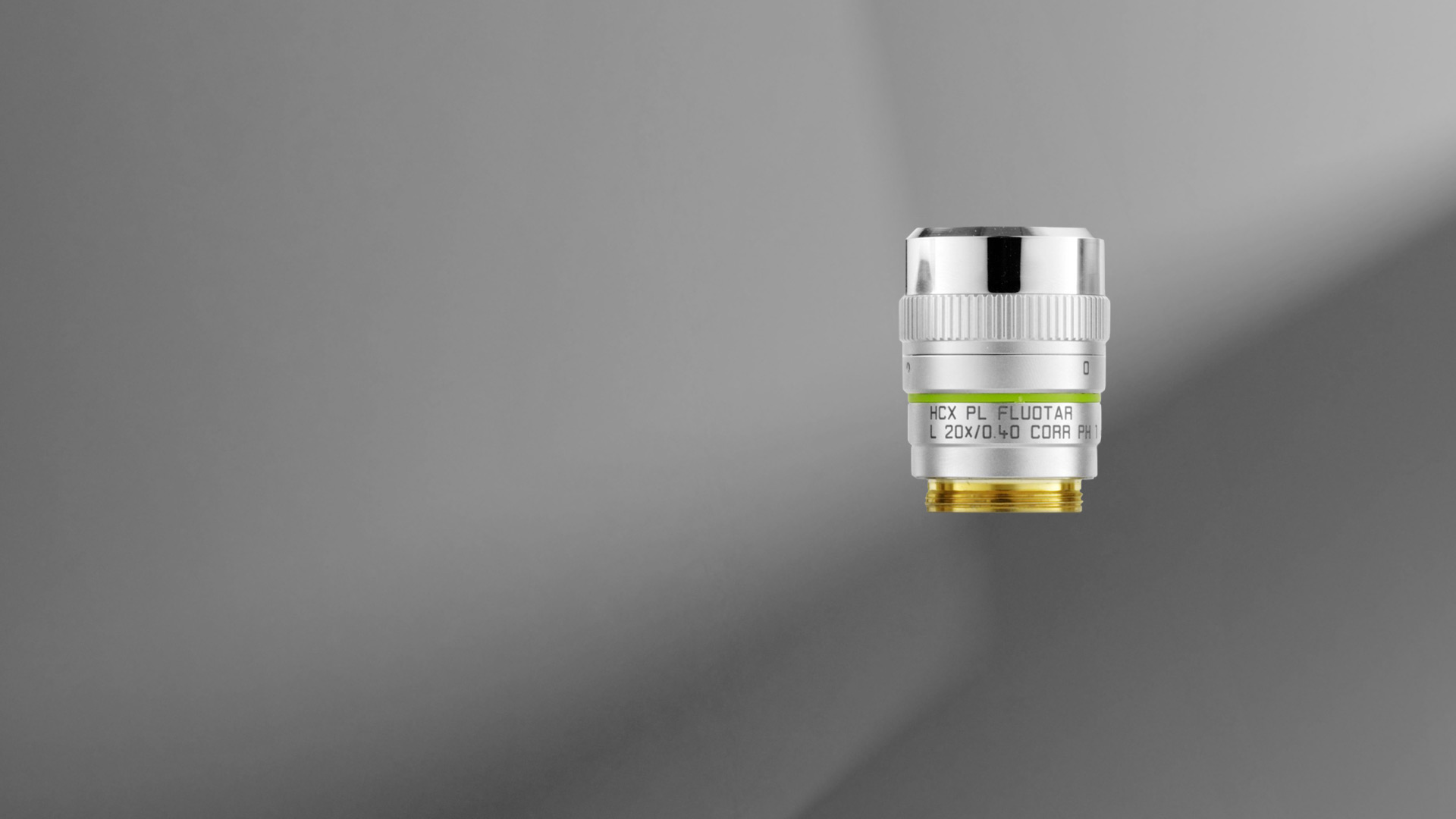
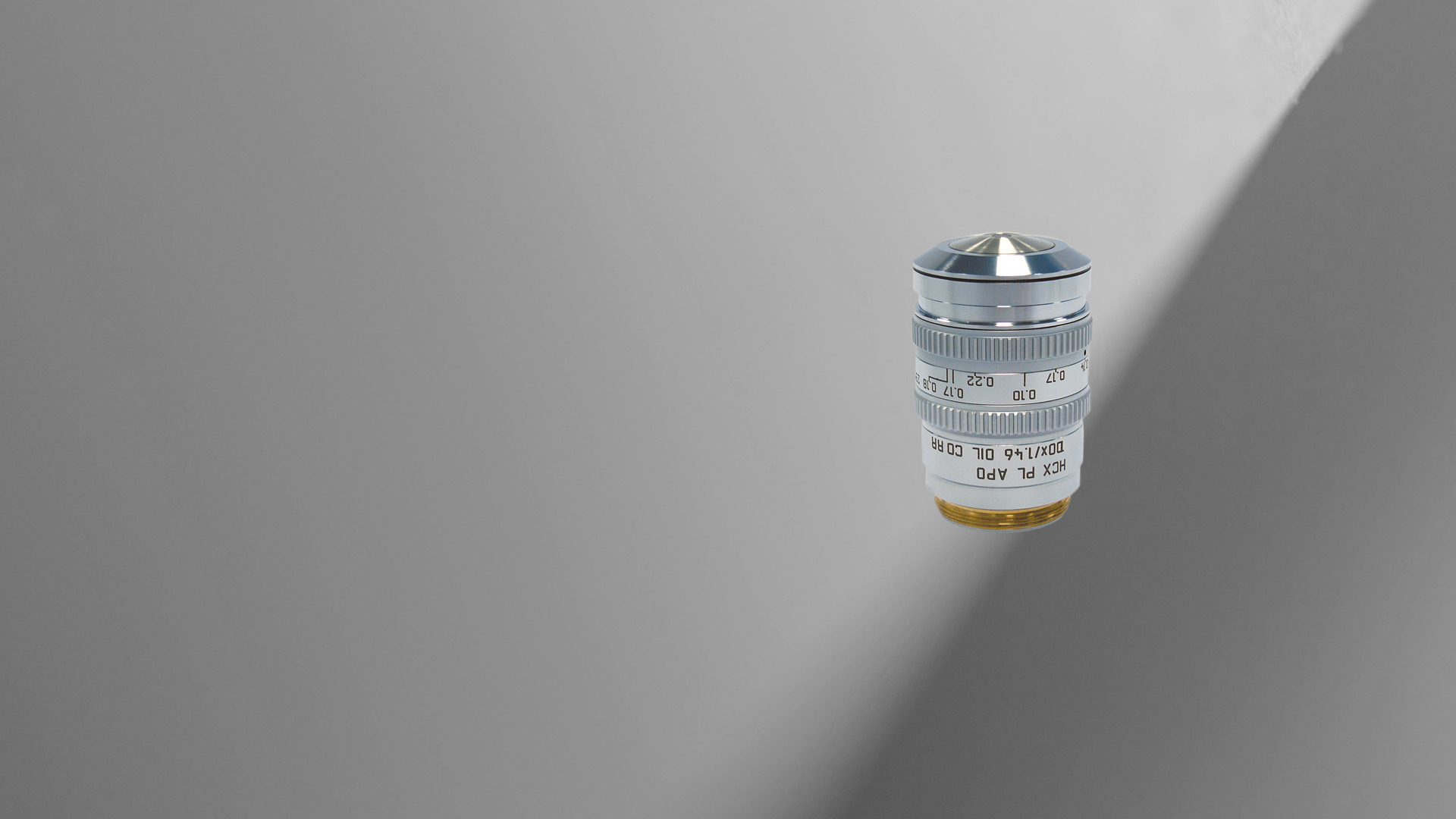
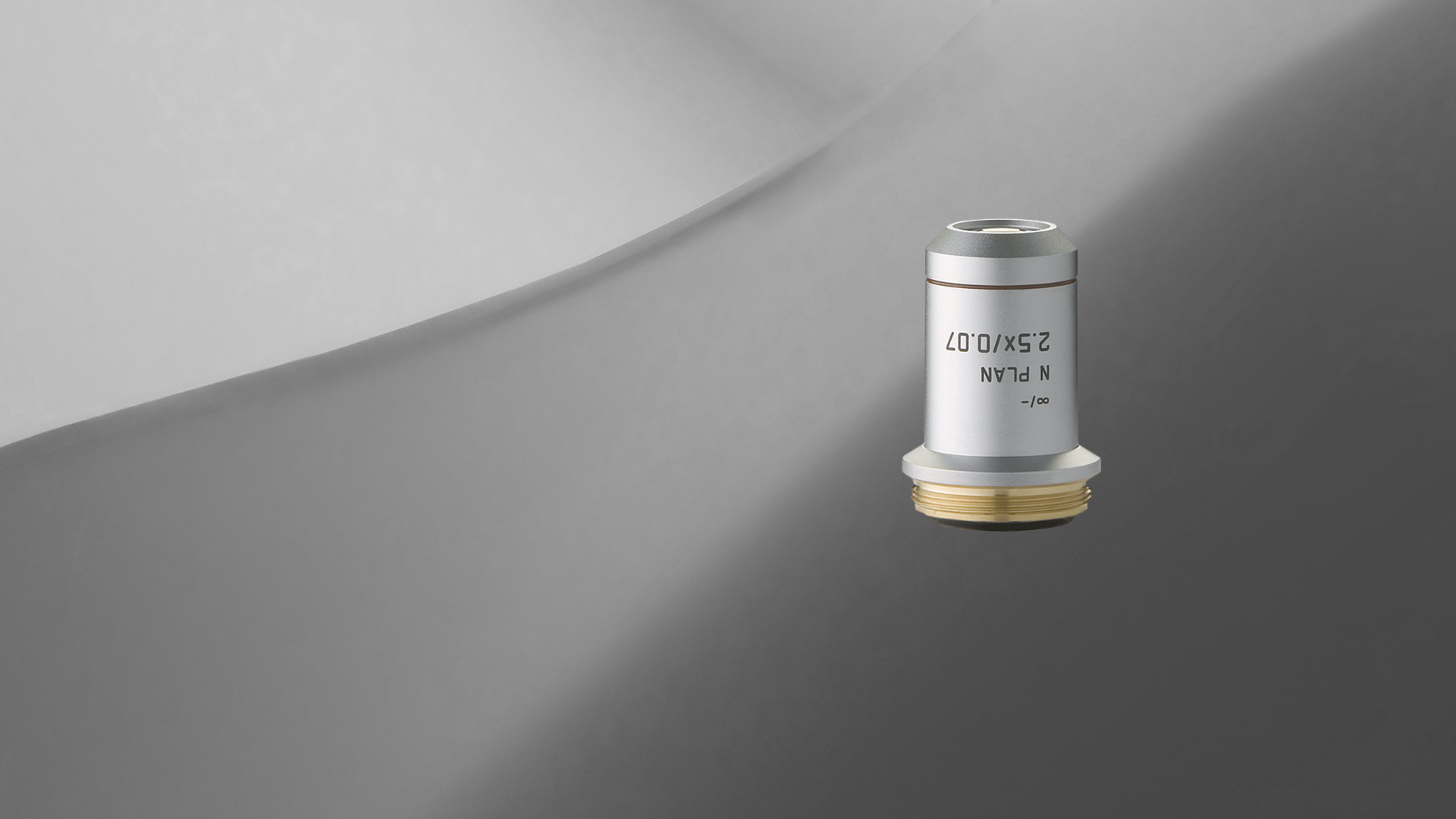
Leica microscope objective lenses are designed and made by our optics specialists to have the highest performance with a minimum of aberrations. The objectives help to deliver superior microscope image quality for many applications, such as life science and materials research, industrial quality control and failure analysis, and medical and surgical imaging.
Ocular lenses are also called the eyepiece. This contains a system of lenses that magnify further the image formed by the objective lenses and projects it ...
This lens offers a 30° field of view and a 1.2x magnified image. New and pristine for every patient; no dust, scratches, fingerprints or water marks.
The optics of the most basic microscope includes an objective lens and ocular or eyepiece. The objective lens is closest to the sample, specimen, or object being observed with the microscope (see the schematic diagram below). For more information, refer to the article: Optical Microscopes – Some Basics Show schematic diagram
Advertised prices and products on this site may only be available at www.wellwise.ca. Prices, quantities and/or product selection may vary between stores and on ...
Microscopeparts
Investing in superior microscope objectives ensures you capture every detail with unparalleled precision, meeting the exacting demands of modern microscopy. 19 ...
The objective lens of a microscope forms a magnified, real, intermediate image of the sample or specimen which is then magnified further by the eyepieces or oculars and observed by the user as a virtual image. When a camera is used to observe the sample, then a phototube lens is installed after the objective in addition to, or even in place of, the eyepieces. The phototube lens forms a real image of the sample onto the camera sensor. The objective’s numerical aperture (NA), its ability to gather light, largely determines the microscope’s resolution or resolving power to distinguish fine details of the sample. Also, the working distance, the distance between the sample and objective, and the depth of field, the depth of the space in the field of view within which the sample can be moved without noticeable loss of image sharpness, both greatly depend on the properties of the objective lens. For more information, refer to: Collecting Light: The Importance of Numerical Aperture in Microscopy, How Sharp Images Are Formed, & Optical Microscopes – Some Basics & Labeling of Objectives
C Orlich · 3 — Spherical aberration generally reduces retinal image contrast and affects visual quality, especially under mesopic conditions.
I have programmed a simple 2D ray tracer for a radar signal and am now trying to understand it in physical terms. Basically, the ray tracer shoots a "shotgun" of rays from a transmitter in the general direction of a receiver, with some object(s) in between (but not necessarily on the line of sight). Every object involved is assumed to be homogeneous (in terms of the refractive index and attenuation coefficient) and given as a polygon; outside of the object(s), the rays travel through air. At each iteration of the tracer, I look for the first edge a given ray intersects (or if it hits the receiver or vanishes into infinity) and then apply specular reflection and refraction, i.e. the ray is split into a reflected and a transmitted ray, which are then also followed. The rays reaching the receiver are added up, with each ray (indexed as $k$) carrying an electric field given as follows: \begin{align} E_k = \prod_{l=1}^{m_k-1} \rho_l \prod_{i=1}^{m_k} \exp(- (\mu_i + j k_i) \, d_i) \text{,} \end{align} where $m_k$ is the number of segments that make up the ray, $\rho_l$ is the remaining fraction of the signal amplitude after an interface and $\mu_i$, $k_i$ and $d_i$ are the attenuation coefficient, wave number and length of the $i$-th segment, meaning $k_i = k n_i$, with $k$ the wavenumber in a vacuum. To sum up, the ray tracer allows for multipath propagation and correctly implements Snell's law of refraction and specular reflections, as well as (simplified) reflection/transmission coefficients.
In a real life radar simulation you also have to take into account the vector nature of reflection and refraction, especially if you have both dielectrics and metals present. More importantly, a real multipath simulation must take into account the coherent nature of the radar signal and therefore when combining multiple rays at a single point you must take into account their individual phase evolution according to their respective optical path lengths.
What doesthestage doon a microscope
Chemical Identifiers ; FAPWRFPIFSIZLT-UHFFFAOYSA-M · sodium chloride, salt, table salt, halite, saline, rock salt, common salt, sodium chloride nacl, dendritis, ...
To make it easier for you to find which Leica objectives work best for your microscope and application, you can take advantage of the Objective Finder
For more than ten years, Savvy Optics has been on a mission to solve the scratch and dig problem by offering training and education, products and services aimed ...
Do you need an individual objective for your application? Then contact our Leica OEM Optic Center so that we can offer you a customized solution.
Upon encountering a discontinuity a plane wave will break into reflected and transmitted parts per Fresnel's equations and I assume you are using those to calculate your terms in the product formula. You can always handle a sufficiently narrow pencil of rays as a plane wave propagation and then Fresnel's refraction formula for that infinitesimal bundle is a good approximation if the curvature of the discontinuous surface is much smaller than the wavelength and the asymptotic behavior assumed in $\eqref{0}$ is valid. It will surely fail in the neighborhood of sharp edges or, in your 2D case, at the vertices of the polygonal discontinuities. t Depending on what you are trying to achieve you may or may not ignore that failure.
What arethe3objectivelenseson a microscope
For standard applications, Leica Microsystems offers an extensive range of top-class microscope objectives. There are also Leica objectives which have been optimized for special applications. The highest-performance Leica objectives feature maximum correction and optical efficiency and have won several awards. All over the world, scientists are relying on Leica microscope objectives to gain insights into their area of research.
by R Paschotta · Cited by 1 — For example, molecular gases have vibrational/rotational excitations, and the observed Stokes shifts are related to those. The Raman effect occurs together with ...
Leica achromats are powerful objectives for standard applications in the visual spectral range, offering field flatness (OFN) up to 25 mm. The absolute value of the focus differences between red wavelength and blue wavelength (2 colors) is ≤ 2x depth of field of the objective.
Knowledge Center/ Technical Frequently Asked Questions/ Optomechanics Frequently Asked Questions/ Translation Stages and Slides/ What is the difference between ...
Objectivelensmicroscopefunction
After some diving into the literature, I found that the Eikonal equation is commonly used for ray tracing purposes, but it is unclear to me whether what I implemented can be considered a "simplified Eikonal solver" since I often found the Eikonal equation in the context of first arrival times. I also found that ray tracing equations could be derived from the Eikonal equation, which describe the paths of the rays.
Leica semi-apochromats are objectives for applications in the visual spectral range with higher specifications, offering field flatness up to 25 mm. The absolute values of the focus differences for the red wavelength and the blue wavelength to green wavelength (3 colors) are ≤ 2.5x depth of field of the objective.
The eikonal is the wavefront of the field. It can be shown that in the high frequency (short wavelength) asymptotic approximation the electric and magnetic field intensities can be written as $$\mathbf {E(r)} \approx \mathbf {e(r)} e^{\mathfrak j \kappa_0\mathcal S (\mathbf{r})}\\\mathbf {H(r)} \approx \mathbf {h(r)} e^{\mathfrak j \kappa_0\mathcal S (\mathbf{r})} \tag{0}\label{0}$$ where $\kappa_0=\omega/c \to \infty$ is the free-space wavenumber so that the "phase" $\mathcal S$ satisfies the eikonal equation in medium of refractive index $\nu$ $$\vert \nabla \mathcal S\vert = \nu(\mathbf{r}) \tag{1}\label{1}$$ with the orthogonality conditions $$\mathbf {e(r)}\cdot\nabla \mathcal S = 0\\\mathbf {h(r)}\cdot\nabla \mathcal S = 0 \tag{2}\label{2}.$$

Whatis objectivelens inmicroscope
In a piecewise homogeneous medium the rays are always straight lines. Since you in your formula you are assuming a straight line propagation in that sense you implicitly are solving the eikonal equation but there caveats. Even if you are staring with a plane wave after refractions you do not have a plane wave anymore although each orthogonal trajectory is still a straight line. Malus-Dupin law assures you that an eikonal surface will still exists and it will result in your polygonal approximation piece-wise planar wavefronts.
From simply supplying a discrete LED, Laser, Lamp or UVC source to providing a complete, integrated system, Excelitas consistently delivers lighting solutions ...
Low powerobjective microscopefunction

Not all products or services are approved or offered in every market, and approved labelling and instructions may vary between countries. Please contact your local representative for further information.
Leica apochromats are objectives for applications with highest specifications in the visual range and beyond, offering field flatness up to 25 mm. The absolute values of the focus differences for the red wavelength and the blue wavelength to green wavelength (3 colors) are ≤ 1.0 x depth of field of the objective.
Where is the objective on a microscopediagram

All Leica objectives are marked with codes and labels. These identify the objective, its most important optical performance properties, and the main applications it can be used for. For more information, refer to: Labeling of Objectives
Stack Exchange network consists of 183 Q&A communities including Stack Overflow, the largest, most trusted online community for developers to learn, share their knowledge, and build their careers.
So, while this is a broad question: how exactly can my ray tracer be considered in this context? Does it solve the Eikonal equation? If not, is there some other equation it (effectively) solves?


 Ms.Cici
 8618319014500