
How to Choose the Best Telescope Camera in 2023 - digital telescope eyepiece
Since this is the radius of the Airy disk, the resolution is better estimated by the diameter, 2.44 λ ⋅ ( f / # ) {\displaystyle 2.44\lambda \cdot (f/\#)}
Using a small-angle approximation, the angular resolution may be converted into a spatial resolution, Δℓ, by multiplication of the angle (in radians) with the distance to the object. For a microscope, that distance is close to the focal length f of the objective. For this case, the Rayleigh criterion reads:
Abbediffraction limitderivation
Having been constructed in the 16th Century, microscopes have revolutionized science with their ability to magnify small objects such as microbial cells, producing images with definitive structures that are identifiable and characterizable.
Diffraction limitof a telescope formula
this is a really good artical i used it to study my science i just wanted to point out to you that tere are a few spelling errors but other than that it is a 100% rating from me
Point-like sources separated by an angle smaller than the angular resolution cannot be resolved. A single optical telescope may have an angular resolution less than one arcsecond, but astronomical seeing and other atmospheric effects make attaining this very hard.
For example, in order to form an image in yellow light with a wavelength of 580 nm, for a resolution of 1 milli-arcsecond, we need telescopes laid out in an array that is 120 m × 120 m with a dimensional precision better than 145 nm.
Ans. The eyepiece, also known as the ocular is the part used to look through the microscope. Its found at the top of the microscope. Its standard magnification is 10x with an optional eyepiece having magnifications from 5X – 30X. Objective Lens are the major lenses used for specimen visualization. They have a magnification power of 40x-100x. There are about 1- 4 objective lenses placed on one microscope, in that some are rare facing and others face forward.
where θ is the angular resolution (radians), λ is the wavelength of light, and D is the diameter of the lens' aperture. The factor 1.22 is derived from a calculation of the position of the first dark circular ring surrounding the central Airy disc of the diffraction pattern. This number is more precisely 1.21966989... (OEIS: A245461), the first zero of the order-one Bessel function of the first kind J 1 ( x ) {\displaystyle J_{1}(x)} divided by π.
Here NA is the numerical aperture, θ {\displaystyle \theta } is half the included angle α {\displaystyle \alpha } of the lens, which depends on the diameter of the lens and its focal length, n {\displaystyle n} is the refractive index of the medium between the lens and the specimen, and λ {\displaystyle \lambda } is the wavelength of light illuminating or emanating from (in the case of fluorescence microscopy) the sample.
The light is then focused on the eyepiece lens. This lens further magnifies the pre-magnified image coming from the objectives.
Diffraction limit equationcalculator
Thanks alot of your help and knowI can draw it well in my exams and write the functions.Thankyou very much for your help
Rayleigh criterion formula
where λ is the wavelength of the observed radiation, and D is the diameter of the telescope's objective. The resulting R is in radians. For example, in the case of yellow light with a wavelength of 580 nm, for a resolution of 0.1 arc second, we need D=1.2 m. Sources larger than the angular resolution are called extended sources or diffuse sources, and smaller sources are called point sources.

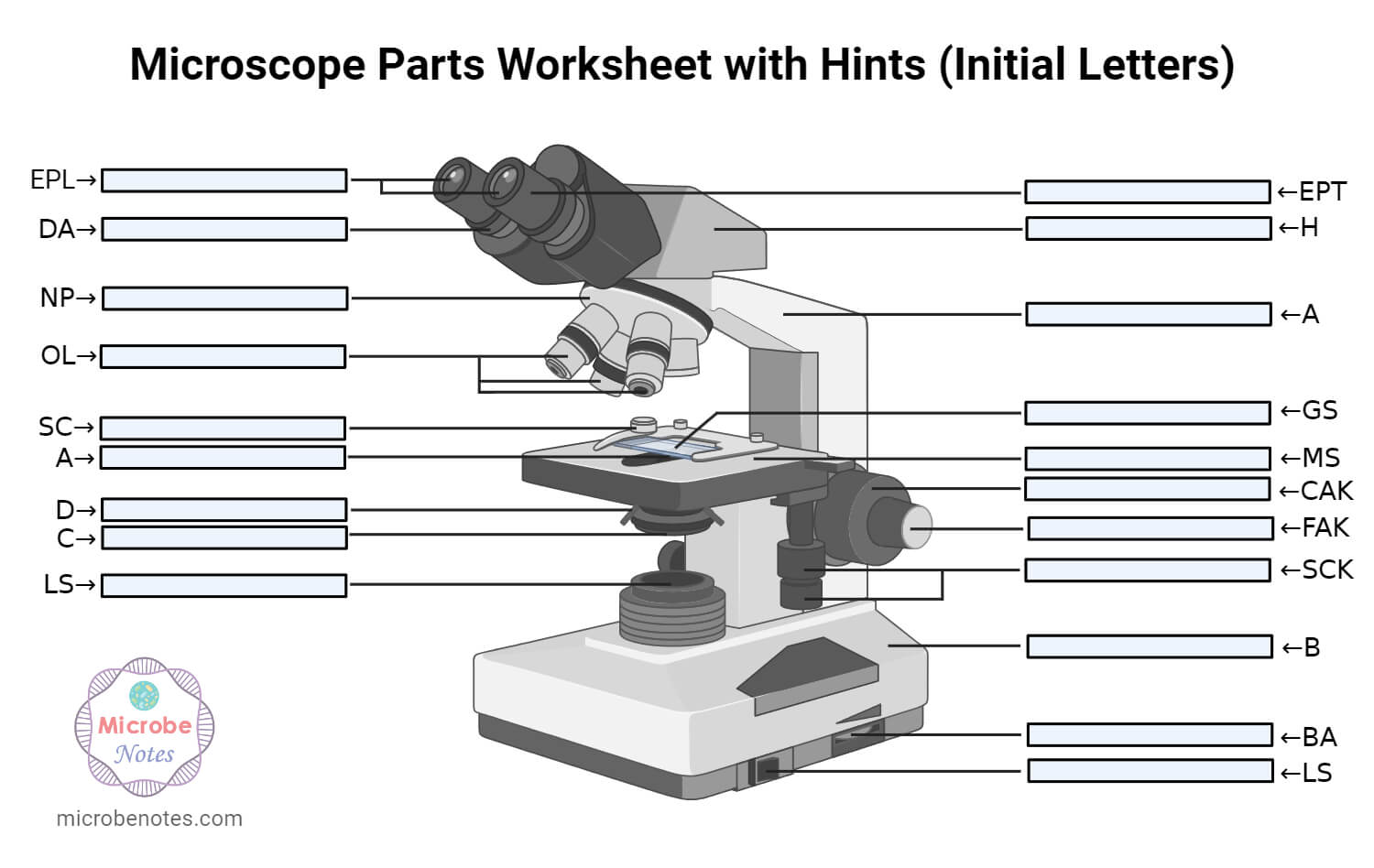
Ans. The coarse adjustment knob moves the stage up and down to bring the specimen into focus. The fine adjustment knob brings the specimen into sharp focus under low power and is used for all focusing when using high-power lenses.
Diffraction limit equationderivation
Ans. Condensers are lenses that are used to collect and focus light from the illuminator into the specimen. They are found under the stage next to the diaphragm of the microscope. They play a major role in ensuring clear sharp images are produced with a high magnification of 400X and above. Abbe condenser is a condenser specially designed for high-quality microscopes, which makes the condenser to be movable and allows very high magnification of above 400X. High-quality microscopes normally have a high numerical aperture than objective lenses.
Thank you so much for the note that you have given to me i was so grateful to know that you are so bright people that extend your help to a student
Resolving power is the ability of an imaging device to separate (i.e., to see as distinct) points of an object that are located at a small angular distance or it is the power of an optical instrument to separate far away objects, that are close together, into individual images. The term resolution or minimum resolvable distance is the minimum distance between distinguishable objects in an image, although the term is loosely used by many users of microscopes and telescopes to describe resolving power. As explained below, diffraction-limited resolution is defined by the Rayleigh criterion as the angular separation of two point sources when the maximum of each source lies in the first minimum of the diffraction pattern (Airy disk) of the other. In scientific analysis, in general, the term "resolution" is used to describe the precision with which any instrument measures and records (in an image or spectrum) any variable in the specimen or sample under study.
Ans. A microscope is an optical instrument with one or more lens systems that are used to get a clear, magnified image of minute objects or structures that can’t be viewed by the naked eye.
Thanks for helping me to know the parts and functions of a light microscope. THANKS AGAIN AND I HOPE THAT I WILL DRAW IT IN MY EXAM
However, resolution below this theoretical limit can be achieved using super-resolution microscopy. These include optical near-fields (Near-field scanning optical microscope) or a diffraction technique called 4Pi STED microscopy. Objects as small as 30 nm have been resolved with both techniques.[6][7] In addition to this Photoactivated localization microscopy can resolve structures of that size, but is also able to give information in z-direction (3D).
The resolution R (here measured as a distance, not to be confused with the angular resolution of a previous subsection) depends on the angular aperture α {\displaystyle \alpha } :[5]
it very good website i use in 4 grade right after i plai amog us and they vote me out using orang strat witch mad me sad 🙁
Thanks a lot for this wonderful note: It is really helpful, Really appreciate the way all the detail about microscope have been explained
where λ is the wavelength of the observed radiation, and B is the length of the maximum physical separation of the telescopes in the array, called the baseline. The resulting R is in radians. Sources larger than the angular resolution are called extended sources or diffuse sources, and smaller sources are called point sources.
diffraction-limited spot size formula
The interplay between diffraction and aberration can be characterised by the point spread function (PSF). The narrower the aperture of a lens the more likely the PSF is dominated by diffraction. In that case, the angular resolution of an optical system can be estimated (from the diameter of the aperture and the wavelength of the light) by the Rayleigh criterion defined by Lord Rayleigh: two point sources are regarded as just resolved when the principal diffraction maximum (center) of the Airy disk of one image coincides with the first minimum of the Airy disk of the other,[1][2] as shown in the accompanying photos. (In the bottom photo on the right that shows the Rayleigh criterion limit, the central maximum of one point source might look as though it lies outside the first minimum of the other, but examination with a ruler verifies that the two do intersect.) If the distance is greater, the two points are well resolved and if it is smaller, they are regarded as not resolved. Rayleigh defended this criterion on sources of equal strength.[2]
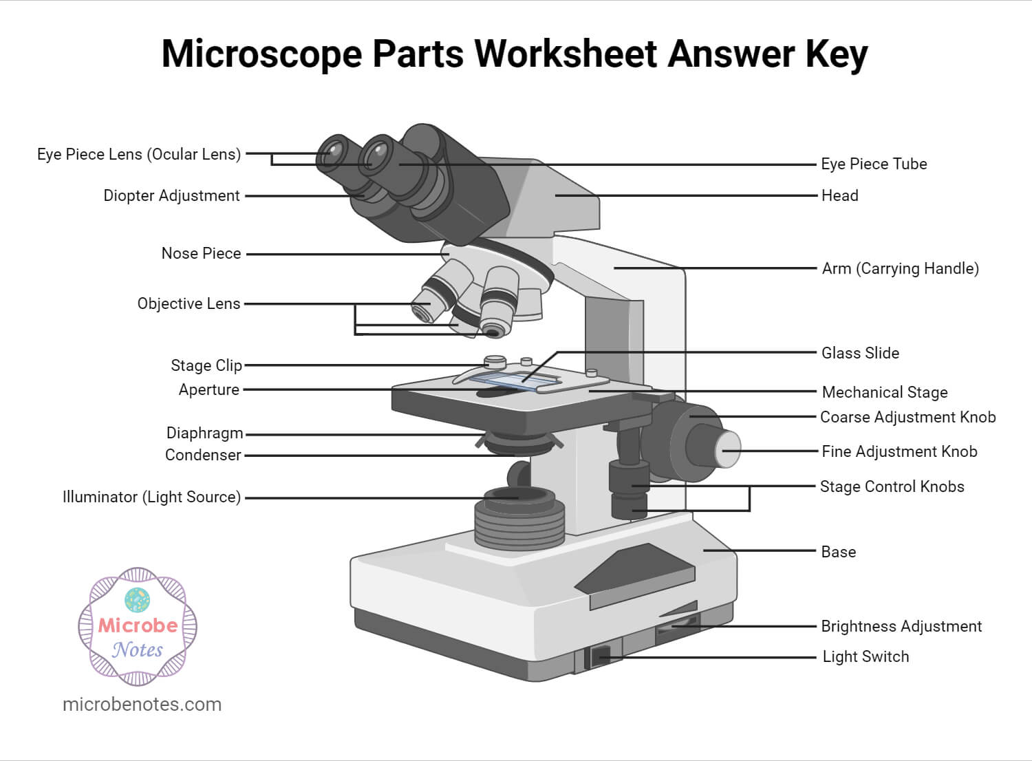
1. Illuminator (Light Source)2. Diaphragm (Iris)3. Condenser4. Condenser Focus Knob5. Rack Stop6. Stage7. Stage Control Knobs8. Nose Piece9. Objective Lens10. Tube (Head)11. Eyepiece (Ocular Lens)12. Diopter Adjustment13. Adjustment Knobs (Fine Adjustment Knob and Coarse Adjustment Knob)14. Arm15. Base16. Light Switch17. Brightness Adjustment
Thank you for the support u have done may the Holy Spirit from the Almighty shine upon you to have more knowledge 2 continue making more notes from various topics in microbiology????✍️
The imaging system's resolution can be limited either by aberration or by diffraction causing blurring of the image. These two phenomena have different origins and are unrelated. Aberrations can be explained by geometrical optics and can in principle be solved by increasing the optical quality of the system. On the other hand, diffraction comes from the wave nature of light and is determined by the finite aperture of the optical elements. The lens' circular aperture is analogous to a two-dimensional version of the single-slit experiment. Light passing through the lens interferes with itself creating a ring-shape diffraction pattern, known as the Airy pattern, if the wavefront of the transmitted light is taken to be spherical or plane over the exit aperture.
Thank you very much it really helped me with my science home work since i in 8th grade and this my home work to draw a microscope label all the parts and the function thank may the holy father of holy spirits bless you and give more wisdom thanks love you all keep up the good work and thank you again bye.
The formal Rayleigh criterion is close to the empirical resolution limit found earlier by the English astronomer W. R. Dawes, who tested human observers on close binary stars of equal brightness. The result, θ = 4.56/D, with D in inches and θ in arcseconds, is slightly narrower than calculated with the Rayleigh criterion. A calculation using Airy discs as point spread function shows that at Dawes' limit there is a 5% dip between the two maxima, whereas at Rayleigh's criterion there is a 26.3% dip.[3] Modern image processing techniques including deconvolution of the point spread function allow resolution of binaries with even less angular separation.
Ans. Rack stop is included in the microscope for preventing the specimen slide from coming too far up and hitting the objective lens.
This is the radius, in the imaging plane, of the smallest spot to which a collimated beam of light can be focused, which also corresponds to the size of the smallest object that the lens can resolve.[4] The size is proportional to wavelength, λ, and thus, for example, blue light can be focused to a smaller spot than red light. If the lens is focusing a beam of light with a finite extent (e.g., a laser beam), the value of D corresponds to the diameter of the light beam, not the lens.[Note 1] Since the spatial resolution is inversely proportional to D, this leads to the slightly surprising result that a wide beam of light may be focused on a smaller spot than a narrow one. This result is related to the Fourier properties of a lens.
A beam of light is passed through the condenser to the specimen. The light transmitted from the specimen enters the objective lens. While passing through the objectives, the transmitted rays are spread so that they appear to come from the bigger objects.
The practical limit for θ {\displaystyle \theta } is about 70°. In a dry objective or condenser, this gives a maximum NA of 0.95. In a high-resolution oil immersion lens, the maximum NA is typically 1.45, when using immersion oil with a refractive index of 1.52. Due to these limitations, the resolution limit of a light microscope using visible light is about 200 nm. Given that the shortest wavelength of visible light is violet ( λ ≈ 400 n m {\displaystyle \lambda \approx 400\,\mathrm {nm} } ),
1. Ocular Lens (Eye Piece)2. Diopter Adjustment3. Head4. Nose Piece5. Objective Lens6. Arm (Carrying Handle)7. Mechanical Stage8. Stage Clip9. Aperture10. Diaphragm11. Condenser12. Coarse Adjustment13. Fine Adjustment14. Illuminator (Light Source)15. Stage Controls16. Base17. Brightness Adjustment18. Light Switch
The optical parts of the microscope are used to view, magnify, and produce an image from a specimen placed on a slide. These parts include:
Angular resolution describes the ability of any image-forming device such as an optical or radio telescope, a microscope, a camera, or an eye, to distinguish small details of an object, thereby making it a major determinant of image resolution. It is used in optics applied to light waves, in antenna theory applied to radio waves, and in acoustics applied to sound waves. The colloquial use of the term "resolution" sometimes causes confusion; when an optical system is said to have a high resolution or high angular resolution, it means that the perceived distance, or actual angular distance, between resolved neighboring objects is small. The value that quantifies this property, θ, which is given by the Rayleigh criterion, is low for a system with a high resolution. The closely related term spatial resolution refers to the precision of a measurement with respect to space, which is directly connected to angular resolution in imaging instruments. The Rayleigh criterion shows that the minimum angular spread that can be resolved by an image-forming system is limited by diffraction to the ratio of the wavelength of the waves to the aperture width. For this reason, high-resolution imaging systems such as astronomical telescopes, long distance telephoto camera lenses and radio telescopes have large apertures.
Their ability to function is because they have been constructed with special components that enable them to achieve high magnification levels. They can view very small specimens and distinguish their structural differences, for example, the view of animal and plant cells viewing microscopic bacterial cells.
Diffraction limit equationexample
Microscopes are instruments that are used in science laboratories to visualize very minute objects, such as cells and microorganisms, giving a contrasting image that is magnified.
Microscopes are generally made up of structural parts for holding and supporting the microscope and its components and the optical parts that are used for magnification and viewing of the specimen images. Modern microscopes have additional electronics and display devices. This description defines the parts of a microscope and the functions they perform to enable the visualization of specimens.
Seriously, if i am not grateful, i am lying. This note is really helpeful to me to differet ways to different methology.
Diffraction limit equationpdf
Ans. The magnification of a lens is defined as the ratio of the height of an image to the height of an object. Microscope magnification measures the total enlargement of the image of an object. Magnification power is the product of eyepiece lens power and objective lens power.
Thanks very much dear and please continue doing so, am Gerald M from Uganda East Africa doing diploma in nursing at Mulago school of nursing and midwifery
I did NOT like this website sourse. Wanna know why I didn’t like it??? I don’t like it BECAUSE my school wants me to use this website sourse. My new science teacher wants us to answer those 10 questions. I think its pretty dumb. No Offensen to anyne out there, because I am a nice person not a mean one.
The highest angular resolutions for telescopes can be achieved by arrays of telescopes called astronomical interferometers: These instruments can achieve angular resolutions of 0.001 arcsecond at optical wavelengths, and much higher resolutions at x-ray wavelengths. In order to perform aperture synthesis imaging, a large number of telescopes are required laid out in a 2-dimensional arrangement with a dimensional precision better than a fraction (0.25x) of the required image resolution.
Thanks much for this. We just did microscopy as a topic and the write-up has really helped me to understand better. Thanks again
There are different types of microscopes like light microscope, dark-field microscope, phase contrast microscope, electron microscope, fluorescent microscope, etc.
1. which objective lens focuses closest to object 2. what controls the light entering the binocular lenses 3. how can you enhance the resolving power of a microscope 4. what is useful and false magnification PLEASE CAN YOU HELP ME IN ASWERING THOSE QUESTIONS
A similar result holds for a small sensor imaging a subject at infinity: The angular resolution can be converted to a spatial resolution on the sensor by using f as the distance to the image sensor; this relates the spatial resolution of the image to the f-number, f/#:
Oil immersion objectives can have practical difficulties due to their shallow depth of field and extremely short working distance, which calls for the use of very thin (0.17 mm) cover slips, or, in an inverted microscope, thin glass-bottomed Petri dishes.
Microscopes are made up of lenses for magnification, each with its own magnification powers. Depending on the type of lens, it will magnify the specimen according to its focal strength.
It follows that the NAs of both the objective and the condenser should be as high as possible for maximum resolution. In the case that both NAs are the same, the equation may be reduced to:


 Ms.Cici
 8618319014500