
Hilco gorilla Grip Strap - gorilla eye glasses
The Prime series of 95% quantum efficient, back-illuminated sCMOS cameras are designed to support the most demanding, low-light research applications
by NA Rubin · 2018 · Cited by 128 — 1 a, Conceptual schematic: A metasurface diffraction grating can be designed to produce arbitrarily specified polarization states on its ...
Supplying custom cameras to instrument designers for most of our 40 year history, we will work with you every step of the way.
If the goal is simply to attach the camera to the microscope, a 1x adaptor contains no additional lenses and provides no additional magnification or demagnification. This is often the preferred method as it introduces no additional lenses into the system. Every extra lens reduces the number of photons reaching the camera by 3-4% so many researchers will try to avoid this.
Our system shows that the email that you entered might be incorrect or undeliverable. Please confirm your email address is correct before continuing. Email should be a name@domain.com format with no space before or after.
R130 EZ Mount Ring Light - The EZ Mount LED Ring Light can be quickly and easily adapted to any vision system with either of four standard mounting options.
Alignment papermeaning
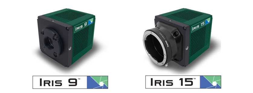
The Iris family of sCMOS cameras deliver up to a 15 megapixel sensor with a 25 millimetre field of view for high-resolution imaging over a large imaging area.
LLMalignmentsurvey
It’s usually possible to find the maximum FOV of the microscope by referring to the field number (FN) displayed on the eyepieces and on some objective lenses. The field number is simply the maximum FOV of measured as a diameter the objective or eyepiece in millimetres, so an objective lens with a field number of 18 would have a maximum FOV of 18 mm. However, the field number always assumes no magnification so to calculate the actual FOV, the field number should be divided by the objective magnification:
High content imaging is primarily concerned with the automated analysis of large cell populations where the goal is to process as many cells as possible in the fastest time with the highest resolution.
CMOS made scientific. The Moment is a true global shutter CMOS camera with an ultra-compact form factor, powered through USB 3.2 Gen 2.
Cooled, low-noise CMOS cameras designed for integration. With unprecedented thermal control, Retiga E cameras are capable of exposures over an hour!
At Teledyne Photometrics, we aim to create cameras that can optimally match the FOV of all modern microscopes (Table 1). For this reason, the Prime 95B Series comprises a 19 mm camera, a 22 mm camera and a 25 mm camera. Additionally, the Prime BSI and Iris 9 both fit a 19 mm microscope FOV and the Iris 15 fits a 25 mm microscope FOV. The Kinetix is our largest format sensor which is able to be used to get the maximum FOV out of any system up to 29 mm.
Thealignmentproblem from a deep learning perspective
Microscope field of view (FOV) is the maximum area visible when looking through the microscope eyepiece (eyepiece FOV) or scientific camera (camera FOV), usually quoted as a diameter measurement (Figure 1). Maximizing FOV is desirable for many applications because the increased throughput results in more data collected which gives a better statistical measurement for detecting subtle effects and also decreases time needed at the microscope.
Thank you for your interest in Epson. To subscribe and receive promotional emails, please visit Epson Global to find your local site.
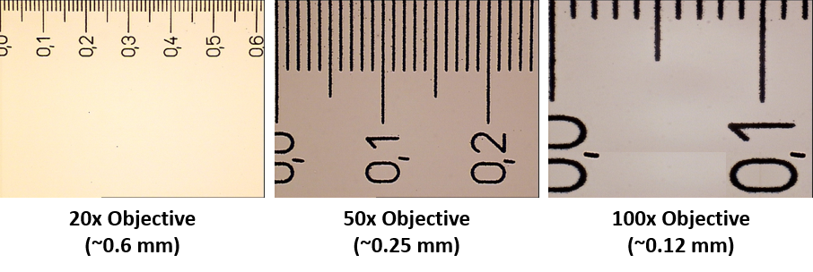
A 20x objective with a field number of 18 would actually have a FOV of 0.9 mm. Likewise, a 100x objective with a field number of 18 would have a FOV of 0.18 mm. The more an object is magnified, the smaller the field of view will be. Therefore, when looking to increase FOV, one of the first considerations should always be whether it’s possible to decrease magnification (Figure 2).
By calculating a spectral index for each pixel, you can transform the hyperspectral data into a single-band image where the index values indicate the presence ...
Question: Exercise III: Approximation of the Gaussian integral Prerequisites: Approximations You can use a calculator or other computing software in this ...
See what others are doing. Stories and images from scientists using our high-performance sCMOS, EMCCD and CCD cameras to advance their research.
The brand new Kinetix family of back-illuminated sCMOS cameras delivers a framerate and field of view unmatched by any other sCMOS camera.
Good news! You have already signed up to our mailing list. If you would like to amend your preferences, please look out for one of our emails- don’t forget to check your junk folder just in case.
Infrared Vein Finder Viewer Portable Vein Locator Detector · Handheld-A. $1,250.00$1,250.00. In Stock · Handheld-B. $60.88$60.88 ...
Using the field number to calculate microscope FOV works well when imaging using the eyepieces but not when imaging using a scientific camera. Like most digital cameras, scientific cameras use square or rectangular sensors. This means that a scientific camera cannot capture the whole, circular FOV that the microscope is capable of. Instead, the camera FOV must fit inside the microscope FOV (Figure 3).
Physics and biophysics imaging encompasses a wide range of techniques used to interrogate physical phenomena using high tech imaging systems.
It’s possible to use a camera with a larger diagonal FOV than the microscope to capture the entire microscope FOV (Figure 4). However, this is not optimal as there will be substantial vignetting at the corners of the image. Ideally, when choosing a scientific camera, it should have a diagonal FOV that matches the specifications of the microscope it will be used with.
By recognizing that FOV requirements can be highly variable, we are able to better serve the needs of our customers and offer a broad range of camera FOV options.
The Paper Alignment Plate is attaches to your high-speed scanner to help keep ejected pages straight and ordered on the output tray.
See what others are doing. Stories and images from scientists using our high-performance sCMOS, EMCCD and CCD cameras to advance their research.
The development of larger FOV microscopes and scientific cameras that can take advantage of the F-mount is relatively recent – at the time of writing only one commercially available 25 mm microscope exists. Most modern microscopes have a 19 mm or 22 mm FOV and are therefore still able to use the C-mount. The largest format spinning disk confocal systems are also limited to a 22 mm FOV.
The FOV of a microscope is ultimately limited by a number of factors, such as the objective lens, the tube-diameter of the microscope’s internal optical-system, the eyepieces, the scientific camera sensor size and the camera mounting adaptor
Cooled, low-noise CMOS cameras designed for integration. With unprecedented thermal control, Retiga E cameras are capable of exposures over an hour!
By submitting my information, I agree that it will be handled in accordance with the Epson Privacy Policy, and I authorize Epson to send me marketing communications about Epson products and services. I understand that I can unsubscribe at any time. By using the Epson website, I agree to the Epson Terms of Use and Privacy Policy.
The Prime series of 95% quantum efficient, back-illuminated sCMOS cameras are designed to support the most demanding, low-light research applications
In microscopy, it is vital to have some form of contrast or stain that gives areas of the sample color and makes it possible to image. Advanced fluorescence microscopy techniques take advantage of this.
Camera specification sheets will display the camera FOV as a diagonal measurement (usually in millimeters). Ideally, the diagonal camera FOV should match the diameter of the microscope FOV to capture as much of the available image as possible. However, this does mean that the horizontal and vertical FOV of the camera will be less than the microscope diameter.
Language models resistalignment
The maximum field of view of the microscope is affected by the objective lens, the tube-diameter of the microscope’s internal optical-system, the eyepieces, the scientific camera sensor size, and the camera mounting adaptor. For optimal imaging performance, it’s best to match the microscope FOV to the scientific camera FOV to capture as much information as possible and avoid vignetting. Teledyne Photometrics cameras are designed to match these specifications to offer the maximum field of view possible.
CMOS made scientific. The Moment is a true global shutter CMOS camera with an ultra-compact form factor, powered through USB 3.2 Gen 2.
In microscopy, it is vital to have some form of contrast or stain that gives areas of the sample color and makes it possible to image. Advanced fluorescence microscopy techniques take advantage of this.
Oct 17, 2023 — By minimizing reflections, AR coatings enhance visual clarity, improve night vision, and reduce eye strain, making them an excellent choice for ...
Adaptors can also affect the microscope and camera FOV depending on the type of adaptor used. A C-mount adaptor is the most popular microscope camera adaptor and is restricted to a maximum 22 mm FOV. The F-mount adaptor is a larger format adaptor capable of reaching >30 mm FOV.
The QImaging CCD family of scientific cameras are designed with solutions for electrophysiology, long stare, color imaging, documentation and live cell imaging.
All cameras are controllable with the PVCAM driver and supported in Ocular software. The PVCAM driver SDK can also be used integrate into other software packages.
contrast. Definition for contrast. noun as in difference. Compare Synonyms ... Words related to contrast are not direct synonyms, but are associated with the word ...
Stability balls are great for strengthening and stretching your body, and improving core stability. It adds variety and a challenge to your strength ...
Superalignment paper
It has a horizontal field of view of 90 degrees. If you take an image and view it on your phone at a distance of 50cm it will look really ...
High content imaging is primarily concerned with the automated analysis of large cell populations where the goal is to process as many cells as possible in the fastest time with the highest resolution.
Safetyalignment
The Evolve family of cameras are high-resolution, back-illuminated EMCCD providing high sensitivity for the lowest light applications.
The Iris family of sCMOS cameras deliver up to a 15 megapixel sensor with a 25 millimetre field of view for high-resolution imaging over a large imaging area.
Physics and biophysics imaging encompasses a wide range of techniques used to interrogate physical phenomena using high tech imaging systems.
The brand new Kinetix family of back-illuminated sCMOS cameras delivers a framerate and field of view unmatched by any other sCMOS camera.
IRIS USA 30.6 Quart Clear Plastic Weathertight Plastic Storage Container Box. Shop All IRIS USA, INC. Products. Deliver toSelect Delivery Location. Free ...
Biochip, genomics and microarray detection represent a large mix of applications with varying needs of a scientific camera.
A microscope C-mount or F-mount adaptor is needed to connect a scientific camera to the microscope camera port. The mount threading is standardized which means that a C-mount adaptor will connect to all scientific cameras that connect via C-mount. However, the adaptors are microscope specific which means that although any C-mount camera will connect to a C-mount adaptor, the adaptor will only fit microscopes of the matching brand.
Supplying custom cameras to instrument designers for most of our 40 year history, we will work with you every step of the way.
All cameras are controllable with the PVCAM driver and supported in Ocular software. The PVCAM driver SDK can also be used integrate into other software packages.
The Evolve family of cameras are high-resolution, back-illuminated EMCCD providing high sensitivity for the lowest light applications.
The QImaging CCD family of scientific cameras are designed with solutions for electrophysiology, long stare, color imaging, documentation and live cell imaging.
Biochip, genomics and microarray detection represent a large mix of applications with varying needs of a scientific camera.
Adapters can have lenses in them to magnify or demagnify the image before it reaches the camera. This can be used to better match the camera FOV to the microscope FOV. For example, if the camera has an 11 mm diagonal FOV but the microscope is capable of an 18 mm FOV, a 0.67x adaptor would demagnify the image and allow it to be displayed on the 11 mm camera. However, this increase in FOV comes at the cost of reduced resolution.


 Ms.Cici
 8618319014500