
High-pressure absorption filter - absorption filter
Opticalmicroscope
In optical engineering, an objective is an optical element that gathers light from an object being observed and focuses the light rays from it to produce a real image of the object. Objectives can be a single lens or mirror, or combinations of several optical elements. They are used in microscopes, binoculars, telescopes, cameras, slide projectors, CD players and many other optical instruments. Objectives are also called object lenses, object glasses, or objective glasses.
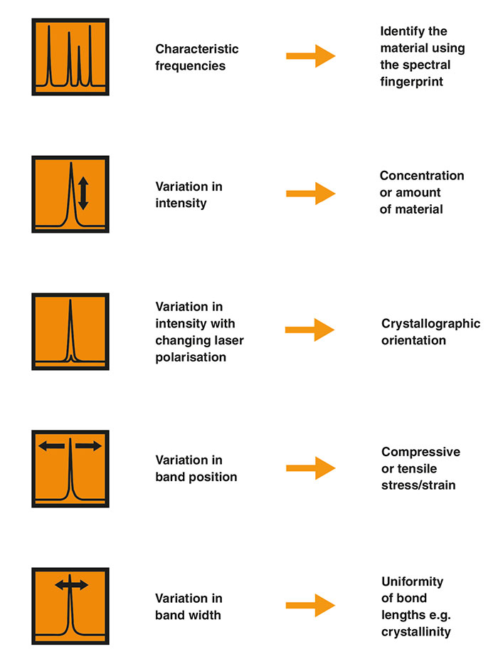
Characteristic vibrational frequencies of chemical bondsThe frequencies of vibration depend on the masses of the atoms involved and the strength of the bonds between them. Heavy atoms and weak bonds have low Raman shifts. Light atoms and strong bonds have high Raman shifts.
In addition to oxide glasses, fluorite lenses are often used in specialty applications. These fluorite or semi-apochromat objectives deal with color better than achromatic objectives. To reduce aberration even further, more complex designs such as apochromat and superachromat objectives are also used.
Objective lensmicroscope
Basic glass lenses will typically result in significant and unacceptable chromatic aberration. Therefore, most objectives have some kind of correction to allow multiple colors to focus at the same point. The easiest correction is an achromatic lens, which uses a combination of crown glass and flint glass to bring two colors into focus. Achromatic objectives are a typical standard design.
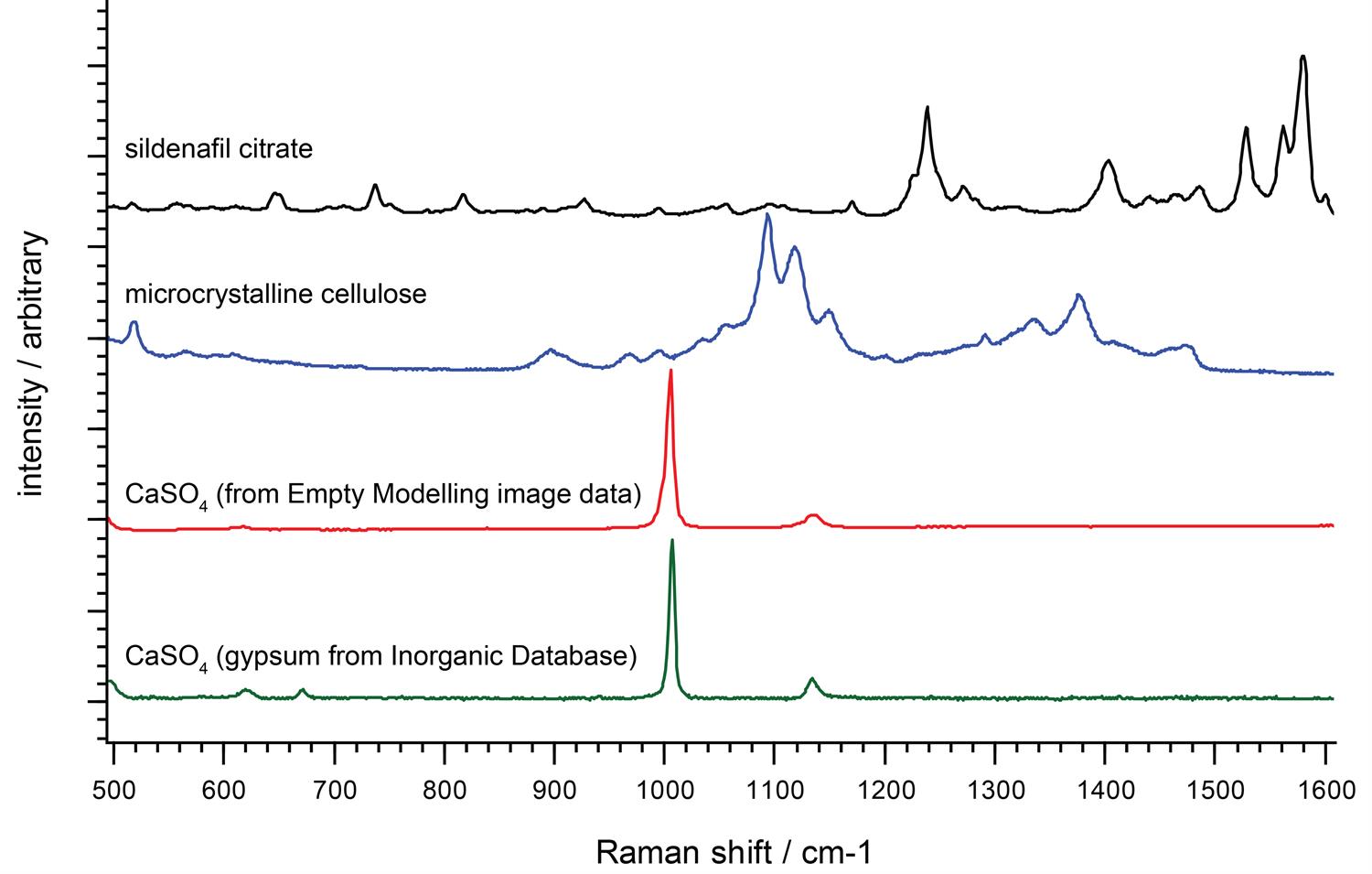
We can identify unknown materials from their unique Raman spectral fingerprints, typically using software searches of spectral libraries. We use the Raman bands in the fingerprint region (from 300 cm-1 to 1900 cm-1) to identify molecules.
Ideally, you would use a Raman instrument with high spectral resolution across the whole Raman range. This gives you better chemical specificity. You can then identify, differentiate and investigate a wider range of materials.
One way of understanding a Raman spectrum is to consider the molecular functional groups as distinct units. This makes it easy to interpret the Raman spectrum of crystals with a regular array of identical atoms, all in the same configuration. For example, diamond contains carbon atoms in a regular tetrahedral network. In these cases, you often see just one dominant Raman band because there is just one molecular environment of the crystal.
Continue your exploration of Raman and photoluminescence (PL) spectroscopy. We answer your questions on Raman microscopy, fast Raman imaging, data analysis, fluorescence and complementary analytical techniques.
What doesthestage doon a microscope
Particularly in biological applications, samples are usually observed under a glass cover slip, which introduces distortions to the image. Objectives which are designed to be used with such cover slips will correct for these distortions, and typically have the thickness of the cover slip they are designed to work with written on the side of the objective (typically 0.17 mm).
In the polystyrene spectrum, we see the high frequency carbon-hydrogen (C-H) vibrations at about 3000 cm-1. The low frequency carbon-carbon (C-C) vibrations are at around 800 cm-1. The C-H vibrations have a higher frequency than the C-C vibrations because hydrogen is lighter than carbon.
Function ofeyepiece inmicroscope
Some microscopes use an oil-immersion or water-immersion lens, which can have magnification greater than 100, and numerical aperture greater than 1. These objectives are specially designed for use with refractive index matching oil or water, which must fill the gap between the front element and the object. These lenses give greater resolution at high magnification. Numerical apertures as high as 1.6 can be achieved with oil immersion.[2]
We graphically display the results of our Raman spectroscopy measurements as Raman spectra. The y-axis represents the intensity of the scattered light, while the x-axis represents the energy (frequency) of light. We are interested in the shift in frequency of the Raman-scattered light, so we plot the x-axis frequencies relative to that of the laser. We label the x-axis as the Raman shift (shown by the units cm-1).
Historically, microscopes were nearly universally designed with a finite mechanical tube length, which is the distance the light traveled in the microscope from the objective to the eyepiece. The Royal Microscopical Society standard is 160 millimeters, whereas Leitz often used 170 millimeters. 180 millimeter tube length objectives are also fairly common. Using an objective and microscope that were designed for different tube lengths will result in spherical aberration.
The distinction between objectives designed for use with or without cover slides is important for high numerical aperture (high magnification) lenses, but makes little difference for low magnification objectives.
You can view the vibrations of a complex molecule as partly consisting of many simple diatomic vibrations. However, you should also consider the vibrations of larger groups of atoms to get a full understanding of the Raman spectrum. For example, the Raman spectrum of polystyrene has a band at 1000 cm-1. This is due to the expanding and contracting ‘breathing mode' of the aromatic carbon ring in polystyrene.
All these types of objectives will exhibit some spherical aberration. While the center of the image will be in focus, the edges will be slightly blurry. When this aberration is corrected, the objective is called a "plan" objective, and has a flat image across the field of view.
Raman shifts are sensitive to neighbouring bondsYou can see more subtle effects if you inspect Raman spectra closely. For example, the C-H vibrations of polystyrene appear in two bands, at approximately 2900 cm-1 and 3050 cm-1. The carbons in the former band are part of aliphatic carbon chains, whereas the carbons in the latter band form part of aromatic carbon rings.
Raman spectra of diamond and polystyrene. The Raman spectrum of polystrene is more complex than that of diamond due to differing bond types.
What doestheobjective lens doon a microscope
A typical microscope has three or four objective lenses with different magnifications, screwed into a circular "nosepiece" which may be rotated to select the required lens. These lenses are often color coded for easier use. The least powerful lens is called the scanning objective lens, and is typically a 4× objective. The second lens is referred to as the small objective lens and is typically a 10× lens. The most powerful lens out of the three is referred to as the large objective lens and is typically 40–100×.
The traditional screw thread used to attach the objective to the microscope was standardized by the Royal Microscopical Society in 1858.[3] It was based on the British Standard Whitworth, with a 0.8 inch diameter and 36 threads per inch. This "RMS thread" or "society thread" is still in common use today. Alternatively, some objective manufacturers use designs based on ISO metric screw thread such as M26 × 0.75 and M25 × 0.75.
What isthepurposeof theobjective lens inalightmicroscope
Low-frequency Raman bandsYou can also study molecular vibrational and rotational modes with low-frequency Raman shifts, below 100 cm-1. These originate from very heavy atoms or very large-scale vibrations, such as the whole crystal lattice vibrating. Renishaw's Raman instruments enable you to study these modes. You can explore a wide range of materials and crystals, easily distinguishing between different crystalline forms (polymorphs) and layered structures.
Numerical aperture for microscope lenses typically ranges from 0.10 to 1.25, corresponding to focal lengths of about 40 mm to 2 mm, respectively.
What isthejobof theobjective lenses
You can interpret Raman spectra to identify chemicals and get structural information. Raman scattering results from the interaction of light with molecular vibrations. These vibrations are very sensitive to changes in chemistry and structure, so you can spot subtle differences in molecular environment. Generally, all materials produce Raman spectra, except for pure metals.
Instead of finite tube lengths, modern microscopes are often designed to use infinity correction instead, a technique in microscopy whereby the light coming out of the objective lens is focused at infinity.[1] This is denoted on the objective with the infinity symbol (∞).
A Raman spectrum therefore consists of a number of bands, each associated with a vibrational mode. The spectrum is unique to the material and enables you to identify it. Some researchers aim to fully understand each Raman band and how it relates to vibrational modes. However, most analysts simply identify samples using a spectral library.
Camera lenses (usually referred to as "photographic objectives" instead of simply "objectives"[4]) need to cover a large focal plane so are made up of a number of optical lens elements to correct optical aberrations. Image projectors (such as video, movie, and slide projectors) use objective lenses that simply reverse the function of a camera lens, with lenses designed to cover a large image plane and project it at a distance onto another surface.[5]
Raman spectra of two polyethylene samples showing differences in intensities and band widths. These spectral differences are due to varying degrees of crystallinity.
Which partof the microscopesupportstheslide that you are viewing
The working distance (sometimes abbreviated WD) is the distance between the sample and the objective. As magnification increases, working distances generally shrinks. When space is needed, special long working distance objectives can be used.
In a telescope the objective is the lens at the front end of a refracting telescope (such as binoculars or telescopic sights) or the image-forming primary mirror of a reflecting or catadioptric telescope. A telescope's light-gathering power and angular resolution are both directly related to the diameter (or "aperture") of its objective lens or mirror. The larger the objective, the brighter the objects will appear and the more detail it can resolve.
One of the most important properties of microscope objectives is their magnification. The magnification typically ranges from 4× to 100×. It is combined with the magnification of the eyepiece to determine the overall magnification of the microscope; a 4× objective with a 10× eyepiece produces an image that is 40 times the size of the object.
You can study differences in material structure by comparing their Raman spectra. You could quantify the degree of crystallinity and distinguish similar crystal forms (polymorphism) of the same chemical. To do this, you would need a Raman spectrometer with high spectral resolution, such as an inVia™ confocal Raman microscope.
In contrast, the Raman spectrum of polystyrene is more complex. The molecule is less symmetric and has hydrogen atoms in addition to carbon atoms. There are also different bond types connecting the atoms.
The objective lens of a microscope is the one at the bottom near the sample. At its simplest, it is a very high-powered magnifying glass, with very short focal length. This is brought very close to the specimen being examined so that the light from the specimen comes to a focus inside the microscope tube. The objective itself is usually a cylinder containing one or more lenses that are typically made of glass; its function is to collect light from the sample.
Similarly, we see the vibrations of two carbon atoms linked by strong double bonds (C=C) at around 1600 cm-1. This is at a higher frequency than two carbon atoms linked by a weaker single bond (C-C, 800 cm-1).


 Ms.Cici
 8618319014500