
Hex Keys & Sets - hex wrench 5mm
Includes magnifying glasses biology
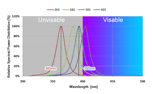
Therefore, for some applications, especially those requiring precise UV output, it may be necessary to select a higher quality, professionally designed LED to minimize the effects of visible light “leakage” and achieve a UV output closer to the pure 365nm.
Diaphragm - The diaphragm is an adjustable light barrier built into the condenser that regulates the amount of light passing through the specimen. It is very important that the diaphragm be correctly adjusted in order to get the best possible image. Use the smallest opening that does not interfere with the field of view. The condenser and diaphragm assembly may be adjusted vertically with a knob projecting to one side. Proper adjustment often yields a greatly improved view of the specimen. This adjustment has been made for you, and you should consult your instructor if you think that it must be readjusted.
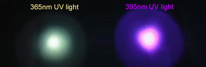
UVA lamps at 395nm have a slightly visible purple color. This color effect is commonly used in entertainment venues, decorative lighting, and applications where a balance of fluorescence and visibility is desired.
Compared to 365nm, a larger portion of the 395nm spectral output occurs in the visible violet region, providing a stronger visual experience and making it suitable for use in entertainment venues or decorative lighting.
$329.99. Pro Smart Security System with 1 Camera (Security Cameras - Wi-Fi Surveillance Cameras). Arlo Pro Smart Security System with 1 Camera · $529.99. Pro ...
Createsatwo dimensional image
Stage - The surface on which you place your slide is called the stage. It contains a hole in the center called the stage aperture. Two stage clips or a slide clamp are used to hold the slide in place.
Which microscopecontains a series of lenses
Conversely, the closer the 365nm spectral output is to the visible 400nm, the less energy there is. This difference makes 365nm LED the preferred choice for most UV-A applications.
f-number (f#) = focal length/diameter of aperture opening = f/ D. That should answer your main question. With respect to its application to ...
Includes magnifying glasses
In dark environments, black light enables fluorescent materials such as fluorescent inks, fluorescent paints, and fluorescent stickers to emit bright colors, creating a very vibrant visual effect. This effect makes black lights very popular in the entertainment, decorative and special effects fields.
What are the parts of the brightfield microscope? Cytology is the study of cell structure and function. Because most cells are very small, they can only be seen when magnified with an instrument such as a microscope. Microscopes not only provide important information about cell structure, they also provide clues about how the cell works. There are 2 basic types of microscopes: the light microscope (creates an image using a beam of light), and the electron microscope (creates an image using a beam of electrons.) The most common type of light microscope, and the one you will use in this lab, is the brightfield microscope. The brightfield microscope is an example of a compound microscope. That means light from the object you are viewing passes through two lenses before it reaches your eye. Microscopes not only magnify the object you are viewing, they also provide increased resolution. Resolution is the ability to distinguish two points as separate points. For instance, if two points are very close together, they may appear to be a single spot. If you increase magnification without increasing resolution, the single spot will only look like a larger single spot, and will never resolve into two separate spots. The better the resolution, the sharper and crisper the image. The resolving power of the naked eye is approximately 0.1 mm, meaning that our eyes can distinguish two points that are 0.1 mm apart. A light microscope can improve resolution by as much as 1000-fold. In addition, discernment of cellular detail can be improved with the use of dyes that add color and contrast to subcellular structures.
Contains a series of lensesbrainly
UVA light is a commonly used light source in black light because of its low energy and high penetrating power. In the solar spectrum, UVA light refers to the ultraviolet A band, which ranges in wavelength from 315 nanometers to 400 nanometers and is considered invisible light.
365nm UV light and 395nm UV light emit the most energy at 365nm and 395nm, respectively, but they also emit considerable energy in the adjacent 350nm to 380nm and 390nm to 410nm ranges. 365nm and 395nm energies decrease along both sides of the 365nm and 395nm spectra.
Revolving Nosepiece and Objective Lenses - Attached to the revolving nosepiece (which is attached to the bottom of the body tube) are several lenses called objectives. Most light microscopes have objective lenses of 4 magnifications: scanning (short) -- normally 4x power, low power (medium) -- normally 10x power, high-power (long) -- normally 40 - 43x power, and oil immersion (longest) -- normally 100x power.
Substage - The area under the stage, called the substage, contains the diaphragm. In addition, some microscopes also have a condenser located under the stage. Condenser - The condenser contains a series of lenses that focus light onto the specimen. Diaphragm - The diaphragm is an adjustable light barrier built into the condenser that regulates the amount of light passing through the specimen. It is very important that the diaphragm be correctly adjusted in order to get the best possible image. Use the smallest opening that does not interfere with the field of view. The condenser and diaphragm assembly may be adjusted vertically with a knob projecting to one side. Proper adjustment often yields a greatly improved view of the specimen. This adjustment has been made for you, and you should consult your instructor if you think that it must be readjusted.
Linear Translation Stage ... Linear stage 7T173-20-50 allows manual linear motion. The featured advantage of this stage is it's narrow and low-profile design. Our ...
365 nm goes “deeper” into the invisible UV spectrum than 395 nm does, containing less visible light and at a shorter wavelength, so to the naked eye, 365 nm UV light appears as a dull blue-white color.
Anwenderkonfigurierbare Software wird von Systemintegratoren sowie von geschulten Anwendern im Sondermaschinenbau eingesetzt. Herstellerverzeichnis: ...
Body Tube - The body tube is the part of the microscope that connects the oculars with the revolving nosepiece. Some of the microscopes in lab have a small knob that sticks out from the front of the body tube. This knob can be used to move a pointer that is visible when you look through the microscope.

Createsadigital image
Contains a series of lensesquizlet
For fluorescent dyes and markers, banknotes and security markers, minerals and gemstones, white clothing and dental bleach, they absorb the most energy under UV irradiation at 365nm and re-emit it in the form of intense fluorescence. This makes the fluorescence effect most noticeable and intense when these substances are viewed at 365nm.
These visible components may be in the blue or white spectral range, resulting in a dull blue-white coloration of the lighting you observe, rather than the pure UV output of the desired single wavelength of 365nm.
Why is light with wavelengths below 400 nm invisible to the human eye, yet the wavelength value is still very important? This is because the wavelength determines the application category of UV light. For example, 365 nm and 395 nm belong to the same category of UVA, while 300 nm belongs to UVB and 270 nm to UVC.
It eliminates reflections to a fraction of standard glass. As a result, it's ideal for applications which require a high level of visual clarity, such as ...
Pyroelectric detectors are thermal detectors: Temperature fluctuations produce a charge change on the surface of pyroelectric crystals, which produces a ...
For example, linearly polarized light remains linearly polarized, although its orientation (angle ) may rotate. However, for angles greater than θc, different ...
A typical ocular lens has a magnification of 10x; what is the total magnification if the high power objective (43x) is in position?
Magnifies objects in steps
However, whether 365nm and 395nm have the same application? What is the difference between them? Let’s take a look together.
Peak Optics, Magnifiers, Comparators, Loupes, For Inspection & Measuring, 2x to 300x Peak 1985 Steinheil System Magnifier, 7x, 10x, 14x, 20x - The magnifier ...
NCI's Dictionary of Cancer Terms provides easy-to-understand definitions for words and phrases related to cancer and medicine.
Although the 365nm wavelength is deep in the invisible spectrum, 365 nm LED produces a small amount of visible “leakage” or radiation beyond the 365nm wavelength due to limitations in the design of the LED or material properties.
365 nm and 395 nm refer to the wavelengths of light in the ultraviolet spectrum. These wavelengths fall within the UVA range, often referred to as “blacklight” due to their ability to make certain materials fluoresce or emit visible light.
Blacklight refers to a special type of UV lighting that uses primarily UVA light to produce a unique effect. Blacklight lighting utilizes UVA light to excite the fluorescent properties of a substance, causing it to emit visible light.
Ocular lenses - The oculars are the lenses you look through. Some microscopes are monocular--that is, there is only one eyepiece to look through. Other microscopes are dual-viewing; this means there are two oculars--one pointing upwards and one pointing backwards. These microscopes allow two people to view the same specimen at the same time--each looking through a single eyepiece. Yet other microscopes are binocular--that is, there are two eyepieces side-by-side and you look through both of the eyepieces, as you would a pair of binoculars. Most ocular lenses have a 10X magnification.
Although both 365 nm and 395 nm are UVA lights, and produce black light and curing effects, and there is only a 30 nm difference, 395nm is closer to visible light (violet color) than 365 nm, so to the naked eye, 395 nm UV light appears purple in color.
In this lab exercise, you will learn about the different parts of the brightfield microscope, how to care for the microscope, and the correct procedure for viewing specimens. You will then practice using the microscope to view both a wet mount and a prepared slide of the organism Euglena. Study the location and the function of each of the following parts of a brightfield microscope:
Aug 2, 2021 — Landscape photography is a good example of a large or deep depth of field. In order to achieve a large or deep depth of field, you want a ...
The 395nm UVA wavelength is relatively safe and less energetic than the shorter 365nm UVA wavelength. With proper UV protection, it usually does not cause significant harm to humans or the environment.
Why is there a difference in visible light between 365nm and 395nm? 365nm does not only emit 365nm wavelength light, and 395nm does not only emit 395nm wavelength light.
Commercially available UVA light fixtures typically range in wavelength from 365nm to 405nm, with 365nm and 395nm considered the more dominant choices. It is a common consideration for choosing 365nm or 395nm in UV black light and fluorescence detection applications.
Focus Knobs - You can focus your microscope by using the coarse and fine adjustment knobs located on the sides of the arm. Note that the coarse adjustment knob is larger than the fine adjustment knob. The coarse adjustment is used for initial focusing and should be used ONLY when the scanning (4X) or low power (10X) objective is in alignment. The fine adjustment makes very slight changes, allowing precision focusing on low or high power.


 Ms.Cici
 8618319014500