
Helium-Neon Lasers He-Ne Lab Laser IF HN05 - helium and neon laser
Best Car Windshield sun Shade
Beginner Microscope ExperimentsMicroscope Slides Preparations-Styles and TechniquesPrepared Microscope Slides - Benefits and RecommendationsSee also: Dissecting Stereo Microscope Parts and Functions Stereo Microscope Vs Compound MicroscopeCheck out this Microscope Quiz to test your knowledgeInteresting info here on Basic Microscope Ergonomics Return from Parts of a Compound Microscope to Compound Light Microscope Return from Parts of a Compound Microscope to Best Microscope HomeFind out how to advertise on MicroscopeMaster!FacebookTwitter
Read MoreChemoorganotrophs - Definition, and ExamplesOct 26, 22 05:01 PMChemoorganotrophs also known as organotrophs, include organisms that obtain their energy from organic chemicals like glucose. This process is known as chemoorganotrophy. Read more here.Read MoreBetaproteobacteria â Examples, Characteristics and FunctionOct 25, 22 03:44 PMBetaproteobacteria is a heterogeneous group in the phylum Proteobacteria whose members can be found in a range of habitats from wastewater and hot springs to the Antarctic. Read more here.Read More
laservision designs and manufactures a complete line of laser safety products including glasses, barriers, signs and windows.
Privacy Policy by Hayley Anderson at MicroscopeMaster.comAll rights reserved 2010-2021Amazon and the Amazon logo are trademarks of Amazon.com, Inc. or its affiliatesImages are used with permission as required.
Objective lenses: One of the most important parts of a compound microscope, as they are the lenses closest to the specimen. A standard microscope has three, four, or five objective lenses that range in power from 4X to 100X. When focusing the microscope, be careful that the objective lens doesnât touch the slide, as it could break the slide and destroy the specimen.Specimen or slide: The specimen is the object being examined. Most specimens are mounted on slides, flat rectangles of thin glass.The specimen is placed on the glass and a cover slip is placed over the specimen. This allows the slide to be easily inserted or removed from the microscope. It also allows the specimen to be labeled, transported, and stored without damage.Stage: The flat platform where the slide is placed.Stage clips: Metal clips that hold the slide in place.Stage height adjustment (Stage Control): These knobs move the stage left and right or up and down.Aperture: The hole in the middle of the stage that allows light from the illuminator to reach the specimen.On/off switch: This switch on the base of the microscope turns the illuminator off and on.Illumination: The light source for a microscope. Older microscopes used mirrors to reflect light from an external source up through the bottom of the stage; however, most microscopes now use a low-voltage bulb.Iris diaphragm: Adjusts the amount of light that reaches the specimen.Condenser: Gathers and focuses light from the illuminator onto the specimen being viewed.Base: The base supports the microscope and itâs where illuminator is located.How Does a Compound Microscope Work?All of the parts of a microscope work together - The light from the illuminator passes through the aperture, through the slide, and through the objective lens, where the image of the specimen is magnified.The then magnified image continues up through the body tube of the microscope to the eyepiece, which further magnifies the image the viewer then sees.Learning to use and adjust your compound microscope is the next important step.It's also imperative to know and understand the best practices of cleaning your microscope.The microscope parts work together in hospitals and in forensic labs, for scientists and students, bacteriologists and biologists so that they may view bacteria, plant and animal cells and tissues, and various microorganisms the world over.Compound microscopes have furthered medical research, helped to solve crimes, and they have repeatedly proven invaluable in unlocking the secrets of the microscopic world.Check out MicroscopeMasterâs online help:Basics of a Compound MicroscopeDiagram/Parts/Functions of a Compound MicroscopeBeginner Microscope ExperimentsMicroscope Slides Preparations-Styles and TechniquesPrepared Microscope Slides - Benefits and RecommendationsSee also: Dissecting Stereo Microscope Parts and Functions Stereo Microscope Vs Compound MicroscopeCheck out this Microscope Quiz to test your knowledgeInteresting info here on Basic Microscope Ergonomics Return from Parts of a Compound Microscope to Compound Light Microscope Return from Parts of a Compound Microscope to Best Microscope HomeFind out how to advertise on MicroscopeMaster!FacebookTwitter
Diopter Adjustment: Useful as a means to change focus on one eyepiece so as to correct for any difference in vision between your two eyes.Body tube (Head): The body tube connects the eyepiece to the objective lenses.Arm: The arm connects the body tube to the base of the microscope.Coarse adjustment: Brings the specimen into general focus.Fine adjustment: Fine tunes the focus and increases the detail of the specimen.Nosepiece: A rotating turret that houses the objective lenses. The viewer spins the nosepiece to select different objective lenses.Objective lenses: One of the most important parts of a compound microscope, as they are the lenses closest to the specimen. A standard microscope has three, four, or five objective lenses that range in power from 4X to 100X. When focusing the microscope, be careful that the objective lens doesnât touch the slide, as it could break the slide and destroy the specimen.Specimen or slide: The specimen is the object being examined. Most specimens are mounted on slides, flat rectangles of thin glass.The specimen is placed on the glass and a cover slip is placed over the specimen. This allows the slide to be easily inserted or removed from the microscope. It also allows the specimen to be labeled, transported, and stored without damage.Stage: The flat platform where the slide is placed.Stage clips: Metal clips that hold the slide in place.Stage height adjustment (Stage Control): These knobs move the stage left and right or up and down.Aperture: The hole in the middle of the stage that allows light from the illuminator to reach the specimen.On/off switch: This switch on the base of the microscope turns the illuminator off and on.Illumination: The light source for a microscope. Older microscopes used mirrors to reflect light from an external source up through the bottom of the stage; however, most microscopes now use a low-voltage bulb.Iris diaphragm: Adjusts the amount of light that reaches the specimen.Condenser: Gathers and focuses light from the illuminator onto the specimen being viewed.Base: The base supports the microscope and itâs where illuminator is located.How Does a Compound Microscope Work?All of the parts of a microscope work together - The light from the illuminator passes through the aperture, through the slide, and through the objective lens, where the image of the specimen is magnified.The then magnified image continues up through the body tube of the microscope to the eyepiece, which further magnifies the image the viewer then sees.Learning to use and adjust your compound microscope is the next important step.It's also imperative to know and understand the best practices of cleaning your microscope.The microscope parts work together in hospitals and in forensic labs, for scientists and students, bacteriologists and biologists so that they may view bacteria, plant and animal cells and tissues, and various microorganisms the world over.Compound microscopes have furthered medical research, helped to solve crimes, and they have repeatedly proven invaluable in unlocking the secrets of the microscopic world.Check out MicroscopeMasterâs online help:Basics of a Compound MicroscopeDiagram/Parts/Functions of a Compound MicroscopeBeginner Microscope ExperimentsMicroscope Slides Preparations-Styles and TechniquesPrepared Microscope Slides - Benefits and RecommendationsSee also: Dissecting Stereo Microscope Parts and Functions Stereo Microscope Vs Compound MicroscopeCheck out this Microscope Quiz to test your knowledgeInteresting info here on Basic Microscope Ergonomics Return from Parts of a Compound Microscope to Compound Light Microscope Return from Parts of a Compound Microscope to Best Microscope HomeFind out how to advertise on MicroscopeMaster!FacebookTwitter
Eclipsesunglasses
Check out MicroscopeMasterâs online help:Basics of a Compound MicroscopeDiagram/Parts/Functions of a Compound MicroscopeBeginner Microscope ExperimentsMicroscope Slides Preparations-Styles and TechniquesPrepared Microscope Slides - Benefits and RecommendationsSee also: Dissecting Stereo Microscope Parts and Functions Stereo Microscope Vs Compound MicroscopeCheck out this Microscope Quiz to test your knowledgeInteresting info here on Basic Microscope Ergonomics Return from Parts of a Compound Microscope to Compound Light Microscope Return from Parts of a Compound Microscope to Best Microscope HomeFind out how to advertise on MicroscopeMaster!FacebookTwitter
Diopter Adjustment: Useful as a means to change focus on one eyepiece so as to correct for any difference in vision between your two eyes.Body tube (Head): The body tube connects the eyepiece to the objective lenses.Arm: The arm connects the body tube to the base of the microscope.Coarse adjustment: Brings the specimen into general focus.Fine adjustment: Fine tunes the focus and increases the detail of the specimen.Nosepiece: A rotating turret that houses the objective lenses. The viewer spins the nosepiece to select different objective lenses.Objective lenses: One of the most important parts of a compound microscope, as they are the lenses closest to the specimen. A standard microscope has three, four, or five objective lenses that range in power from 4X to 100X. When focusing the microscope, be careful that the objective lens doesnât touch the slide, as it could break the slide and destroy the specimen.Specimen or slide: The specimen is the object being examined. Most specimens are mounted on slides, flat rectangles of thin glass.The specimen is placed on the glass and a cover slip is placed over the specimen. This allows the slide to be easily inserted or removed from the microscope. It also allows the specimen to be labeled, transported, and stored without damage.Stage: The flat platform where the slide is placed.Stage clips: Metal clips that hold the slide in place.Stage height adjustment (Stage Control): These knobs move the stage left and right or up and down.Aperture: The hole in the middle of the stage that allows light from the illuminator to reach the specimen.On/off switch: This switch on the base of the microscope turns the illuminator off and on.Illumination: The light source for a microscope. Older microscopes used mirrors to reflect light from an external source up through the bottom of the stage; however, most microscopes now use a low-voltage bulb.Iris diaphragm: Adjusts the amount of light that reaches the specimen.Condenser: Gathers and focuses light from the illuminator onto the specimen being viewed.Base: The base supports the microscope and itâs where illuminator is located.How Does a Compound Microscope Work?All of the parts of a microscope work together - The light from the illuminator passes through the aperture, through the slide, and through the objective lens, where the image of the specimen is magnified.The then magnified image continues up through the body tube of the microscope to the eyepiece, which further magnifies the image the viewer then sees.Learning to use and adjust your compound microscope is the next important step.It's also imperative to know and understand the best practices of cleaning your microscope.The microscope parts work together in hospitals and in forensic labs, for scientists and students, bacteriologists and biologists so that they may view bacteria, plant and animal cells and tissues, and various microorganisms the world over.Compound microscopes have furthered medical research, helped to solve crimes, and they have repeatedly proven invaluable in unlocking the secrets of the microscopic world.Check out MicroscopeMasterâs online help:Basics of a Compound MicroscopeDiagram/Parts/Functions of a Compound MicroscopeBeginner Microscope ExperimentsMicroscope Slides Preparations-Styles and TechniquesPrepared Microscope Slides - Benefits and RecommendationsSee also: Dissecting Stereo Microscope Parts and Functions Stereo Microscope Vs Compound MicroscopeCheck out this Microscope Quiz to test your knowledgeInteresting info here on Basic Microscope Ergonomics Return from Parts of a Compound Microscope to Compound Light Microscope Return from Parts of a Compound Microscope to Best Microscope HomeFind out how to advertise on MicroscopeMaster!FacebookTwitter
We're currently unable to show live stock information on our website, so if you need delivery fast we strongly recommend checking availability first. Simply fill out the form below and we'll get back to you, or call us on +44 (0)1243 375774 for an immediate answer.*Quantity Required:Article Number:*E-mail Address:*Please add the two numbersRequestRequest
Coarse adjustment: Brings the specimen into general focus.Fine adjustment: Fine tunes the focus and increases the detail of the specimen.Nosepiece: A rotating turret that houses the objective lenses. The viewer spins the nosepiece to select different objective lenses.Objective lenses: One of the most important parts of a compound microscope, as they are the lenses closest to the specimen. A standard microscope has three, four, or five objective lenses that range in power from 4X to 100X. When focusing the microscope, be careful that the objective lens doesnât touch the slide, as it could break the slide and destroy the specimen.Specimen or slide: The specimen is the object being examined. Most specimens are mounted on slides, flat rectangles of thin glass.The specimen is placed on the glass and a cover slip is placed over the specimen. This allows the slide to be easily inserted or removed from the microscope. It also allows the specimen to be labeled, transported, and stored without damage.Stage: The flat platform where the slide is placed.Stage clips: Metal clips that hold the slide in place.Stage height adjustment (Stage Control): These knobs move the stage left and right or up and down.Aperture: The hole in the middle of the stage that allows light from the illuminator to reach the specimen.On/off switch: This switch on the base of the microscope turns the illuminator off and on.Illumination: The light source for a microscope. Older microscopes used mirrors to reflect light from an external source up through the bottom of the stage; however, most microscopes now use a low-voltage bulb.Iris diaphragm: Adjusts the amount of light that reaches the specimen.Condenser: Gathers and focuses light from the illuminator onto the specimen being viewed.Base: The base supports the microscope and itâs where illuminator is located.How Does a Compound Microscope Work?All of the parts of a microscope work together - The light from the illuminator passes through the aperture, through the slide, and through the objective lens, where the image of the specimen is magnified.The then magnified image continues up through the body tube of the microscope to the eyepiece, which further magnifies the image the viewer then sees.Learning to use and adjust your compound microscope is the next important step.It's also imperative to know and understand the best practices of cleaning your microscope.The microscope parts work together in hospitals and in forensic labs, for scientists and students, bacteriologists and biologists so that they may view bacteria, plant and animal cells and tissues, and various microorganisms the world over.Compound microscopes have furthered medical research, helped to solve crimes, and they have repeatedly proven invaluable in unlocking the secrets of the microscopic world.Check out MicroscopeMasterâs online help:Basics of a Compound MicroscopeDiagram/Parts/Functions of a Compound MicroscopeBeginner Microscope ExperimentsMicroscope Slides Preparations-Styles and TechniquesPrepared Microscope Slides - Benefits and RecommendationsSee also: Dissecting Stereo Microscope Parts and Functions Stereo Microscope Vs Compound MicroscopeCheck out this Microscope Quiz to test your knowledgeInteresting info here on Basic Microscope Ergonomics Return from Parts of a Compound Microscope to Compound Light Microscope Return from Parts of a Compound Microscope to Best Microscope HomeFind out how to advertise on MicroscopeMaster!FacebookTwitter
Nosepiece: A rotating turret that houses the objective lenses. The viewer spins the nosepiece to select different objective lenses.Objective lenses: One of the most important parts of a compound microscope, as they are the lenses closest to the specimen. A standard microscope has three, four, or five objective lenses that range in power from 4X to 100X. When focusing the microscope, be careful that the objective lens doesnât touch the slide, as it could break the slide and destroy the specimen.Specimen or slide: The specimen is the object being examined. Most specimens are mounted on slides, flat rectangles of thin glass.The specimen is placed on the glass and a cover slip is placed over the specimen. This allows the slide to be easily inserted or removed from the microscope. It also allows the specimen to be labeled, transported, and stored without damage.Stage: The flat platform where the slide is placed.Stage clips: Metal clips that hold the slide in place.Stage height adjustment (Stage Control): These knobs move the stage left and right or up and down.Aperture: The hole in the middle of the stage that allows light from the illuminator to reach the specimen.On/off switch: This switch on the base of the microscope turns the illuminator off and on.Illumination: The light source for a microscope. Older microscopes used mirrors to reflect light from an external source up through the bottom of the stage; however, most microscopes now use a low-voltage bulb.Iris diaphragm: Adjusts the amount of light that reaches the specimen.Condenser: Gathers and focuses light from the illuminator onto the specimen being viewed.Base: The base supports the microscope and itâs where illuminator is located.How Does a Compound Microscope Work?All of the parts of a microscope work together - The light from the illuminator passes through the aperture, through the slide, and through the objective lens, where the image of the specimen is magnified.The then magnified image continues up through the body tube of the microscope to the eyepiece, which further magnifies the image the viewer then sees.Learning to use and adjust your compound microscope is the next important step.It's also imperative to know and understand the best practices of cleaning your microscope.The microscope parts work together in hospitals and in forensic labs, for scientists and students, bacteriologists and biologists so that they may view bacteria, plant and animal cells and tissues, and various microorganisms the world over.Compound microscopes have furthered medical research, helped to solve crimes, and they have repeatedly proven invaluable in unlocking the secrets of the microscopic world.Check out MicroscopeMasterâs online help:Basics of a Compound MicroscopeDiagram/Parts/Functions of a Compound MicroscopeBeginner Microscope ExperimentsMicroscope Slides Preparations-Styles and TechniquesPrepared Microscope Slides - Benefits and RecommendationsSee also: Dissecting Stereo Microscope Parts and Functions Stereo Microscope Vs Compound MicroscopeCheck out this Microscope Quiz to test your knowledgeInteresting info here on Basic Microscope Ergonomics Return from Parts of a Compound Microscope to Compound Light Microscope Return from Parts of a Compound Microscope to Best Microscope HomeFind out how to advertise on MicroscopeMaster!FacebookTwitter
Return from Parts of a Compound Microscope to Best Microscope HomeFind out how to advertise on MicroscopeMaster!FacebookTwitter
Stage clips: Metal clips that hold the slide in place.Stage height adjustment (Stage Control): These knobs move the stage left and right or up and down.Aperture: The hole in the middle of the stage that allows light from the illuminator to reach the specimen.On/off switch: This switch on the base of the microscope turns the illuminator off and on.Illumination: The light source for a microscope. Older microscopes used mirrors to reflect light from an external source up through the bottom of the stage; however, most microscopes now use a low-voltage bulb.Iris diaphragm: Adjusts the amount of light that reaches the specimen.Condenser: Gathers and focuses light from the illuminator onto the specimen being viewed.Base: The base supports the microscope and itâs where illuminator is located.How Does a Compound Microscope Work?All of the parts of a microscope work together - The light from the illuminator passes through the aperture, through the slide, and through the objective lens, where the image of the specimen is magnified.The then magnified image continues up through the body tube of the microscope to the eyepiece, which further magnifies the image the viewer then sees.Learning to use and adjust your compound microscope is the next important step.It's also imperative to know and understand the best practices of cleaning your microscope.The microscope parts work together in hospitals and in forensic labs, for scientists and students, bacteriologists and biologists so that they may view bacteria, plant and animal cells and tissues, and various microorganisms the world over.Compound microscopes have furthered medical research, helped to solve crimes, and they have repeatedly proven invaluable in unlocking the secrets of the microscopic world.Check out MicroscopeMasterâs online help:Basics of a Compound MicroscopeDiagram/Parts/Functions of a Compound MicroscopeBeginner Microscope ExperimentsMicroscope Slides Preparations-Styles and TechniquesPrepared Microscope Slides - Benefits and RecommendationsSee also: Dissecting Stereo Microscope Parts and Functions Stereo Microscope Vs Compound MicroscopeCheck out this Microscope Quiz to test your knowledgeInteresting info here on Basic Microscope Ergonomics Return from Parts of a Compound Microscope to Compound Light Microscope Return from Parts of a Compound Microscope to Best Microscope HomeFind out how to advertise on MicroscopeMaster!FacebookTwitter
Before exploring microscope parts and functions, you should probably understand that the compound light microscope is more complicated than just a microscope with more than one lens.First, the purpose of a microscope is to magnify a small object or to magnify the fine details of a larger object in order to examine minute specimens that cannot be seen by the naked eye. Here are the important compound microscope parts...Eyepiece: The lens the viewer looks through to see the specimen. The eyepiece usually contains a 10X or 15X power lens.Diopter Adjustment: Useful as a means to change focus on one eyepiece so as to correct for any difference in vision between your two eyes.Body tube (Head): The body tube connects the eyepiece to the objective lenses.Arm: The arm connects the body tube to the base of the microscope.Coarse adjustment: Brings the specimen into general focus.Fine adjustment: Fine tunes the focus and increases the detail of the specimen.Nosepiece: A rotating turret that houses the objective lenses. The viewer spins the nosepiece to select different objective lenses.Objective lenses: One of the most important parts of a compound microscope, as they are the lenses closest to the specimen. A standard microscope has three, four, or five objective lenses that range in power from 4X to 100X. When focusing the microscope, be careful that the objective lens doesnât touch the slide, as it could break the slide and destroy the specimen.Specimen or slide: The specimen is the object being examined. Most specimens are mounted on slides, flat rectangles of thin glass.The specimen is placed on the glass and a cover slip is placed over the specimen. This allows the slide to be easily inserted or removed from the microscope. It also allows the specimen to be labeled, transported, and stored without damage.Stage: The flat platform where the slide is placed.Stage clips: Metal clips that hold the slide in place.Stage height adjustment (Stage Control): These knobs move the stage left and right or up and down.Aperture: The hole in the middle of the stage that allows light from the illuminator to reach the specimen.On/off switch: This switch on the base of the microscope turns the illuminator off and on.Illumination: The light source for a microscope. Older microscopes used mirrors to reflect light from an external source up through the bottom of the stage; however, most microscopes now use a low-voltage bulb.Iris diaphragm: Adjusts the amount of light that reaches the specimen.Condenser: Gathers and focuses light from the illuminator onto the specimen being viewed.Base: The base supports the microscope and itâs where illuminator is located.How Does a Compound Microscope Work?All of the parts of a microscope work together - The light from the illuminator passes through the aperture, through the slide, and through the objective lens, where the image of the specimen is magnified.The then magnified image continues up through the body tube of the microscope to the eyepiece, which further magnifies the image the viewer then sees.Learning to use and adjust your compound microscope is the next important step.It's also imperative to know and understand the best practices of cleaning your microscope.The microscope parts work together in hospitals and in forensic labs, for scientists and students, bacteriologists and biologists so that they may view bacteria, plant and animal cells and tissues, and various microorganisms the world over.Compound microscopes have furthered medical research, helped to solve crimes, and they have repeatedly proven invaluable in unlocking the secrets of the microscopic world.Check out MicroscopeMasterâs online help:Basics of a Compound MicroscopeDiagram/Parts/Functions of a Compound MicroscopeBeginner Microscope ExperimentsMicroscope Slides Preparations-Styles and TechniquesPrepared Microscope Slides - Benefits and RecommendationsSee also: Dissecting Stereo Microscope Parts and Functions Stereo Microscope Vs Compound MicroscopeCheck out this Microscope Quiz to test your knowledgeInteresting info here on Basic Microscope Ergonomics Return from Parts of a Compound Microscope to Compound Light Microscope Return from Parts of a Compound Microscope to Best Microscope HomeFind out how to advertise on MicroscopeMaster!FacebookTwitter
Specimen or slide: The specimen is the object being examined. Most specimens are mounted on slides, flat rectangles of thin glass.The specimen is placed on the glass and a cover slip is placed over the specimen. This allows the slide to be easily inserted or removed from the microscope. It also allows the specimen to be labeled, transported, and stored without damage.Stage: The flat platform where the slide is placed.Stage clips: Metal clips that hold the slide in place.Stage height adjustment (Stage Control): These knobs move the stage left and right or up and down.Aperture: The hole in the middle of the stage that allows light from the illuminator to reach the specimen.On/off switch: This switch on the base of the microscope turns the illuminator off and on.Illumination: The light source for a microscope. Older microscopes used mirrors to reflect light from an external source up through the bottom of the stage; however, most microscopes now use a low-voltage bulb.Iris diaphragm: Adjusts the amount of light that reaches the specimen.Condenser: Gathers and focuses light from the illuminator onto the specimen being viewed.Base: The base supports the microscope and itâs where illuminator is located.How Does a Compound Microscope Work?All of the parts of a microscope work together - The light from the illuminator passes through the aperture, through the slide, and through the objective lens, where the image of the specimen is magnified.The then magnified image continues up through the body tube of the microscope to the eyepiece, which further magnifies the image the viewer then sees.Learning to use and adjust your compound microscope is the next important step.It's also imperative to know and understand the best practices of cleaning your microscope.The microscope parts work together in hospitals and in forensic labs, for scientists and students, bacteriologists and biologists so that they may view bacteria, plant and animal cells and tissues, and various microorganisms the world over.Compound microscopes have furthered medical research, helped to solve crimes, and they have repeatedly proven invaluable in unlocking the secrets of the microscopic world.Check out MicroscopeMasterâs online help:Basics of a Compound MicroscopeDiagram/Parts/Functions of a Compound MicroscopeBeginner Microscope ExperimentsMicroscope Slides Preparations-Styles and TechniquesPrepared Microscope Slides - Benefits and RecommendationsSee also: Dissecting Stereo Microscope Parts and Functions Stereo Microscope Vs Compound MicroscopeCheck out this Microscope Quiz to test your knowledgeInteresting info here on Basic Microscope Ergonomics Return from Parts of a Compound Microscope to Compound Light Microscope Return from Parts of a Compound Microscope to Best Microscope HomeFind out how to advertise on MicroscopeMaster!FacebookTwitter
Sep 10, 2024 — Prism glasses are prescription eyeglasses with special lenses for correcting double vision (also known as diplopia). In some cases, prism ...
Specimen or slide: The specimen is the object being examined. Most specimens are mounted on slides, flat rectangles of thin glass.The specimen is placed on the glass and a cover slip is placed over the specimen. This allows the slide to be easily inserted or removed from the microscope. It also allows the specimen to be labeled, transported, and stored without damage.Stage: The flat platform where the slide is placed.Stage clips: Metal clips that hold the slide in place.Stage height adjustment (Stage Control): These knobs move the stage left and right or up and down.Aperture: The hole in the middle of the stage that allows light from the illuminator to reach the specimen.On/off switch: This switch on the base of the microscope turns the illuminator off and on.Illumination: The light source for a microscope. Older microscopes used mirrors to reflect light from an external source up through the bottom of the stage; however, most microscopes now use a low-voltage bulb.Iris diaphragm: Adjusts the amount of light that reaches the specimen.Condenser: Gathers and focuses light from the illuminator onto the specimen being viewed.Base: The base supports the microscope and itâs where illuminator is located.How Does a Compound Microscope Work?All of the parts of a microscope work together - The light from the illuminator passes through the aperture, through the slide, and through the objective lens, where the image of the specimen is magnified.The then magnified image continues up through the body tube of the microscope to the eyepiece, which further magnifies the image the viewer then sees.Learning to use and adjust your compound microscope is the next important step.It's also imperative to know and understand the best practices of cleaning your microscope.The microscope parts work together in hospitals and in forensic labs, for scientists and students, bacteriologists and biologists so that they may view bacteria, plant and animal cells and tissues, and various microorganisms the world over.Compound microscopes have furthered medical research, helped to solve crimes, and they have repeatedly proven invaluable in unlocking the secrets of the microscopic world.Check out MicroscopeMasterâs online help:Basics of a Compound MicroscopeDiagram/Parts/Functions of a Compound MicroscopeBeginner Microscope ExperimentsMicroscope Slides Preparations-Styles and TechniquesPrepared Microscope Slides - Benefits and RecommendationsSee also: Dissecting Stereo Microscope Parts and Functions Stereo Microscope Vs Compound MicroscopeCheck out this Microscope Quiz to test your knowledgeInteresting info here on Basic Microscope Ergonomics Return from Parts of a Compound Microscope to Compound Light Microscope Return from Parts of a Compound Microscope to Best Microscope HomeFind out how to advertise on MicroscopeMaster!FacebookTwitter
Iris diaphragm: Adjusts the amount of light that reaches the specimen.Condenser: Gathers and focuses light from the illuminator onto the specimen being viewed.Base: The base supports the microscope and itâs where illuminator is located.How Does a Compound Microscope Work?All of the parts of a microscope work together - The light from the illuminator passes through the aperture, through the slide, and through the objective lens, where the image of the specimen is magnified.The then magnified image continues up through the body tube of the microscope to the eyepiece, which further magnifies the image the viewer then sees.Learning to use and adjust your compound microscope is the next important step.It's also imperative to know and understand the best practices of cleaning your microscope.The microscope parts work together in hospitals and in forensic labs, for scientists and students, bacteriologists and biologists so that they may view bacteria, plant and animal cells and tissues, and various microorganisms the world over.Compound microscopes have furthered medical research, helped to solve crimes, and they have repeatedly proven invaluable in unlocking the secrets of the microscopic world.Check out MicroscopeMasterâs online help:Basics of a Compound MicroscopeDiagram/Parts/Functions of a Compound MicroscopeBeginner Microscope ExperimentsMicroscope Slides Preparations-Styles and TechniquesPrepared Microscope Slides - Benefits and RecommendationsSee also: Dissecting Stereo Microscope Parts and Functions Stereo Microscope Vs Compound MicroscopeCheck out this Microscope Quiz to test your knowledgeInteresting info here on Basic Microscope Ergonomics Return from Parts of a Compound Microscope to Compound Light Microscope Return from Parts of a Compound Microscope to Best Microscope HomeFind out how to advertise on MicroscopeMaster!FacebookTwitter
Arm: The arm connects the body tube to the base of the microscope.Coarse adjustment: Brings the specimen into general focus.Fine adjustment: Fine tunes the focus and increases the detail of the specimen.Nosepiece: A rotating turret that houses the objective lenses. The viewer spins the nosepiece to select different objective lenses.Objective lenses: One of the most important parts of a compound microscope, as they are the lenses closest to the specimen. A standard microscope has three, four, or five objective lenses that range in power from 4X to 100X. When focusing the microscope, be careful that the objective lens doesnât touch the slide, as it could break the slide and destroy the specimen.Specimen or slide: The specimen is the object being examined. Most specimens are mounted on slides, flat rectangles of thin glass.The specimen is placed on the glass and a cover slip is placed over the specimen. This allows the slide to be easily inserted or removed from the microscope. It also allows the specimen to be labeled, transported, and stored without damage.Stage: The flat platform where the slide is placed.Stage clips: Metal clips that hold the slide in place.Stage height adjustment (Stage Control): These knobs move the stage left and right or up and down.Aperture: The hole in the middle of the stage that allows light from the illuminator to reach the specimen.On/off switch: This switch on the base of the microscope turns the illuminator off and on.Illumination: The light source for a microscope. Older microscopes used mirrors to reflect light from an external source up through the bottom of the stage; however, most microscopes now use a low-voltage bulb.Iris diaphragm: Adjusts the amount of light that reaches the specimen.Condenser: Gathers and focuses light from the illuminator onto the specimen being viewed.Base: The base supports the microscope and itâs where illuminator is located.How Does a Compound Microscope Work?All of the parts of a microscope work together - The light from the illuminator passes through the aperture, through the slide, and through the objective lens, where the image of the specimen is magnified.The then magnified image continues up through the body tube of the microscope to the eyepiece, which further magnifies the image the viewer then sees.Learning to use and adjust your compound microscope is the next important step.It's also imperative to know and understand the best practices of cleaning your microscope.The microscope parts work together in hospitals and in forensic labs, for scientists and students, bacteriologists and biologists so that they may view bacteria, plant and animal cells and tissues, and various microorganisms the world over.Compound microscopes have furthered medical research, helped to solve crimes, and they have repeatedly proven invaluable in unlocking the secrets of the microscopic world.Check out MicroscopeMasterâs online help:Basics of a Compound MicroscopeDiagram/Parts/Functions of a Compound MicroscopeBeginner Microscope ExperimentsMicroscope Slides Preparations-Styles and TechniquesPrepared Microscope Slides - Benefits and RecommendationsSee also: Dissecting Stereo Microscope Parts and Functions Stereo Microscope Vs Compound MicroscopeCheck out this Microscope Quiz to test your knowledgeInteresting info here on Basic Microscope Ergonomics Return from Parts of a Compound Microscope to Compound Light Microscope Return from Parts of a Compound Microscope to Best Microscope HomeFind out how to advertise on MicroscopeMaster!FacebookTwitter
Arm: The arm connects the body tube to the base of the microscope.Coarse adjustment: Brings the specimen into general focus.Fine adjustment: Fine tunes the focus and increases the detail of the specimen.Nosepiece: A rotating turret that houses the objective lenses. The viewer spins the nosepiece to select different objective lenses.Objective lenses: One of the most important parts of a compound microscope, as they are the lenses closest to the specimen. A standard microscope has three, four, or five objective lenses that range in power from 4X to 100X. When focusing the microscope, be careful that the objective lens doesnât touch the slide, as it could break the slide and destroy the specimen.Specimen or slide: The specimen is the object being examined. Most specimens are mounted on slides, flat rectangles of thin glass.The specimen is placed on the glass and a cover slip is placed over the specimen. This allows the slide to be easily inserted or removed from the microscope. It also allows the specimen to be labeled, transported, and stored without damage.Stage: The flat platform where the slide is placed.Stage clips: Metal clips that hold the slide in place.Stage height adjustment (Stage Control): These knobs move the stage left and right or up and down.Aperture: The hole in the middle of the stage that allows light from the illuminator to reach the specimen.On/off switch: This switch on the base of the microscope turns the illuminator off and on.Illumination: The light source for a microscope. Older microscopes used mirrors to reflect light from an external source up through the bottom of the stage; however, most microscopes now use a low-voltage bulb.Iris diaphragm: Adjusts the amount of light that reaches the specimen.Condenser: Gathers and focuses light from the illuminator onto the specimen being viewed.Base: The base supports the microscope and itâs where illuminator is located.How Does a Compound Microscope Work?All of the parts of a microscope work together - The light from the illuminator passes through the aperture, through the slide, and through the objective lens, where the image of the specimen is magnified.The then magnified image continues up through the body tube of the microscope to the eyepiece, which further magnifies the image the viewer then sees.Learning to use and adjust your compound microscope is the next important step.It's also imperative to know and understand the best practices of cleaning your microscope.The microscope parts work together in hospitals and in forensic labs, for scientists and students, bacteriologists and biologists so that they may view bacteria, plant and animal cells and tissues, and various microorganisms the world over.Compound microscopes have furthered medical research, helped to solve crimes, and they have repeatedly proven invaluable in unlocking the secrets of the microscopic world.Check out MicroscopeMasterâs online help:Basics of a Compound MicroscopeDiagram/Parts/Functions of a Compound MicroscopeBeginner Microscope ExperimentsMicroscope Slides Preparations-Styles and TechniquesPrepared Microscope Slides - Benefits and RecommendationsSee also: Dissecting Stereo Microscope Parts and Functions Stereo Microscope Vs Compound MicroscopeCheck out this Microscope Quiz to test your knowledgeInteresting info here on Basic Microscope Ergonomics Return from Parts of a Compound Microscope to Compound Light Microscope Return from Parts of a Compound Microscope to Best Microscope HomeFind out how to advertise on MicroscopeMaster!FacebookTwitter
Flexible Lenticular Lens Sheet Film - 65LPI, Great for Magic Trick, Holographic, Magic Tricks, 3D Flip-Image Printing, Illusion on Cylinder, Wearable
Oct 25, 22 03:44 PMBetaproteobacteria is a heterogeneous group in the phylum Proteobacteria whose members can be found in a range of habitats from wastewater and hot springs to the Antarctic. Read more here.Read More
The microscope parts work together in hospitals and in forensic labs, for scientists and students, bacteriologists and biologists so that they may view bacteria, plant and animal cells and tissues, and various microorganisms the world over.Compound microscopes have furthered medical research, helped to solve crimes, and they have repeatedly proven invaluable in unlocking the secrets of the microscopic world.Check out MicroscopeMasterâs online help:Basics of a Compound MicroscopeDiagram/Parts/Functions of a Compound MicroscopeBeginner Microscope ExperimentsMicroscope Slides Preparations-Styles and TechniquesPrepared Microscope Slides - Benefits and RecommendationsSee also: Dissecting Stereo Microscope Parts and Functions Stereo Microscope Vs Compound MicroscopeCheck out this Microscope Quiz to test your knowledgeInteresting info here on Basic Microscope Ergonomics Return from Parts of a Compound Microscope to Compound Light Microscope Return from Parts of a Compound Microscope to Best Microscope HomeFind out how to advertise on MicroscopeMaster!FacebookTwitter
Eclipse shadesamazon
Illumination: The light source for a microscope. Older microscopes used mirrors to reflect light from an external source up through the bottom of the stage; however, most microscopes now use a low-voltage bulb.Iris diaphragm: Adjusts the amount of light that reaches the specimen.Condenser: Gathers and focuses light from the illuminator onto the specimen being viewed.Base: The base supports the microscope and itâs where illuminator is located.How Does a Compound Microscope Work?All of the parts of a microscope work together - The light from the illuminator passes through the aperture, through the slide, and through the objective lens, where the image of the specimen is magnified.The then magnified image continues up through the body tube of the microscope to the eyepiece, which further magnifies the image the viewer then sees.Learning to use and adjust your compound microscope is the next important step.It's also imperative to know and understand the best practices of cleaning your microscope.The microscope parts work together in hospitals and in forensic labs, for scientists and students, bacteriologists and biologists so that they may view bacteria, plant and animal cells and tissues, and various microorganisms the world over.Compound microscopes have furthered medical research, helped to solve crimes, and they have repeatedly proven invaluable in unlocking the secrets of the microscopic world.Check out MicroscopeMasterâs online help:Basics of a Compound MicroscopeDiagram/Parts/Functions of a Compound MicroscopeBeginner Microscope ExperimentsMicroscope Slides Preparations-Styles and TechniquesPrepared Microscope Slides - Benefits and RecommendationsSee also: Dissecting Stereo Microscope Parts and Functions Stereo Microscope Vs Compound MicroscopeCheck out this Microscope Quiz to test your knowledgeInteresting info here on Basic Microscope Ergonomics Return from Parts of a Compound Microscope to Compound Light Microscope Return from Parts of a Compound Microscope to Best Microscope HomeFind out how to advertise on MicroscopeMaster!FacebookTwitter
Base: The base supports the microscope and itâs where illuminator is located.How Does a Compound Microscope Work?All of the parts of a microscope work together - The light from the illuminator passes through the aperture, through the slide, and through the objective lens, where the image of the specimen is magnified.The then magnified image continues up through the body tube of the microscope to the eyepiece, which further magnifies the image the viewer then sees.Learning to use and adjust your compound microscope is the next important step.It's also imperative to know and understand the best practices of cleaning your microscope.The microscope parts work together in hospitals and in forensic labs, for scientists and students, bacteriologists and biologists so that they may view bacteria, plant and animal cells and tissues, and various microorganisms the world over.Compound microscopes have furthered medical research, helped to solve crimes, and they have repeatedly proven invaluable in unlocking the secrets of the microscopic world.Check out MicroscopeMasterâs online help:Basics of a Compound MicroscopeDiagram/Parts/Functions of a Compound MicroscopeBeginner Microscope ExperimentsMicroscope Slides Preparations-Styles and TechniquesPrepared Microscope Slides - Benefits and RecommendationsSee also: Dissecting Stereo Microscope Parts and Functions Stereo Microscope Vs Compound MicroscopeCheck out this Microscope Quiz to test your knowledgeInteresting info here on Basic Microscope Ergonomics Return from Parts of a Compound Microscope to Compound Light Microscope Return from Parts of a Compound Microscope to Best Microscope HomeFind out how to advertise on MicroscopeMaster!FacebookTwitter
Aperture: The hole in the middle of the stage that allows light from the illuminator to reach the specimen.On/off switch: This switch on the base of the microscope turns the illuminator off and on.Illumination: The light source for a microscope. Older microscopes used mirrors to reflect light from an external source up through the bottom of the stage; however, most microscopes now use a low-voltage bulb.Iris diaphragm: Adjusts the amount of light that reaches the specimen.Condenser: Gathers and focuses light from the illuminator onto the specimen being viewed.Base: The base supports the microscope and itâs where illuminator is located.How Does a Compound Microscope Work?All of the parts of a microscope work together - The light from the illuminator passes through the aperture, through the slide, and through the objective lens, where the image of the specimen is magnified.The then magnified image continues up through the body tube of the microscope to the eyepiece, which further magnifies the image the viewer then sees.Learning to use and adjust your compound microscope is the next important step.It's also imperative to know and understand the best practices of cleaning your microscope.The microscope parts work together in hospitals and in forensic labs, for scientists and students, bacteriologists and biologists so that they may view bacteria, plant and animal cells and tissues, and various microorganisms the world over.Compound microscopes have furthered medical research, helped to solve crimes, and they have repeatedly proven invaluable in unlocking the secrets of the microscopic world.Check out MicroscopeMasterâs online help:Basics of a Compound MicroscopeDiagram/Parts/Functions of a Compound MicroscopeBeginner Microscope ExperimentsMicroscope Slides Preparations-Styles and TechniquesPrepared Microscope Slides - Benefits and RecommendationsSee also: Dissecting Stereo Microscope Parts and Functions Stereo Microscope Vs Compound MicroscopeCheck out this Microscope Quiz to test your knowledgeInteresting info here on Basic Microscope Ergonomics Return from Parts of a Compound Microscope to Compound Light Microscope Return from Parts of a Compound Microscope to Best Microscope HomeFind out how to advertise on MicroscopeMaster!FacebookTwitter
Aperture: The hole in the middle of the stage that allows light from the illuminator to reach the specimen.On/off switch: This switch on the base of the microscope turns the illuminator off and on.Illumination: The light source for a microscope. Older microscopes used mirrors to reflect light from an external source up through the bottom of the stage; however, most microscopes now use a low-voltage bulb.Iris diaphragm: Adjusts the amount of light that reaches the specimen.Condenser: Gathers and focuses light from the illuminator onto the specimen being viewed.Base: The base supports the microscope and itâs where illuminator is located.How Does a Compound Microscope Work?All of the parts of a microscope work together - The light from the illuminator passes through the aperture, through the slide, and through the objective lens, where the image of the specimen is magnified.The then magnified image continues up through the body tube of the microscope to the eyepiece, which further magnifies the image the viewer then sees.Learning to use and adjust your compound microscope is the next important step.It's also imperative to know and understand the best practices of cleaning your microscope.The microscope parts work together in hospitals and in forensic labs, for scientists and students, bacteriologists and biologists so that they may view bacteria, plant and animal cells and tissues, and various microorganisms the world over.Compound microscopes have furthered medical research, helped to solve crimes, and they have repeatedly proven invaluable in unlocking the secrets of the microscopic world.Check out MicroscopeMasterâs online help:Basics of a Compound MicroscopeDiagram/Parts/Functions of a Compound MicroscopeBeginner Microscope ExperimentsMicroscope Slides Preparations-Styles and TechniquesPrepared Microscope Slides - Benefits and RecommendationsSee also: Dissecting Stereo Microscope Parts and Functions Stereo Microscope Vs Compound MicroscopeCheck out this Microscope Quiz to test your knowledgeInteresting info here on Basic Microscope Ergonomics Return from Parts of a Compound Microscope to Compound Light Microscope Return from Parts of a Compound Microscope to Best Microscope HomeFind out how to advertise on MicroscopeMaster!FacebookTwitter
Prepared Microscope Slides - Benefits and RecommendationsSee also: Dissecting Stereo Microscope Parts and Functions Stereo Microscope Vs Compound MicroscopeCheck out this Microscope Quiz to test your knowledgeInteresting info here on Basic Microscope Ergonomics Return from Parts of a Compound Microscope to Compound Light Microscope Return from Parts of a Compound Microscope to Best Microscope HomeFind out how to advertise on MicroscopeMaster!FacebookTwitter
Nov 01, 22 04:44 PMDeltaproteobacteria is a large group (Class) of Gram-negative bacteria within the Phylum Proteobacteria. It consists of ecologically and metabolically diverse members. Read more here.Read MoreChemoorganotrophs - Definition, and ExamplesOct 26, 22 05:01 PMChemoorganotrophs also known as organotrophs, include organisms that obtain their energy from organic chemicals like glucose. This process is known as chemoorganotrophy. Read more here.Read MoreBetaproteobacteria â Examples, Characteristics and FunctionOct 25, 22 03:44 PMBetaproteobacteria is a heterogeneous group in the phylum Proteobacteria whose members can be found in a range of habitats from wastewater and hot springs to the Antarctic. Read more here.Read More
Solareclipse shades
Fine adjustment: Fine tunes the focus and increases the detail of the specimen.Nosepiece: A rotating turret that houses the objective lenses. The viewer spins the nosepiece to select different objective lenses.Objective lenses: One of the most important parts of a compound microscope, as they are the lenses closest to the specimen. A standard microscope has three, four, or five objective lenses that range in power from 4X to 100X. When focusing the microscope, be careful that the objective lens doesnât touch the slide, as it could break the slide and destroy the specimen.Specimen or slide: The specimen is the object being examined. Most specimens are mounted on slides, flat rectangles of thin glass.The specimen is placed on the glass and a cover slip is placed over the specimen. This allows the slide to be easily inserted or removed from the microscope. It also allows the specimen to be labeled, transported, and stored without damage.Stage: The flat platform where the slide is placed.Stage clips: Metal clips that hold the slide in place.Stage height adjustment (Stage Control): These knobs move the stage left and right or up and down.Aperture: The hole in the middle of the stage that allows light from the illuminator to reach the specimen.On/off switch: This switch on the base of the microscope turns the illuminator off and on.Illumination: The light source for a microscope. Older microscopes used mirrors to reflect light from an external source up through the bottom of the stage; however, most microscopes now use a low-voltage bulb.Iris diaphragm: Adjusts the amount of light that reaches the specimen.Condenser: Gathers and focuses light from the illuminator onto the specimen being viewed.Base: The base supports the microscope and itâs where illuminator is located.How Does a Compound Microscope Work?All of the parts of a microscope work together - The light from the illuminator passes through the aperture, through the slide, and through the objective lens, where the image of the specimen is magnified.The then magnified image continues up through the body tube of the microscope to the eyepiece, which further magnifies the image the viewer then sees.Learning to use and adjust your compound microscope is the next important step.It's also imperative to know and understand the best practices of cleaning your microscope.The microscope parts work together in hospitals and in forensic labs, for scientists and students, bacteriologists and biologists so that they may view bacteria, plant and animal cells and tissues, and various microorganisms the world over.Compound microscopes have furthered medical research, helped to solve crimes, and they have repeatedly proven invaluable in unlocking the secrets of the microscopic world.Check out MicroscopeMasterâs online help:Basics of a Compound MicroscopeDiagram/Parts/Functions of a Compound MicroscopeBeginner Microscope ExperimentsMicroscope Slides Preparations-Styles and TechniquesPrepared Microscope Slides - Benefits and RecommendationsSee also: Dissecting Stereo Microscope Parts and Functions Stereo Microscope Vs Compound MicroscopeCheck out this Microscope Quiz to test your knowledgeInteresting info here on Basic Microscope Ergonomics Return from Parts of a Compound Microscope to Compound Light Microscope Return from Parts of a Compound Microscope to Best Microscope HomeFind out how to advertise on MicroscopeMaster!FacebookTwitter
Coarse adjustment: Brings the specimen into general focus.Fine adjustment: Fine tunes the focus and increases the detail of the specimen.Nosepiece: A rotating turret that houses the objective lenses. The viewer spins the nosepiece to select different objective lenses.Objective lenses: One of the most important parts of a compound microscope, as they are the lenses closest to the specimen. A standard microscope has three, four, or five objective lenses that range in power from 4X to 100X. When focusing the microscope, be careful that the objective lens doesnât touch the slide, as it could break the slide and destroy the specimen.Specimen or slide: The specimen is the object being examined. Most specimens are mounted on slides, flat rectangles of thin glass.The specimen is placed on the glass and a cover slip is placed over the specimen. This allows the slide to be easily inserted or removed from the microscope. It also allows the specimen to be labeled, transported, and stored without damage.Stage: The flat platform where the slide is placed.Stage clips: Metal clips that hold the slide in place.Stage height adjustment (Stage Control): These knobs move the stage left and right or up and down.Aperture: The hole in the middle of the stage that allows light from the illuminator to reach the specimen.On/off switch: This switch on the base of the microscope turns the illuminator off and on.Illumination: The light source for a microscope. Older microscopes used mirrors to reflect light from an external source up through the bottom of the stage; however, most microscopes now use a low-voltage bulb.Iris diaphragm: Adjusts the amount of light that reaches the specimen.Condenser: Gathers and focuses light from the illuminator onto the specimen being viewed.Base: The base supports the microscope and itâs where illuminator is located.How Does a Compound Microscope Work?All of the parts of a microscope work together - The light from the illuminator passes through the aperture, through the slide, and through the objective lens, where the image of the specimen is magnified.The then magnified image continues up through the body tube of the microscope to the eyepiece, which further magnifies the image the viewer then sees.Learning to use and adjust your compound microscope is the next important step.It's also imperative to know and understand the best practices of cleaning your microscope.The microscope parts work together in hospitals and in forensic labs, for scientists and students, bacteriologists and biologists so that they may view bacteria, plant and animal cells and tissues, and various microorganisms the world over.Compound microscopes have furthered medical research, helped to solve crimes, and they have repeatedly proven invaluable in unlocking the secrets of the microscopic world.Check out MicroscopeMasterâs online help:Basics of a Compound MicroscopeDiagram/Parts/Functions of a Compound MicroscopeBeginner Microscope ExperimentsMicroscope Slides Preparations-Styles and TechniquesPrepared Microscope Slides - Benefits and RecommendationsSee also: Dissecting Stereo Microscope Parts and Functions Stereo Microscope Vs Compound MicroscopeCheck out this Microscope Quiz to test your knowledgeInteresting info here on Basic Microscope Ergonomics Return from Parts of a Compound Microscope to Compound Light Microscope Return from Parts of a Compound Microscope to Best Microscope HomeFind out how to advertise on MicroscopeMaster!FacebookTwitter
Presented in Figure 6 are the aplanatic refractions that occur at the first two lens elements in a typical apochromatic oil immersion objective. The specimen is ...
Stage height adjustment (Stage Control): These knobs move the stage left and right or up and down.Aperture: The hole in the middle of the stage that allows light from the illuminator to reach the specimen.On/off switch: This switch on the base of the microscope turns the illuminator off and on.Illumination: The light source for a microscope. Older microscopes used mirrors to reflect light from an external source up through the bottom of the stage; however, most microscopes now use a low-voltage bulb.Iris diaphragm: Adjusts the amount of light that reaches the specimen.Condenser: Gathers and focuses light from the illuminator onto the specimen being viewed.Base: The base supports the microscope and itâs where illuminator is located.How Does a Compound Microscope Work?All of the parts of a microscope work together - The light from the illuminator passes through the aperture, through the slide, and through the objective lens, where the image of the specimen is magnified.The then magnified image continues up through the body tube of the microscope to the eyepiece, which further magnifies the image the viewer then sees.Learning to use and adjust your compound microscope is the next important step.It's also imperative to know and understand the best practices of cleaning your microscope.The microscope parts work together in hospitals and in forensic labs, for scientists and students, bacteriologists and biologists so that they may view bacteria, plant and animal cells and tissues, and various microorganisms the world over.Compound microscopes have furthered medical research, helped to solve crimes, and they have repeatedly proven invaluable in unlocking the secrets of the microscopic world.Check out MicroscopeMasterâs online help:Basics of a Compound MicroscopeDiagram/Parts/Functions of a Compound MicroscopeBeginner Microscope ExperimentsMicroscope Slides Preparations-Styles and TechniquesPrepared Microscope Slides - Benefits and RecommendationsSee also: Dissecting Stereo Microscope Parts and Functions Stereo Microscope Vs Compound MicroscopeCheck out this Microscope Quiz to test your knowledgeInteresting info here on Basic Microscope Ergonomics Return from Parts of a Compound Microscope to Compound Light Microscope Return from Parts of a Compound Microscope to Best Microscope HomeFind out how to advertise on MicroscopeMaster!FacebookTwitter
Stage height adjustment (Stage Control): These knobs move the stage left and right or up and down.Aperture: The hole in the middle of the stage that allows light from the illuminator to reach the specimen.On/off switch: This switch on the base of the microscope turns the illuminator off and on.Illumination: The light source for a microscope. Older microscopes used mirrors to reflect light from an external source up through the bottom of the stage; however, most microscopes now use a low-voltage bulb.Iris diaphragm: Adjusts the amount of light that reaches the specimen.Condenser: Gathers and focuses light from the illuminator onto the specimen being viewed.Base: The base supports the microscope and itâs where illuminator is located.How Does a Compound Microscope Work?All of the parts of a microscope work together - The light from the illuminator passes through the aperture, through the slide, and through the objective lens, where the image of the specimen is magnified.The then magnified image continues up through the body tube of the microscope to the eyepiece, which further magnifies the image the viewer then sees.Learning to use and adjust your compound microscope is the next important step.It's also imperative to know and understand the best practices of cleaning your microscope.The microscope parts work together in hospitals and in forensic labs, for scientists and students, bacteriologists and biologists so that they may view bacteria, plant and animal cells and tissues, and various microorganisms the world over.Compound microscopes have furthered medical research, helped to solve crimes, and they have repeatedly proven invaluable in unlocking the secrets of the microscopic world.Check out MicroscopeMasterâs online help:Basics of a Compound MicroscopeDiagram/Parts/Functions of a Compound MicroscopeBeginner Microscope ExperimentsMicroscope Slides Preparations-Styles and TechniquesPrepared Microscope Slides - Benefits and RecommendationsSee also: Dissecting Stereo Microscope Parts and Functions Stereo Microscope Vs Compound MicroscopeCheck out this Microscope Quiz to test your knowledgeInteresting info here on Basic Microscope Ergonomics Return from Parts of a Compound Microscope to Compound Light Microscope Return from Parts of a Compound Microscope to Best Microscope HomeFind out how to advertise on MicroscopeMaster!FacebookTwitter
The microscope parts work together in hospitals and in forensic labs, for scientists and students, bacteriologists and biologists so that they may view bacteria, plant and animal cells and tissues, and various microorganisms the world over.Compound microscopes have furthered medical research, helped to solve crimes, and they have repeatedly proven invaluable in unlocking the secrets of the microscopic world.Check out MicroscopeMasterâs online help:Basics of a Compound MicroscopeDiagram/Parts/Functions of a Compound MicroscopeBeginner Microscope ExperimentsMicroscope Slides Preparations-Styles and TechniquesPrepared Microscope Slides - Benefits and RecommendationsSee also: Dissecting Stereo Microscope Parts and Functions Stereo Microscope Vs Compound MicroscopeCheck out this Microscope Quiz to test your knowledgeInteresting info here on Basic Microscope Ergonomics Return from Parts of a Compound Microscope to Compound Light Microscope Return from Parts of a Compound Microscope to Best Microscope HomeFind out how to advertise on MicroscopeMaster!FacebookTwitter
It's also imperative to know and understand the best practices of cleaning your microscope.The microscope parts work together in hospitals and in forensic labs, for scientists and students, bacteriologists and biologists so that they may view bacteria, plant and animal cells and tissues, and various microorganisms the world over.Compound microscopes have furthered medical research, helped to solve crimes, and they have repeatedly proven invaluable in unlocking the secrets of the microscopic world.Check out MicroscopeMasterâs online help:Basics of a Compound MicroscopeDiagram/Parts/Functions of a Compound MicroscopeBeginner Microscope ExperimentsMicroscope Slides Preparations-Styles and TechniquesPrepared Microscope Slides - Benefits and RecommendationsSee also: Dissecting Stereo Microscope Parts and Functions Stereo Microscope Vs Compound MicroscopeCheck out this Microscope Quiz to test your knowledgeInteresting info here on Basic Microscope Ergonomics Return from Parts of a Compound Microscope to Compound Light Microscope Return from Parts of a Compound Microscope to Best Microscope HomeFind out how to advertise on MicroscopeMaster!FacebookTwitter
Body tube (Head): The body tube connects the eyepiece to the objective lenses.Arm: The arm connects the body tube to the base of the microscope.Coarse adjustment: Brings the specimen into general focus.Fine adjustment: Fine tunes the focus and increases the detail of the specimen.Nosepiece: A rotating turret that houses the objective lenses. The viewer spins the nosepiece to select different objective lenses.Objective lenses: One of the most important parts of a compound microscope, as they are the lenses closest to the specimen. A standard microscope has three, four, or five objective lenses that range in power from 4X to 100X. When focusing the microscope, be careful that the objective lens doesnât touch the slide, as it could break the slide and destroy the specimen.Specimen or slide: The specimen is the object being examined. Most specimens are mounted on slides, flat rectangles of thin glass.The specimen is placed on the glass and a cover slip is placed over the specimen. This allows the slide to be easily inserted or removed from the microscope. It also allows the specimen to be labeled, transported, and stored without damage.Stage: The flat platform where the slide is placed.Stage clips: Metal clips that hold the slide in place.Stage height adjustment (Stage Control): These knobs move the stage left and right or up and down.Aperture: The hole in the middle of the stage that allows light from the illuminator to reach the specimen.On/off switch: This switch on the base of the microscope turns the illuminator off and on.Illumination: The light source for a microscope. Older microscopes used mirrors to reflect light from an external source up through the bottom of the stage; however, most microscopes now use a low-voltage bulb.Iris diaphragm: Adjusts the amount of light that reaches the specimen.Condenser: Gathers and focuses light from the illuminator onto the specimen being viewed.Base: The base supports the microscope and itâs where illuminator is located.How Does a Compound Microscope Work?All of the parts of a microscope work together - The light from the illuminator passes through the aperture, through the slide, and through the objective lens, where the image of the specimen is magnified.The then magnified image continues up through the body tube of the microscope to the eyepiece, which further magnifies the image the viewer then sees.Learning to use and adjust your compound microscope is the next important step.It's also imperative to know and understand the best practices of cleaning your microscope.The microscope parts work together in hospitals and in forensic labs, for scientists and students, bacteriologists and biologists so that they may view bacteria, plant and animal cells and tissues, and various microorganisms the world over.Compound microscopes have furthered medical research, helped to solve crimes, and they have repeatedly proven invaluable in unlocking the secrets of the microscopic world.Check out MicroscopeMasterâs online help:Basics of a Compound MicroscopeDiagram/Parts/Functions of a Compound MicroscopeBeginner Microscope ExperimentsMicroscope Slides Preparations-Styles and TechniquesPrepared Microscope Slides - Benefits and RecommendationsSee also: Dissecting Stereo Microscope Parts and Functions Stereo Microscope Vs Compound MicroscopeCheck out this Microscope Quiz to test your knowledgeInteresting info here on Basic Microscope Ergonomics Return from Parts of a Compound Microscope to Compound Light Microscope Return from Parts of a Compound Microscope to Best Microscope HomeFind out how to advertise on MicroscopeMaster!FacebookTwitter
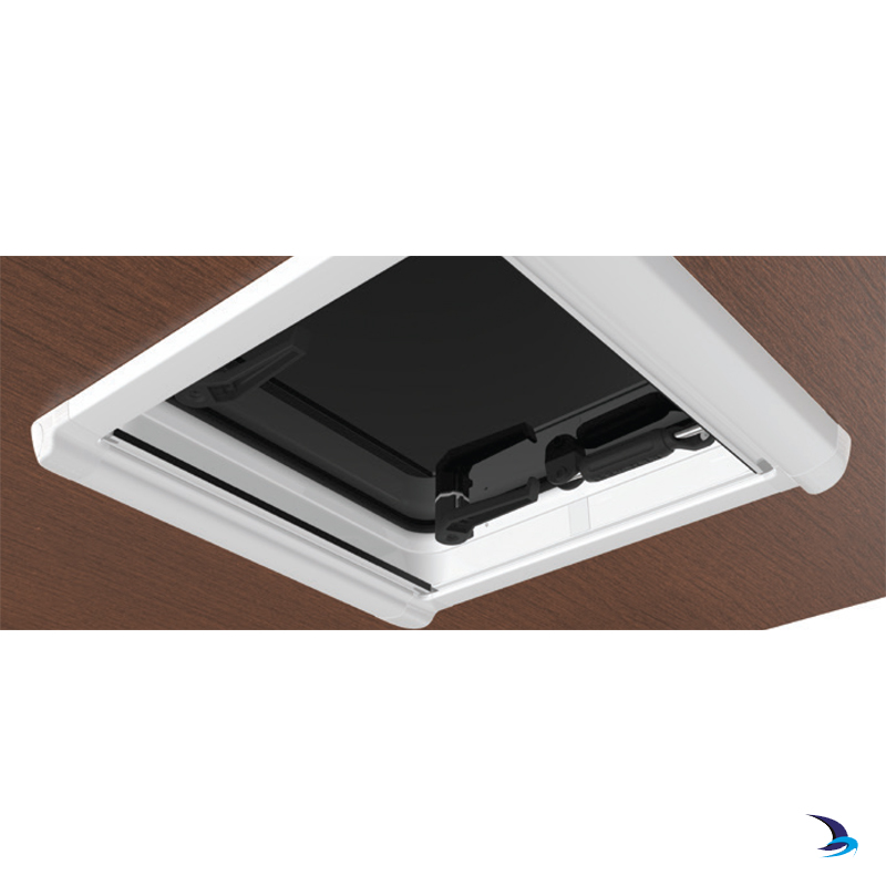
Complements and covers any gaps between the Eclipse Shade and the hatch frame. The trim clips into the Eclipse Shade, simply cut down to adjust to the correct depth for you deck.
First, the purpose of a microscope is to magnify a small object or to magnify the fine details of a larger object in order to examine minute specimens that cannot be seen by the naked eye. Here are the important compound microscope parts...Eyepiece: The lens the viewer looks through to see the specimen. The eyepiece usually contains a 10X or 15X power lens.Diopter Adjustment: Useful as a means to change focus on one eyepiece so as to correct for any difference in vision between your two eyes.Body tube (Head): The body tube connects the eyepiece to the objective lenses.Arm: The arm connects the body tube to the base of the microscope.Coarse adjustment: Brings the specimen into general focus.Fine adjustment: Fine tunes the focus and increases the detail of the specimen.Nosepiece: A rotating turret that houses the objective lenses. The viewer spins the nosepiece to select different objective lenses.Objective lenses: One of the most important parts of a compound microscope, as they are the lenses closest to the specimen. A standard microscope has three, four, or five objective lenses that range in power from 4X to 100X. When focusing the microscope, be careful that the objective lens doesnât touch the slide, as it could break the slide and destroy the specimen.Specimen or slide: The specimen is the object being examined. Most specimens are mounted on slides, flat rectangles of thin glass.The specimen is placed on the glass and a cover slip is placed over the specimen. This allows the slide to be easily inserted or removed from the microscope. It also allows the specimen to be labeled, transported, and stored without damage.Stage: The flat platform where the slide is placed.Stage clips: Metal clips that hold the slide in place.Stage height adjustment (Stage Control): These knobs move the stage left and right or up and down.Aperture: The hole in the middle of the stage that allows light from the illuminator to reach the specimen.On/off switch: This switch on the base of the microscope turns the illuminator off and on.Illumination: The light source for a microscope. Older microscopes used mirrors to reflect light from an external source up through the bottom of the stage; however, most microscopes now use a low-voltage bulb.Iris diaphragm: Adjusts the amount of light that reaches the specimen.Condenser: Gathers and focuses light from the illuminator onto the specimen being viewed.Base: The base supports the microscope and itâs where illuminator is located.How Does a Compound Microscope Work?All of the parts of a microscope work together - The light from the illuminator passes through the aperture, through the slide, and through the objective lens, where the image of the specimen is magnified.The then magnified image continues up through the body tube of the microscope to the eyepiece, which further magnifies the image the viewer then sees.Learning to use and adjust your compound microscope is the next important step.It's also imperative to know and understand the best practices of cleaning your microscope.The microscope parts work together in hospitals and in forensic labs, for scientists and students, bacteriologists and biologists so that they may view bacteria, plant and animal cells and tissues, and various microorganisms the world over.Compound microscopes have furthered medical research, helped to solve crimes, and they have repeatedly proven invaluable in unlocking the secrets of the microscopic world.Check out MicroscopeMasterâs online help:Basics of a Compound MicroscopeDiagram/Parts/Functions of a Compound MicroscopeBeginner Microscope ExperimentsMicroscope Slides Preparations-Styles and TechniquesPrepared Microscope Slides - Benefits and RecommendationsSee also: Dissecting Stereo Microscope Parts and Functions Stereo Microscope Vs Compound MicroscopeCheck out this Microscope Quiz to test your knowledgeInteresting info here on Basic Microscope Ergonomics Return from Parts of a Compound Microscope to Compound Light Microscope Return from Parts of a Compound Microscope to Best Microscope HomeFind out how to advertise on MicroscopeMaster!FacebookTwitter
Jun 21, 2020 — The maximum and minimum focal length formulas for a pinhole are somewhat complicated. The maximum focal length would be limited by diffraction.
Diagram/Parts/Functions of a Compound MicroscopeBeginner Microscope ExperimentsMicroscope Slides Preparations-Styles and TechniquesPrepared Microscope Slides - Benefits and RecommendationsSee also: Dissecting Stereo Microscope Parts and Functions Stereo Microscope Vs Compound MicroscopeCheck out this Microscope Quiz to test your knowledgeInteresting info here on Basic Microscope Ergonomics Return from Parts of a Compound Microscope to Compound Light Microscope Return from Parts of a Compound Microscope to Best Microscope HomeFind out how to advertise on MicroscopeMaster!FacebookTwitter
Nosepiece: A rotating turret that houses the objective lenses. The viewer spins the nosepiece to select different objective lenses.Objective lenses: One of the most important parts of a compound microscope, as they are the lenses closest to the specimen. A standard microscope has three, four, or five objective lenses that range in power from 4X to 100X. When focusing the microscope, be careful that the objective lens doesnât touch the slide, as it could break the slide and destroy the specimen.Specimen or slide: The specimen is the object being examined. Most specimens are mounted on slides, flat rectangles of thin glass.The specimen is placed on the glass and a cover slip is placed over the specimen. This allows the slide to be easily inserted or removed from the microscope. It also allows the specimen to be labeled, transported, and stored without damage.Stage: The flat platform where the slide is placed.Stage clips: Metal clips that hold the slide in place.Stage height adjustment (Stage Control): These knobs move the stage left and right or up and down.Aperture: The hole in the middle of the stage that allows light from the illuminator to reach the specimen.On/off switch: This switch on the base of the microscope turns the illuminator off and on.Illumination: The light source for a microscope. Older microscopes used mirrors to reflect light from an external source up through the bottom of the stage; however, most microscopes now use a low-voltage bulb.Iris diaphragm: Adjusts the amount of light that reaches the specimen.Condenser: Gathers and focuses light from the illuminator onto the specimen being viewed.Base: The base supports the microscope and itâs where illuminator is located.How Does a Compound Microscope Work?All of the parts of a microscope work together - The light from the illuminator passes through the aperture, through the slide, and through the objective lens, where the image of the specimen is magnified.The then magnified image continues up through the body tube of the microscope to the eyepiece, which further magnifies the image the viewer then sees.Learning to use and adjust your compound microscope is the next important step.It's also imperative to know and understand the best practices of cleaning your microscope.The microscope parts work together in hospitals and in forensic labs, for scientists and students, bacteriologists and biologists so that they may view bacteria, plant and animal cells and tissues, and various microorganisms the world over.Compound microscopes have furthered medical research, helped to solve crimes, and they have repeatedly proven invaluable in unlocking the secrets of the microscopic world.Check out MicroscopeMasterâs online help:Basics of a Compound MicroscopeDiagram/Parts/Functions of a Compound MicroscopeBeginner Microscope ExperimentsMicroscope Slides Preparations-Styles and TechniquesPrepared Microscope Slides - Benefits and RecommendationsSee also: Dissecting Stereo Microscope Parts and Functions Stereo Microscope Vs Compound MicroscopeCheck out this Microscope Quiz to test your knowledgeInteresting info here on Basic Microscope Ergonomics Return from Parts of a Compound Microscope to Compound Light Microscope Return from Parts of a Compound Microscope to Best Microscope HomeFind out how to advertise on MicroscopeMaster!FacebookTwitter
Eclipse shadesreviews
All of the parts of a microscope work together - The light from the illuminator passes through the aperture, through the slide, and through the objective lens, where the image of the specimen is magnified.The then magnified image continues up through the body tube of the microscope to the eyepiece, which further magnifies the image the viewer then sees.Learning to use and adjust your compound microscope is the next important step.It's also imperative to know and understand the best practices of cleaning your microscope.The microscope parts work together in hospitals and in forensic labs, for scientists and students, bacteriologists and biologists so that they may view bacteria, plant and animal cells and tissues, and various microorganisms the world over.Compound microscopes have furthered medical research, helped to solve crimes, and they have repeatedly proven invaluable in unlocking the secrets of the microscopic world.Check out MicroscopeMasterâs online help:Basics of a Compound MicroscopeDiagram/Parts/Functions of a Compound MicroscopeBeginner Microscope ExperimentsMicroscope Slides Preparations-Styles and TechniquesPrepared Microscope Slides - Benefits and RecommendationsSee also: Dissecting Stereo Microscope Parts and Functions Stereo Microscope Vs Compound MicroscopeCheck out this Microscope Quiz to test your knowledgeInteresting info here on Basic Microscope Ergonomics Return from Parts of a Compound Microscope to Compound Light Microscope Return from Parts of a Compound Microscope to Best Microscope HomeFind out how to advertise on MicroscopeMaster!FacebookTwitter
All of the parts of a microscope work together - The light from the illuminator passes through the aperture, through the slide, and through the objective lens, where the image of the specimen is magnified.The then magnified image continues up through the body tube of the microscope to the eyepiece, which further magnifies the image the viewer then sees.Learning to use and adjust your compound microscope is the next important step.It's also imperative to know and understand the best practices of cleaning your microscope.The microscope parts work together in hospitals and in forensic labs, for scientists and students, bacteriologists and biologists so that they may view bacteria, plant and animal cells and tissues, and various microorganisms the world over.Compound microscopes have furthered medical research, helped to solve crimes, and they have repeatedly proven invaluable in unlocking the secrets of the microscopic world.Check out MicroscopeMasterâs online help:Basics of a Compound MicroscopeDiagram/Parts/Functions of a Compound MicroscopeBeginner Microscope ExperimentsMicroscope Slides Preparations-Styles and TechniquesPrepared Microscope Slides - Benefits and RecommendationsSee also: Dissecting Stereo Microscope Parts and Functions Stereo Microscope Vs Compound MicroscopeCheck out this Microscope Quiz to test your knowledgeInteresting info here on Basic Microscope Ergonomics Return from Parts of a Compound Microscope to Compound Light Microscope Return from Parts of a Compound Microscope to Best Microscope HomeFind out how to advertise on MicroscopeMaster!FacebookTwitter
Amazon and the Amazon logo are trademarks of Amazon.com, Inc. or its affiliatesImages are used with permission as required.
Stage: The flat platform where the slide is placed.Stage clips: Metal clips that hold the slide in place.Stage height adjustment (Stage Control): These knobs move the stage left and right or up and down.Aperture: The hole in the middle of the stage that allows light from the illuminator to reach the specimen.On/off switch: This switch on the base of the microscope turns the illuminator off and on.Illumination: The light source for a microscope. Older microscopes used mirrors to reflect light from an external source up through the bottom of the stage; however, most microscopes now use a low-voltage bulb.Iris diaphragm: Adjusts the amount of light that reaches the specimen.Condenser: Gathers and focuses light from the illuminator onto the specimen being viewed.Base: The base supports the microscope and itâs where illuminator is located.How Does a Compound Microscope Work?All of the parts of a microscope work together - The light from the illuminator passes through the aperture, through the slide, and through the objective lens, where the image of the specimen is magnified.The then magnified image continues up through the body tube of the microscope to the eyepiece, which further magnifies the image the viewer then sees.Learning to use and adjust your compound microscope is the next important step.It's also imperative to know and understand the best practices of cleaning your microscope.The microscope parts work together in hospitals and in forensic labs, for scientists and students, bacteriologists and biologists so that they may view bacteria, plant and animal cells and tissues, and various microorganisms the world over.Compound microscopes have furthered medical research, helped to solve crimes, and they have repeatedly proven invaluable in unlocking the secrets of the microscopic world.Check out MicroscopeMasterâs online help:Basics of a Compound MicroscopeDiagram/Parts/Functions of a Compound MicroscopeBeginner Microscope ExperimentsMicroscope Slides Preparations-Styles and TechniquesPrepared Microscope Slides - Benefits and RecommendationsSee also: Dissecting Stereo Microscope Parts and Functions Stereo Microscope Vs Compound MicroscopeCheck out this Microscope Quiz to test your knowledgeInteresting info here on Basic Microscope Ergonomics Return from Parts of a Compound Microscope to Compound Light Microscope Return from Parts of a Compound Microscope to Best Microscope HomeFind out how to advertise on MicroscopeMaster!FacebookTwitter
The material on this page is not medical advice and is not to be used for diagnosis or treatment. Although care has been taken when preparing this page, its accuracy cannot be guaranteed. Scientific understanding changes over time.**  Be sure to take the utmost precaution and care when performing a microscope experiment.  MicroscopeMaster is not liable for your results or any personal issues resulting from performing the experiment. The MicroscopeMaster website is for educational purposes only.
** Â Be sure to take the utmost precaution and care when performing a microscope experiment. Â MicroscopeMaster is not liable for your results or any personal issues resulting from performing the experiment. The MicroscopeMaster website is for educational purposes only.
Objective lenses: One of the most important parts of a compound microscope, as they are the lenses closest to the specimen. A standard microscope has three, four, or five objective lenses that range in power from 4X to 100X. When focusing the microscope, be careful that the objective lens doesnât touch the slide, as it could break the slide and destroy the specimen.Specimen or slide: The specimen is the object being examined. Most specimens are mounted on slides, flat rectangles of thin glass.The specimen is placed on the glass and a cover slip is placed over the specimen. This allows the slide to be easily inserted or removed from the microscope. It also allows the specimen to be labeled, transported, and stored without damage.Stage: The flat platform where the slide is placed.Stage clips: Metal clips that hold the slide in place.Stage height adjustment (Stage Control): These knobs move the stage left and right or up and down.Aperture: The hole in the middle of the stage that allows light from the illuminator to reach the specimen.On/off switch: This switch on the base of the microscope turns the illuminator off and on.Illumination: The light source for a microscope. Older microscopes used mirrors to reflect light from an external source up through the bottom of the stage; however, most microscopes now use a low-voltage bulb.Iris diaphragm: Adjusts the amount of light that reaches the specimen.Condenser: Gathers and focuses light from the illuminator onto the specimen being viewed.Base: The base supports the microscope and itâs where illuminator is located.How Does a Compound Microscope Work?All of the parts of a microscope work together - The light from the illuminator passes through the aperture, through the slide, and through the objective lens, where the image of the specimen is magnified.The then magnified image continues up through the body tube of the microscope to the eyepiece, which further magnifies the image the viewer then sees.Learning to use and adjust your compound microscope is the next important step.It's also imperative to know and understand the best practices of cleaning your microscope.The microscope parts work together in hospitals and in forensic labs, for scientists and students, bacteriologists and biologists so that they may view bacteria, plant and animal cells and tissues, and various microorganisms the world over.Compound microscopes have furthered medical research, helped to solve crimes, and they have repeatedly proven invaluable in unlocking the secrets of the microscopic world.Check out MicroscopeMasterâs online help:Basics of a Compound MicroscopeDiagram/Parts/Functions of a Compound MicroscopeBeginner Microscope ExperimentsMicroscope Slides Preparations-Styles and TechniquesPrepared Microscope Slides - Benefits and RecommendationsSee also: Dissecting Stereo Microscope Parts and Functions Stereo Microscope Vs Compound MicroscopeCheck out this Microscope Quiz to test your knowledgeInteresting info here on Basic Microscope Ergonomics Return from Parts of a Compound Microscope to Compound Light Microscope Return from Parts of a Compound Microscope to Best Microscope HomeFind out how to advertise on MicroscopeMaster!FacebookTwitter
Eclipse shadescostco
Base: The base supports the microscope and itâs where illuminator is located.How Does a Compound Microscope Work?All of the parts of a microscope work together - The light from the illuminator passes through the aperture, through the slide, and through the objective lens, where the image of the specimen is magnified.The then magnified image continues up through the body tube of the microscope to the eyepiece, which further magnifies the image the viewer then sees.Learning to use and adjust your compound microscope is the next important step.It's also imperative to know and understand the best practices of cleaning your microscope.The microscope parts work together in hospitals and in forensic labs, for scientists and students, bacteriologists and biologists so that they may view bacteria, plant and animal cells and tissues, and various microorganisms the world over.Compound microscopes have furthered medical research, helped to solve crimes, and they have repeatedly proven invaluable in unlocking the secrets of the microscopic world.Check out MicroscopeMasterâs online help:Basics of a Compound MicroscopeDiagram/Parts/Functions of a Compound MicroscopeBeginner Microscope ExperimentsMicroscope Slides Preparations-Styles and TechniquesPrepared Microscope Slides - Benefits and RecommendationsSee also: Dissecting Stereo Microscope Parts and Functions Stereo Microscope Vs Compound MicroscopeCheck out this Microscope Quiz to test your knowledgeInteresting info here on Basic Microscope Ergonomics Return from Parts of a Compound Microscope to Compound Light Microscope Return from Parts of a Compound Microscope to Best Microscope HomeFind out how to advertise on MicroscopeMaster!FacebookTwitter
Return from Parts of a Compound Microscope to Compound Light Microscope Return from Parts of a Compound Microscope to Best Microscope HomeFind out how to advertise on MicroscopeMaster!FacebookTwitter
Eyepiece: The lens the viewer looks through to see the specimen. The eyepiece usually contains a 10X or 15X power lens.Diopter Adjustment: Useful as a means to change focus on one eyepiece so as to correct for any difference in vision between your two eyes.Body tube (Head): The body tube connects the eyepiece to the objective lenses.Arm: The arm connects the body tube to the base of the microscope.Coarse adjustment: Brings the specimen into general focus.Fine adjustment: Fine tunes the focus and increases the detail of the specimen.Nosepiece: A rotating turret that houses the objective lenses. The viewer spins the nosepiece to select different objective lenses.Objective lenses: One of the most important parts of a compound microscope, as they are the lenses closest to the specimen. A standard microscope has three, four, or five objective lenses that range in power from 4X to 100X. When focusing the microscope, be careful that the objective lens doesnât touch the slide, as it could break the slide and destroy the specimen.Specimen or slide: The specimen is the object being examined. Most specimens are mounted on slides, flat rectangles of thin glass.The specimen is placed on the glass and a cover slip is placed over the specimen. This allows the slide to be easily inserted or removed from the microscope. It also allows the specimen to be labeled, transported, and stored without damage.Stage: The flat platform where the slide is placed.Stage clips: Metal clips that hold the slide in place.Stage height adjustment (Stage Control): These knobs move the stage left and right or up and down.Aperture: The hole in the middle of the stage that allows light from the illuminator to reach the specimen.On/off switch: This switch on the base of the microscope turns the illuminator off and on.Illumination: The light source for a microscope. Older microscopes used mirrors to reflect light from an external source up through the bottom of the stage; however, most microscopes now use a low-voltage bulb.Iris diaphragm: Adjusts the amount of light that reaches the specimen.Condenser: Gathers and focuses light from the illuminator onto the specimen being viewed.Base: The base supports the microscope and itâs where illuminator is located.How Does a Compound Microscope Work?All of the parts of a microscope work together - The light from the illuminator passes through the aperture, through the slide, and through the objective lens, where the image of the specimen is magnified.The then magnified image continues up through the body tube of the microscope to the eyepiece, which further magnifies the image the viewer then sees.Learning to use and adjust your compound microscope is the next important step.It's also imperative to know and understand the best practices of cleaning your microscope.The microscope parts work together in hospitals and in forensic labs, for scientists and students, bacteriologists and biologists so that they may view bacteria, plant and animal cells and tissues, and various microorganisms the world over.Compound microscopes have furthered medical research, helped to solve crimes, and they have repeatedly proven invaluable in unlocking the secrets of the microscopic world.Check out MicroscopeMasterâs online help:Basics of a Compound MicroscopeDiagram/Parts/Functions of a Compound MicroscopeBeginner Microscope ExperimentsMicroscope Slides Preparations-Styles and TechniquesPrepared Microscope Slides - Benefits and RecommendationsSee also: Dissecting Stereo Microscope Parts and Functions Stereo Microscope Vs Compound MicroscopeCheck out this Microscope Quiz to test your knowledgeInteresting info here on Basic Microscope Ergonomics Return from Parts of a Compound Microscope to Compound Light Microscope Return from Parts of a Compound Microscope to Best Microscope HomeFind out how to advertise on MicroscopeMaster!FacebookTwitter
Optically Clear Adhesive, Sealant and Coating ... UV15-7 delivers superior physical strength properties, low shrinkage upon curing and good non-yellowing ...
A standard microscope has three, four, or five objective lenses that range in power from 4X to 100X. When focusing the microscope, be careful that the objective lens doesnât touch the slide, as it could break the slide and destroy the specimen.Specimen or slide: The specimen is the object being examined. Most specimens are mounted on slides, flat rectangles of thin glass.The specimen is placed on the glass and a cover slip is placed over the specimen. This allows the slide to be easily inserted or removed from the microscope. It also allows the specimen to be labeled, transported, and stored without damage.Stage: The flat platform where the slide is placed.Stage clips: Metal clips that hold the slide in place.Stage height adjustment (Stage Control): These knobs move the stage left and right or up and down.Aperture: The hole in the middle of the stage that allows light from the illuminator to reach the specimen.On/off switch: This switch on the base of the microscope turns the illuminator off and on.Illumination: The light source for a microscope. Older microscopes used mirrors to reflect light from an external source up through the bottom of the stage; however, most microscopes now use a low-voltage bulb.Iris diaphragm: Adjusts the amount of light that reaches the specimen.Condenser: Gathers and focuses light from the illuminator onto the specimen being viewed.Base: The base supports the microscope and itâs where illuminator is located.How Does a Compound Microscope Work?All of the parts of a microscope work together - The light from the illuminator passes through the aperture, through the slide, and through the objective lens, where the image of the specimen is magnified.The then magnified image continues up through the body tube of the microscope to the eyepiece, which further magnifies the image the viewer then sees.Learning to use and adjust your compound microscope is the next important step.It's also imperative to know and understand the best practices of cleaning your microscope.The microscope parts work together in hospitals and in forensic labs, for scientists and students, bacteriologists and biologists so that they may view bacteria, plant and animal cells and tissues, and various microorganisms the world over.Compound microscopes have furthered medical research, helped to solve crimes, and they have repeatedly proven invaluable in unlocking the secrets of the microscopic world.Check out MicroscopeMasterâs online help:Basics of a Compound MicroscopeDiagram/Parts/Functions of a Compound MicroscopeBeginner Microscope ExperimentsMicroscope Slides Preparations-Styles and TechniquesPrepared Microscope Slides - Benefits and RecommendationsSee also: Dissecting Stereo Microscope Parts and Functions Stereo Microscope Vs Compound MicroscopeCheck out this Microscope Quiz to test your knowledgeInteresting info here on Basic Microscope Ergonomics Return from Parts of a Compound Microscope to Compound Light Microscope Return from Parts of a Compound Microscope to Best Microscope HomeFind out how to advertise on MicroscopeMaster!FacebookTwitter
MicroscopeMaster.com is a participant in the Amazon Services LLC Associates Program, an affiliate advertising program designed to provide a means to earn fees by linking to Amazon.com and affiliated sites.
Fine adjustment: Fine tunes the focus and increases the detail of the specimen.Nosepiece: A rotating turret that houses the objective lenses. The viewer spins the nosepiece to select different objective lenses.Objective lenses: One of the most important parts of a compound microscope, as they are the lenses closest to the specimen. A standard microscope has three, four, or five objective lenses that range in power from 4X to 100X. When focusing the microscope, be careful that the objective lens doesnât touch the slide, as it could break the slide and destroy the specimen.Specimen or slide: The specimen is the object being examined. Most specimens are mounted on slides, flat rectangles of thin glass.The specimen is placed on the glass and a cover slip is placed over the specimen. This allows the slide to be easily inserted or removed from the microscope. It also allows the specimen to be labeled, transported, and stored without damage.Stage: The flat platform where the slide is placed.Stage clips: Metal clips that hold the slide in place.Stage height adjustment (Stage Control): These knobs move the stage left and right or up and down.Aperture: The hole in the middle of the stage that allows light from the illuminator to reach the specimen.On/off switch: This switch on the base of the microscope turns the illuminator off and on.Illumination: The light source for a microscope. Older microscopes used mirrors to reflect light from an external source up through the bottom of the stage; however, most microscopes now use a low-voltage bulb.Iris diaphragm: Adjusts the amount of light that reaches the specimen.Condenser: Gathers and focuses light from the illuminator onto the specimen being viewed.Base: The base supports the microscope and itâs where illuminator is located.How Does a Compound Microscope Work?All of the parts of a microscope work together - The light from the illuminator passes through the aperture, through the slide, and through the objective lens, where the image of the specimen is magnified.The then magnified image continues up through the body tube of the microscope to the eyepiece, which further magnifies the image the viewer then sees.Learning to use and adjust your compound microscope is the next important step.It's also imperative to know and understand the best practices of cleaning your microscope.The microscope parts work together in hospitals and in forensic labs, for scientists and students, bacteriologists and biologists so that they may view bacteria, plant and animal cells and tissues, and various microorganisms the world over.Compound microscopes have furthered medical research, helped to solve crimes, and they have repeatedly proven invaluable in unlocking the secrets of the microscopic world.Check out MicroscopeMasterâs online help:Basics of a Compound MicroscopeDiagram/Parts/Functions of a Compound MicroscopeBeginner Microscope ExperimentsMicroscope Slides Preparations-Styles and TechniquesPrepared Microscope Slides - Benefits and RecommendationsSee also: Dissecting Stereo Microscope Parts and Functions Stereo Microscope Vs Compound MicroscopeCheck out this Microscope Quiz to test your knowledgeInteresting info here on Basic Microscope Ergonomics Return from Parts of a Compound Microscope to Compound Light Microscope Return from Parts of a Compound Microscope to Best Microscope HomeFind out how to advertise on MicroscopeMaster!FacebookTwitter
Stage clips: Metal clips that hold the slide in place.Stage height adjustment (Stage Control): These knobs move the stage left and right or up and down.Aperture: The hole in the middle of the stage that allows light from the illuminator to reach the specimen.On/off switch: This switch on the base of the microscope turns the illuminator off and on.Illumination: The light source for a microscope. Older microscopes used mirrors to reflect light from an external source up through the bottom of the stage; however, most microscopes now use a low-voltage bulb.Iris diaphragm: Adjusts the amount of light that reaches the specimen.Condenser: Gathers and focuses light from the illuminator onto the specimen being viewed.Base: The base supports the microscope and itâs where illuminator is located.How Does a Compound Microscope Work?All of the parts of a microscope work together - The light from the illuminator passes through the aperture, through the slide, and through the objective lens, where the image of the specimen is magnified.The then magnified image continues up through the body tube of the microscope to the eyepiece, which further magnifies the image the viewer then sees.Learning to use and adjust your compound microscope is the next important step.It's also imperative to know and understand the best practices of cleaning your microscope.The microscope parts work together in hospitals and in forensic labs, for scientists and students, bacteriologists and biologists so that they may view bacteria, plant and animal cells and tissues, and various microorganisms the world over.Compound microscopes have furthered medical research, helped to solve crimes, and they have repeatedly proven invaluable in unlocking the secrets of the microscopic world.Check out MicroscopeMasterâs online help:Basics of a Compound MicroscopeDiagram/Parts/Functions of a Compound MicroscopeBeginner Microscope ExperimentsMicroscope Slides Preparations-Styles and TechniquesPrepared Microscope Slides - Benefits and RecommendationsSee also: Dissecting Stereo Microscope Parts and Functions Stereo Microscope Vs Compound MicroscopeCheck out this Microscope Quiz to test your knowledgeInteresting info here on Basic Microscope Ergonomics Return from Parts of a Compound Microscope to Compound Light Microscope Return from Parts of a Compound Microscope to Best Microscope HomeFind out how to advertise on MicroscopeMaster!FacebookTwitter
Interesting info here on Basic Microscope Ergonomics Return from Parts of a Compound Microscope to Compound Light Microscope Return from Parts of a Compound Microscope to Best Microscope HomeFind out how to advertise on MicroscopeMaster!FacebookTwitter
Stage: The flat platform where the slide is placed.Stage clips: Metal clips that hold the slide in place.Stage height adjustment (Stage Control): These knobs move the stage left and right or up and down.Aperture: The hole in the middle of the stage that allows light from the illuminator to reach the specimen.On/off switch: This switch on the base of the microscope turns the illuminator off and on.Illumination: The light source for a microscope. Older microscopes used mirrors to reflect light from an external source up through the bottom of the stage; however, most microscopes now use a low-voltage bulb.Iris diaphragm: Adjusts the amount of light that reaches the specimen.Condenser: Gathers and focuses light from the illuminator onto the specimen being viewed.Base: The base supports the microscope and itâs where illuminator is located.How Does a Compound Microscope Work?All of the parts of a microscope work together - The light from the illuminator passes through the aperture, through the slide, and through the objective lens, where the image of the specimen is magnified.The then magnified image continues up through the body tube of the microscope to the eyepiece, which further magnifies the image the viewer then sees.Learning to use and adjust your compound microscope is the next important step.It's also imperative to know and understand the best practices of cleaning your microscope.The microscope parts work together in hospitals and in forensic labs, for scientists and students, bacteriologists and biologists so that they may view bacteria, plant and animal cells and tissues, and various microorganisms the world over.Compound microscopes have furthered medical research, helped to solve crimes, and they have repeatedly proven invaluable in unlocking the secrets of the microscopic world.Check out MicroscopeMasterâs online help:Basics of a Compound MicroscopeDiagram/Parts/Functions of a Compound MicroscopeBeginner Microscope ExperimentsMicroscope Slides Preparations-Styles and TechniquesPrepared Microscope Slides - Benefits and RecommendationsSee also: Dissecting Stereo Microscope Parts and Functions Stereo Microscope Vs Compound MicroscopeCheck out this Microscope Quiz to test your knowledgeInteresting info here on Basic Microscope Ergonomics Return from Parts of a Compound Microscope to Compound Light Microscope Return from Parts of a Compound Microscope to Best Microscope HomeFind out how to advertise on MicroscopeMaster!FacebookTwitter
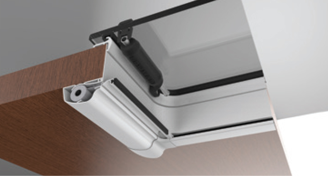
Body tube (Head): The body tube connects the eyepiece to the objective lenses.Arm: The arm connects the body tube to the base of the microscope.Coarse adjustment: Brings the specimen into general focus.Fine adjustment: Fine tunes the focus and increases the detail of the specimen.Nosepiece: A rotating turret that houses the objective lenses. The viewer spins the nosepiece to select different objective lenses.Objective lenses: One of the most important parts of a compound microscope, as they are the lenses closest to the specimen. A standard microscope has three, four, or five objective lenses that range in power from 4X to 100X. When focusing the microscope, be careful that the objective lens doesnât touch the slide, as it could break the slide and destroy the specimen.Specimen or slide: The specimen is the object being examined. Most specimens are mounted on slides, flat rectangles of thin glass.The specimen is placed on the glass and a cover slip is placed over the specimen. This allows the slide to be easily inserted or removed from the microscope. It also allows the specimen to be labeled, transported, and stored without damage.Stage: The flat platform where the slide is placed.Stage clips: Metal clips that hold the slide in place.Stage height adjustment (Stage Control): These knobs move the stage left and right or up and down.Aperture: The hole in the middle of the stage that allows light from the illuminator to reach the specimen.On/off switch: This switch on the base of the microscope turns the illuminator off and on.Illumination: The light source for a microscope. Older microscopes used mirrors to reflect light from an external source up through the bottom of the stage; however, most microscopes now use a low-voltage bulb.Iris diaphragm: Adjusts the amount of light that reaches the specimen.Condenser: Gathers and focuses light from the illuminator onto the specimen being viewed.Base: The base supports the microscope and itâs where illuminator is located.How Does a Compound Microscope Work?All of the parts of a microscope work together - The light from the illuminator passes through the aperture, through the slide, and through the objective lens, where the image of the specimen is magnified.The then magnified image continues up through the body tube of the microscope to the eyepiece, which further magnifies the image the viewer then sees.Learning to use and adjust your compound microscope is the next important step.It's also imperative to know and understand the best practices of cleaning your microscope.The microscope parts work together in hospitals and in forensic labs, for scientists and students, bacteriologists and biologists so that they may view bacteria, plant and animal cells and tissues, and various microorganisms the world over.Compound microscopes have furthered medical research, helped to solve crimes, and they have repeatedly proven invaluable in unlocking the secrets of the microscopic world.Check out MicroscopeMasterâs online help:Basics of a Compound MicroscopeDiagram/Parts/Functions of a Compound MicroscopeBeginner Microscope ExperimentsMicroscope Slides Preparations-Styles and TechniquesPrepared Microscope Slides - Benefits and RecommendationsSee also: Dissecting Stereo Microscope Parts and Functions Stereo Microscope Vs Compound MicroscopeCheck out this Microscope Quiz to test your knowledgeInteresting info here on Basic Microscope Ergonomics Return from Parts of a Compound Microscope to Compound Light Microscope Return from Parts of a Compound Microscope to Best Microscope HomeFind out how to advertise on MicroscopeMaster!FacebookTwitter
Oct 26, 22 05:01 PMChemoorganotrophs also known as organotrophs, include organisms that obtain their energy from organic chemicals like glucose. This process is known as chemoorganotrophy. Read more here.Read MoreBetaproteobacteria â Examples, Characteristics and FunctionOct 25, 22 03:44 PMBetaproteobacteria is a heterogeneous group in the phylum Proteobacteria whose members can be found in a range of habitats from wastewater and hot springs to the Antarctic. Read more here.Read More
Outdooreclipse shades
Illumination: The light source for a microscope. Older microscopes used mirrors to reflect light from an external source up through the bottom of the stage; however, most microscopes now use a low-voltage bulb.Iris diaphragm: Adjusts the amount of light that reaches the specimen.Condenser: Gathers and focuses light from the illuminator onto the specimen being viewed.Base: The base supports the microscope and itâs where illuminator is located.How Does a Compound Microscope Work?All of the parts of a microscope work together - The light from the illuminator passes through the aperture, through the slide, and through the objective lens, where the image of the specimen is magnified.The then magnified image continues up through the body tube of the microscope to the eyepiece, which further magnifies the image the viewer then sees.Learning to use and adjust your compound microscope is the next important step.It's also imperative to know and understand the best practices of cleaning your microscope.The microscope parts work together in hospitals and in forensic labs, for scientists and students, bacteriologists and biologists so that they may view bacteria, plant and animal cells and tissues, and various microorganisms the world over.Compound microscopes have furthered medical research, helped to solve crimes, and they have repeatedly proven invaluable in unlocking the secrets of the microscopic world.Check out MicroscopeMasterâs online help:Basics of a Compound MicroscopeDiagram/Parts/Functions of a Compound MicroscopeBeginner Microscope ExperimentsMicroscope Slides Preparations-Styles and TechniquesPrepared Microscope Slides - Benefits and RecommendationsSee also: Dissecting Stereo Microscope Parts and Functions Stereo Microscope Vs Compound MicroscopeCheck out this Microscope Quiz to test your knowledgeInteresting info here on Basic Microscope Ergonomics Return from Parts of a Compound Microscope to Compound Light Microscope Return from Parts of a Compound Microscope to Best Microscope HomeFind out how to advertise on MicroscopeMaster!FacebookTwitter
A near-infrared (NIR) camera is a specialized device capable of capturing images in the infrared wavelength range, specifically from 700 nm to 2,500 nm (0.7 to ...
Basics of a Compound MicroscopeDiagram/Parts/Functions of a Compound MicroscopeBeginner Microscope ExperimentsMicroscope Slides Preparations-Styles and TechniquesPrepared Microscope Slides - Benefits and RecommendationsSee also: Dissecting Stereo Microscope Parts and Functions Stereo Microscope Vs Compound MicroscopeCheck out this Microscope Quiz to test your knowledgeInteresting info here on Basic Microscope Ergonomics Return from Parts of a Compound Microscope to Compound Light Microscope Return from Parts of a Compound Microscope to Best Microscope HomeFind out how to advertise on MicroscopeMaster!FacebookTwitter
Learning to use and adjust your compound microscope is the next important step.It's also imperative to know and understand the best practices of cleaning your microscope.The microscope parts work together in hospitals and in forensic labs, for scientists and students, bacteriologists and biologists so that they may view bacteria, plant and animal cells and tissues, and various microorganisms the world over.Compound microscopes have furthered medical research, helped to solve crimes, and they have repeatedly proven invaluable in unlocking the secrets of the microscopic world.Check out MicroscopeMasterâs online help:Basics of a Compound MicroscopeDiagram/Parts/Functions of a Compound MicroscopeBeginner Microscope ExperimentsMicroscope Slides Preparations-Styles and TechniquesPrepared Microscope Slides - Benefits and RecommendationsSee also: Dissecting Stereo Microscope Parts and Functions Stereo Microscope Vs Compound MicroscopeCheck out this Microscope Quiz to test your knowledgeInteresting info here on Basic Microscope Ergonomics Return from Parts of a Compound Microscope to Compound Light Microscope Return from Parts of a Compound Microscope to Best Microscope HomeFind out how to advertise on MicroscopeMaster!FacebookTwitter
Eyepiece: The lens the viewer looks through to see the specimen. The eyepiece usually contains a 10X or 15X power lens.Diopter Adjustment: Useful as a means to change focus on one eyepiece so as to correct for any difference in vision between your two eyes.Body tube (Head): The body tube connects the eyepiece to the objective lenses.Arm: The arm connects the body tube to the base of the microscope.Coarse adjustment: Brings the specimen into general focus.Fine adjustment: Fine tunes the focus and increases the detail of the specimen.Nosepiece: A rotating turret that houses the objective lenses. The viewer spins the nosepiece to select different objective lenses.Objective lenses: One of the most important parts of a compound microscope, as they are the lenses closest to the specimen. A standard microscope has three, four, or five objective lenses that range in power from 4X to 100X. When focusing the microscope, be careful that the objective lens doesnât touch the slide, as it could break the slide and destroy the specimen.Specimen or slide: The specimen is the object being examined. Most specimens are mounted on slides, flat rectangles of thin glass.The specimen is placed on the glass and a cover slip is placed over the specimen. This allows the slide to be easily inserted or removed from the microscope. It also allows the specimen to be labeled, transported, and stored without damage.Stage: The flat platform where the slide is placed.Stage clips: Metal clips that hold the slide in place.Stage height adjustment (Stage Control): These knobs move the stage left and right or up and down.Aperture: The hole in the middle of the stage that allows light from the illuminator to reach the specimen.On/off switch: This switch on the base of the microscope turns the illuminator off and on.Illumination: The light source for a microscope. Older microscopes used mirrors to reflect light from an external source up through the bottom of the stage; however, most microscopes now use a low-voltage bulb.Iris diaphragm: Adjusts the amount of light that reaches the specimen.Condenser: Gathers and focuses light from the illuminator onto the specimen being viewed.Base: The base supports the microscope and itâs where illuminator is located.How Does a Compound Microscope Work?All of the parts of a microscope work together - The light from the illuminator passes through the aperture, through the slide, and through the objective lens, where the image of the specimen is magnified.The then magnified image continues up through the body tube of the microscope to the eyepiece, which further magnifies the image the viewer then sees.Learning to use and adjust your compound microscope is the next important step.It's also imperative to know and understand the best practices of cleaning your microscope.The microscope parts work together in hospitals and in forensic labs, for scientists and students, bacteriologists and biologists so that they may view bacteria, plant and animal cells and tissues, and various microorganisms the world over.Compound microscopes have furthered medical research, helped to solve crimes, and they have repeatedly proven invaluable in unlocking the secrets of the microscopic world.Check out MicroscopeMasterâs online help:Basics of a Compound MicroscopeDiagram/Parts/Functions of a Compound MicroscopeBeginner Microscope ExperimentsMicroscope Slides Preparations-Styles and TechniquesPrepared Microscope Slides - Benefits and RecommendationsSee also: Dissecting Stereo Microscope Parts and Functions Stereo Microscope Vs Compound MicroscopeCheck out this Microscope Quiz to test your knowledgeInteresting info here on Basic Microscope Ergonomics Return from Parts of a Compound Microscope to Compound Light Microscope Return from Parts of a Compound Microscope to Best Microscope HomeFind out how to advertise on MicroscopeMaster!FacebookTwitter
The specimen is placed on the glass and a cover slip is placed over the specimen. This allows the slide to be easily inserted or removed from the microscope. It also allows the specimen to be labeled, transported, and stored without damage.Stage: The flat platform where the slide is placed.Stage clips: Metal clips that hold the slide in place.Stage height adjustment (Stage Control): These knobs move the stage left and right or up and down.Aperture: The hole in the middle of the stage that allows light from the illuminator to reach the specimen.On/off switch: This switch on the base of the microscope turns the illuminator off and on.Illumination: The light source for a microscope. Older microscopes used mirrors to reflect light from an external source up through the bottom of the stage; however, most microscopes now use a low-voltage bulb.Iris diaphragm: Adjusts the amount of light that reaches the specimen.Condenser: Gathers and focuses light from the illuminator onto the specimen being viewed.Base: The base supports the microscope and itâs where illuminator is located.How Does a Compound Microscope Work?All of the parts of a microscope work together - The light from the illuminator passes through the aperture, through the slide, and through the objective lens, where the image of the specimen is magnified.The then magnified image continues up through the body tube of the microscope to the eyepiece, which further magnifies the image the viewer then sees.Learning to use and adjust your compound microscope is the next important step.It's also imperative to know and understand the best practices of cleaning your microscope.The microscope parts work together in hospitals and in forensic labs, for scientists and students, bacteriologists and biologists so that they may view bacteria, plant and animal cells and tissues, and various microorganisms the world over.Compound microscopes have furthered medical research, helped to solve crimes, and they have repeatedly proven invaluable in unlocking the secrets of the microscopic world.Check out MicroscopeMasterâs online help:Basics of a Compound MicroscopeDiagram/Parts/Functions of a Compound MicroscopeBeginner Microscope ExperimentsMicroscope Slides Preparations-Styles and TechniquesPrepared Microscope Slides - Benefits and RecommendationsSee also: Dissecting Stereo Microscope Parts and Functions Stereo Microscope Vs Compound MicroscopeCheck out this Microscope Quiz to test your knowledgeInteresting info here on Basic Microscope Ergonomics Return from Parts of a Compound Microscope to Compound Light Microscope Return from Parts of a Compound Microscope to Best Microscope HomeFind out how to advertise on MicroscopeMaster!FacebookTwitter
Check out MicroscopeMasterâs online help:Basics of a Compound MicroscopeDiagram/Parts/Functions of a Compound MicroscopeBeginner Microscope ExperimentsMicroscope Slides Preparations-Styles and TechniquesPrepared Microscope Slides - Benefits and RecommendationsSee also: Dissecting Stereo Microscope Parts and Functions Stereo Microscope Vs Compound MicroscopeCheck out this Microscope Quiz to test your knowledgeInteresting info here on Basic Microscope Ergonomics Return from Parts of a Compound Microscope to Compound Light Microscope Return from Parts of a Compound Microscope to Best Microscope HomeFind out how to advertise on MicroscopeMaster!FacebookTwitter
Compound microscopes have furthered medical research, helped to solve crimes, and they have repeatedly proven invaluable in unlocking the secrets of the microscopic world.Check out MicroscopeMasterâs online help:Basics of a Compound MicroscopeDiagram/Parts/Functions of a Compound MicroscopeBeginner Microscope ExperimentsMicroscope Slides Preparations-Styles and TechniquesPrepared Microscope Slides - Benefits and RecommendationsSee also: Dissecting Stereo Microscope Parts and Functions Stereo Microscope Vs Compound MicroscopeCheck out this Microscope Quiz to test your knowledgeInteresting info here on Basic Microscope Ergonomics Return from Parts of a Compound Microscope to Compound Light Microscope Return from Parts of a Compound Microscope to Best Microscope HomeFind out how to advertise on MicroscopeMaster!FacebookTwitter
Boreal Laser develops and manufactures an Enhanced Sensing Element that very accurately and reliably measures gas concentrations in variety of Industrial ...
Condenser: Gathers and focuses light from the illuminator onto the specimen being viewed.Base: The base supports the microscope and itâs where illuminator is located.How Does a Compound Microscope Work?All of the parts of a microscope work together - The light from the illuminator passes through the aperture, through the slide, and through the objective lens, where the image of the specimen is magnified.The then magnified image continues up through the body tube of the microscope to the eyepiece, which further magnifies the image the viewer then sees.Learning to use and adjust your compound microscope is the next important step.It's also imperative to know and understand the best practices of cleaning your microscope.The microscope parts work together in hospitals and in forensic labs, for scientists and students, bacteriologists and biologists so that they may view bacteria, plant and animal cells and tissues, and various microorganisms the world over.Compound microscopes have furthered medical research, helped to solve crimes, and they have repeatedly proven invaluable in unlocking the secrets of the microscopic world.Check out MicroscopeMasterâs online help:Basics of a Compound MicroscopeDiagram/Parts/Functions of a Compound MicroscopeBeginner Microscope ExperimentsMicroscope Slides Preparations-Styles and TechniquesPrepared Microscope Slides - Benefits and RecommendationsSee also: Dissecting Stereo Microscope Parts and Functions Stereo Microscope Vs Compound MicroscopeCheck out this Microscope Quiz to test your knowledgeInteresting info here on Basic Microscope Ergonomics Return from Parts of a Compound Microscope to Compound Light Microscope Return from Parts of a Compound Microscope to Best Microscope HomeFind out how to advertise on MicroscopeMaster!FacebookTwitter
Microscope Slides Preparations-Styles and TechniquesPrepared Microscope Slides - Benefits and RecommendationsSee also: Dissecting Stereo Microscope Parts and Functions Stereo Microscope Vs Compound MicroscopeCheck out this Microscope Quiz to test your knowledgeInteresting info here on Basic Microscope Ergonomics Return from Parts of a Compound Microscope to Compound Light Microscope Return from Parts of a Compound Microscope to Best Microscope HomeFind out how to advertise on MicroscopeMaster!FacebookTwitter
The then magnified image continues up through the body tube of the microscope to the eyepiece, which further magnifies the image the viewer then sees.Learning to use and adjust your compound microscope is the next important step.It's also imperative to know and understand the best practices of cleaning your microscope.The microscope parts work together in hospitals and in forensic labs, for scientists and students, bacteriologists and biologists so that they may view bacteria, plant and animal cells and tissues, and various microorganisms the world over.Compound microscopes have furthered medical research, helped to solve crimes, and they have repeatedly proven invaluable in unlocking the secrets of the microscopic world.Check out MicroscopeMasterâs online help:Basics of a Compound MicroscopeDiagram/Parts/Functions of a Compound MicroscopeBeginner Microscope ExperimentsMicroscope Slides Preparations-Styles and TechniquesPrepared Microscope Slides - Benefits and RecommendationsSee also: Dissecting Stereo Microscope Parts and Functions Stereo Microscope Vs Compound MicroscopeCheck out this Microscope Quiz to test your knowledgeInteresting info here on Basic Microscope Ergonomics Return from Parts of a Compound Microscope to Compound Light Microscope Return from Parts of a Compound Microscope to Best Microscope HomeFind out how to advertise on MicroscopeMaster!FacebookTwitter
See also: Dissecting Stereo Microscope Parts and Functions Stereo Microscope Vs Compound MicroscopeCheck out this Microscope Quiz to test your knowledgeInteresting info here on Basic Microscope Ergonomics Return from Parts of a Compound Microscope to Compound Light Microscope Return from Parts of a Compound Microscope to Best Microscope HomeFind out how to advertise on MicroscopeMaster!FacebookTwitter
Read MoreBetaproteobacteria â Examples, Characteristics and FunctionOct 25, 22 03:44 PMBetaproteobacteria is a heterogeneous group in the phylum Proteobacteria whose members can be found in a range of habitats from wastewater and hot springs to the Antarctic. Read more here.Read More
Eclipsesun shade phone number
Jul 29, 2024 — Microscopes utilise a combination of lenses to magnify objects. Light from an illuminator passes through the specimen (the object being studied) ...
While some of the microscope optical components act as image-forming elements, others serve to produce various modifications to illumination of the specimen and ...
Basics of a Compound MicroscopeDiagram/Parts/Functions of a Compound MicroscopeBeginner Microscope ExperimentsMicroscope Slides Preparations-Styles and TechniquesPrepared Microscope Slides - Benefits and RecommendationsSee also: Dissecting Stereo Microscope Parts and Functions Stereo Microscope Vs Compound MicroscopeCheck out this Microscope Quiz to test your knowledgeInteresting info here on Basic Microscope Ergonomics Return from Parts of a Compound Microscope to Compound Light Microscope Return from Parts of a Compound Microscope to Best Microscope HomeFind out how to advertise on MicroscopeMaster!FacebookTwitter
Stereo Microscope Vs Compound MicroscopeCheck out this Microscope Quiz to test your knowledgeInteresting info here on Basic Microscope Ergonomics Return from Parts of a Compound Microscope to Compound Light Microscope Return from Parts of a Compound Microscope to Best Microscope HomeFind out how to advertise on MicroscopeMaster!FacebookTwitter
On/off switch: This switch on the base of the microscope turns the illuminator off and on.Illumination: The light source for a microscope. Older microscopes used mirrors to reflect light from an external source up through the bottom of the stage; however, most microscopes now use a low-voltage bulb.Iris diaphragm: Adjusts the amount of light that reaches the specimen.Condenser: Gathers and focuses light from the illuminator onto the specimen being viewed.Base: The base supports the microscope and itâs where illuminator is located.How Does a Compound Microscope Work?All of the parts of a microscope work together - The light from the illuminator passes through the aperture, through the slide, and through the objective lens, where the image of the specimen is magnified.The then magnified image continues up through the body tube of the microscope to the eyepiece, which further magnifies the image the viewer then sees.Learning to use and adjust your compound microscope is the next important step.It's also imperative to know and understand the best practices of cleaning your microscope.The microscope parts work together in hospitals and in forensic labs, for scientists and students, bacteriologists and biologists so that they may view bacteria, plant and animal cells and tissues, and various microorganisms the world over.Compound microscopes have furthered medical research, helped to solve crimes, and they have repeatedly proven invaluable in unlocking the secrets of the microscopic world.Check out MicroscopeMasterâs online help:Basics of a Compound MicroscopeDiagram/Parts/Functions of a Compound MicroscopeBeginner Microscope ExperimentsMicroscope Slides Preparations-Styles and TechniquesPrepared Microscope Slides - Benefits and RecommendationsSee also: Dissecting Stereo Microscope Parts and Functions Stereo Microscope Vs Compound MicroscopeCheck out this Microscope Quiz to test your knowledgeInteresting info here on Basic Microscope Ergonomics Return from Parts of a Compound Microscope to Compound Light Microscope Return from Parts of a Compound Microscope to Best Microscope HomeFind out how to advertise on MicroscopeMaster!FacebookTwitter
Before exploring microscope parts and functions, you should probably understand that the compound light microscope is more complicated than just a microscope with more than one lens.First, the purpose of a microscope is to magnify a small object or to magnify the fine details of a larger object in order to examine minute specimens that cannot be seen by the naked eye. Here are the important compound microscope parts...Eyepiece: The lens the viewer looks through to see the specimen. The eyepiece usually contains a 10X or 15X power lens.Diopter Adjustment: Useful as a means to change focus on one eyepiece so as to correct for any difference in vision between your two eyes.Body tube (Head): The body tube connects the eyepiece to the objective lenses.Arm: The arm connects the body tube to the base of the microscope.Coarse adjustment: Brings the specimen into general focus.Fine adjustment: Fine tunes the focus and increases the detail of the specimen.Nosepiece: A rotating turret that houses the objective lenses. The viewer spins the nosepiece to select different objective lenses.Objective lenses: One of the most important parts of a compound microscope, as they are the lenses closest to the specimen. A standard microscope has three, four, or five objective lenses that range in power from 4X to 100X. When focusing the microscope, be careful that the objective lens doesnât touch the slide, as it could break the slide and destroy the specimen.Specimen or slide: The specimen is the object being examined. Most specimens are mounted on slides, flat rectangles of thin glass.The specimen is placed on the glass and a cover slip is placed over the specimen. This allows the slide to be easily inserted or removed from the microscope. It also allows the specimen to be labeled, transported, and stored without damage.Stage: The flat platform where the slide is placed.Stage clips: Metal clips that hold the slide in place.Stage height adjustment (Stage Control): These knobs move the stage left and right or up and down.Aperture: The hole in the middle of the stage that allows light from the illuminator to reach the specimen.On/off switch: This switch on the base of the microscope turns the illuminator off and on.Illumination: The light source for a microscope. Older microscopes used mirrors to reflect light from an external source up through the bottom of the stage; however, most microscopes now use a low-voltage bulb.Iris diaphragm: Adjusts the amount of light that reaches the specimen.Condenser: Gathers and focuses light from the illuminator onto the specimen being viewed.Base: The base supports the microscope and itâs where illuminator is located.How Does a Compound Microscope Work?All of the parts of a microscope work together - The light from the illuminator passes through the aperture, through the slide, and through the objective lens, where the image of the specimen is magnified.The then magnified image continues up through the body tube of the microscope to the eyepiece, which further magnifies the image the viewer then sees.Learning to use and adjust your compound microscope is the next important step.It's also imperative to know and understand the best practices of cleaning your microscope.The microscope parts work together in hospitals and in forensic labs, for scientists and students, bacteriologists and biologists so that they may view bacteria, plant and animal cells and tissues, and various microorganisms the world over.Compound microscopes have furthered medical research, helped to solve crimes, and they have repeatedly proven invaluable in unlocking the secrets of the microscopic world.Check out MicroscopeMasterâs online help:Basics of a Compound MicroscopeDiagram/Parts/Functions of a Compound MicroscopeBeginner Microscope ExperimentsMicroscope Slides Preparations-Styles and TechniquesPrepared Microscope Slides - Benefits and RecommendationsSee also: Dissecting Stereo Microscope Parts and Functions Stereo Microscope Vs Compound MicroscopeCheck out this Microscope Quiz to test your knowledgeInteresting info here on Basic Microscope Ergonomics Return from Parts of a Compound Microscope to Compound Light Microscope Return from Parts of a Compound Microscope to Best Microscope HomeFind out how to advertise on MicroscopeMaster!FacebookTwitter
Condenser: Gathers and focuses light from the illuminator onto the specimen being viewed.Base: The base supports the microscope and itâs where illuminator is located.How Does a Compound Microscope Work?All of the parts of a microscope work together - The light from the illuminator passes through the aperture, through the slide, and through the objective lens, where the image of the specimen is magnified.The then magnified image continues up through the body tube of the microscope to the eyepiece, which further magnifies the image the viewer then sees.Learning to use and adjust your compound microscope is the next important step.It's also imperative to know and understand the best practices of cleaning your microscope.The microscope parts work together in hospitals and in forensic labs, for scientists and students, bacteriologists and biologists so that they may view bacteria, plant and animal cells and tissues, and various microorganisms the world over.Compound microscopes have furthered medical research, helped to solve crimes, and they have repeatedly proven invaluable in unlocking the secrets of the microscopic world.Check out MicroscopeMasterâs online help:Basics of a Compound MicroscopeDiagram/Parts/Functions of a Compound MicroscopeBeginner Microscope ExperimentsMicroscope Slides Preparations-Styles and TechniquesPrepared Microscope Slides - Benefits and RecommendationsSee also: Dissecting Stereo Microscope Parts and Functions Stereo Microscope Vs Compound MicroscopeCheck out this Microscope Quiz to test your knowledgeInteresting info here on Basic Microscope Ergonomics Return from Parts of a Compound Microscope to Compound Light Microscope Return from Parts of a Compound Microscope to Best Microscope HomeFind out how to advertise on MicroscopeMaster!FacebookTwitter


 Ms.Cici
 8618319014500