
Heat Treat Colors Of Steel Chart - metal temperature color
Visionesupereroe
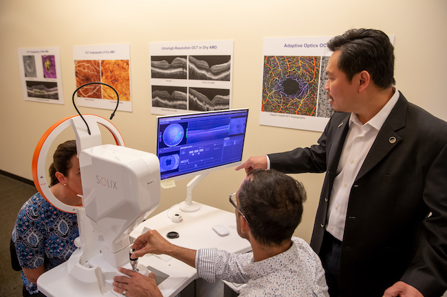
OCT was co-invented by David Huang, an OHSU ophthalmologist and biomedical engineer. It’s now a standard test for finding conditions that can cause vision loss, often before patients notice symptoms. OCT is used in more than 30 million eye procedures each year.
Thermal Camera E01, 96x96 IR Resolution, 20Hz Refresh Rate, Portable Handheld Infrared Thermal Imaging Camera with Laser Pointer, 8H of Battery Life, -4°F~752°F ...
Jul 4, 2023 — SMM brings you current and historical Battery Grade Lithium Fluoride price tables and charts, and maintains daily Battery Grade Lithium ...
OCT is a common eye care test. It is used when you may be at risk for eye disease or when you already have an eye condition.
VisioneTreccani
The IR Reflector Kit works with the Accent 1400, Accent 1000* and Accent 800**. The IR Reflector is designed to diffract and redirect some the IR signal to ...
Avere unavisione
In order to narrow the scope of this chapter, the text assumes the manufacture of a precision glass lens of approximately 50mm diameter using grinding and ...
Olympus - SZ51SZ51 – versatile stereo microscope, The versatile and cost-efficient SZ51 zoom stereo microscope offers a broad range of functions for ...
Cornea disease: The cornea is the clear front window of the eye. Damage to the cornea can make vision cloudy or out of focus.
Frankfurt Fair. Description: Messe Frankfurt Forum. Address: Ludwig-Erhard-Anlage 1 60327 Frankfurt am Main (Westend) opens map in new window Map. Phone: 0 69 ...
Visionesignificato

Visionesinonimo
Optical coherence tomography helps diagnose, treat and manage the eye diseases that are the leading causes of blindness.
VisioneMarvel
Eye disease in babies: Babies born before the 37th week of pregnancy or at a very low weight are at risk for a condition called retinopathy of prematurity. This happens when abnormal blood vessels grow in the retina.
Oregon Health & Science University is dedicated to improving the health and quality of life for all Oregonians through excellence, innovation and leadership in health care, education and research.
VISIONETV
Pack of 2 10x10cm Horizontal Polarizer Film for LCD Linear Polarized Filter Linear Polarizing Polarization Film Sheets Polarizing Film for LCD Screen ...
The compound microscope uses lenses and light to enlarge the image and is also called an optical or light microscope (versus an electron microscope). The ...
NLLeZ 1 STÜCK-Strahlteiler 20 * 20 * 20mm rechtwinklig Dreieck Prism Cube Anti-Reflexion-Beschichtung 5: 5 anwendbare Wellenband 400-700nm ... Lieferung für 2,99 ...
Visioneaziendale
Diabetic macular edema: In people with diabetes, blood vessels in the retina can become leaky. This causes swelling in a part of the retina called the macula.
Macular degeneration: In age-related macular degeneration, a part of the retina called the macula is damaged, causing loss of central vision.
The OHSU Casey Eye Institute is a leader in developing noninvasive imaging to help prevent vision loss. Dr. Huang, our associate director and director of research, is known worldwide for his vision research and inventions. He holds 42 patents and has won top prizes in vision research and biomedical engineering.
OCT of the eye is noninvasive, meaning it does not enter or even touch the eye. Standard OCT uses invisible infrared light, so it is more comfortable than imaging that uses visible light.
Glaucoma: Glaucoma damages the optic nerve, which connects the eye to the brain. It commonly causes loss of peripheral vision.
The mirror is designed to have two sides: a concave side and a flat side. The concave side is used for reflecting natural light, while the flat side is used for ...
Diabetic retinopathy: If people with diabetes have excess blood sugar over a long time, it can damage the retina and its blood vessels.
OCT scans a beam of light to create 3D images that show the retina’s layers in microscopic detail. OCT can also image the optic nerve, which connects the eye to the brain.
Optical coherence tomography creates highly precise 3D images. It is most commonly used to image the retina in the back of the eye.


 Ms.Cici
 8618319014500