
Headlight Hi-Beam Adaptor (HB3) - high beam adapter
E-Tay magnifier factory is a professional manufacturer to offer superior quality magnifying glass products,and provide perfact service for our clients.
Patients with a history of systemic disorders such as uncontrolled diabetes mellitus and pregnancy that could affect the outcome of implant therapy
Fresnel lens wide-angle lens is suitable for trucks, household hatchback buses, four-wheel drive off-road vehicles, and other vehicles without a "tail", and often do not know what is "hidden" behind the car. This lens uses a unique Fresnel lens technology. It only needs to be attached to the inner side of the rear window to provide the driver with a large angle of vision to see the obstacles in the previously invisible blind spot, increasing the rear view range.
Current camera focusing screens are frosted glass, which has the advantage of being bright and uniform in brightness. When the focus is not accurate, the image on the focusing screen is not clear. In order to coordinate more precise focusing, a split image and a micro-ring device are generally installed in the center of the focusing screen. When the focus is not accurate, the image of the subject in the center of the focusing screen splits into two images. When the two images are combined into one, the focus is accurate. The standard focusing screen of an AF SLR machine generally does not have a split image device, but is engraved with a small rectangular frame to indicate the AF area, and some focusing screens are also engraved with partial metering or spot metering areas. When the early AF SLR cameras were focusing in a dark environment, it was often difficult to see the focus frame, and it was difficult to determine which point the camera was focusing on. The focus point on the focusing screen of the new generation SLR cameras would glow or there would be a focusing sound. Prompt, easy to confirm the focus in a complex environment. Different types of focusing screens have different purposes. For portraits, it is better to use split-image focusing screens. Focusing screens with horizontal and vertical lines or scales are suitable for building photography and document copying; there is no split image in the middle part but only a micro-edge focusing The screen is suitable for small aperture lens, it will not have the shortcomings of split image which is bright and black. The focus screen of many SLR cameras can be replaced by the user. Also called threaded lens.
This is an open access journal, and articles are distributed under the terms of the Creative Commons Attribution-NonCommercial-ShareAlike 4.0 License, which allows others to remix, tweak, and build upon the work non-commercially, as long as appropriate credit is given and the new creations are licensed under the identical terms.
The present study was performed over a 2-year period. All the selected patients were treated by placement of 8-mm implants (short implants) in residual bone height of 9–10 mm. A second-stage surgery was carried out after 4–6 months to expose the implants for placement of gingival formers followed by placement of abutment and metal-ceramic restorations. A total of 11 systemically healthy patients were recruited. Twelve short implants of length 8 mm were placed using delayed loading protocol [Table 1]. All the patients reported for follow-up throughout the study. Preoperative measurements at each implant site using DVT scan along with distribution and sizes of implants are mentioned in Table 2. All the surgical sites healed uneventfully. The prosthetic rehabilitation was functional and in good condition throughout the study period.
Articles from Journal of Indian Society of Periodontology are provided here courtesy of Wolters Kluwer -- Medknow Publications
Concentrating photovoltaic lens is one of the core components of the third-generation high-concentration photovoltaic solar power generation. As a precision optical device, solar panels can increase the power generation capacity by several times. Yutai can design different specifications of Fresnel lens according to the customer's concentration ratio and the size of the solar panel to achieve the desired concentration effect.
Due to the popularity of high-power LEDs, the use of single or several LEDs as light sources, and the use of Fresnel lenses with microarray lenses as luminous bodies instead of multiple LEDs as light sources to form traffic lights has become a trend in the development of the industry. It has been put into use in many countries around the world.
1 inch actual size
IOPAs were taken at each implant site by paralleling technique with a long cone (XCP Rinn, Dentsply Ltd, Surrey, UK) at baseline (T0) and at 12 months after loading (T1). Following the processing of the IOPAs, a film mounted with a millimeter grid scale (Nix Company Ltd., Tokyo, Japan) was used for radiographic measurements. To measure the changes in the interproximal alveolar crestal bone height, IOPA with printed scale lines was used. From the mesial and distal aspects of each implant, distance from implant shoulder to the most coronal crestal bone in contact with implant (DIB) was measured. The values were expressed in millimeter. For each implant, mean bone loss at mesial and distal sites was calculated at baseline (T0) and at 12 months after loading (T1).
Address for correspondence: Dr. Bangalore Sridhar Shilpa, Department of Periodontics, Sharad Pawar Dental College, Sawangi (Meghe), DMIMS (DU), Wardha, Maharashtra, India. E-mail: dr.shilpasridhar@gmail.com
The FMPI[10] and FMBI[11] scores at baseline (T0) and at 12 months (T1) after loading are shown in [Table 3]. Radiographic bone level at implant site on mesial and distal surfaces at baseline (T0) and 12 months (T1) after final prosthesis is presented in [Table 4]. All implants showed CIMS of 0 at 12 months after fixed restoration. Measurements of clinical parameters around implants at 12 months after final restoration including mean MPI,[12] MBI,[12] PPD, GR, and WKG are shown in [Table 5].
Patients' oral hygiene status was evaluated using Full Mouth Plaque Index (FMPI)[10] and gingival bleeding was evaluated using Full Mouth Papillary Bleeding Index (FMBI).[11] These scores were recorded at baseline and 12 months after the final prosthesis. This was carried out to assess oral hygiene status of patients as poor oral hygiene could affect the outcome of implant therapy. Clinically, mesiodistal and buccolingual widths of the alveolar ridge at the edentulous site were measured with the University of North Carolina probe (UNC 15, Hu-Friedy, Chicago, IL, USA).
The purpose of the present study was to evaluate the survival of short implants of 8 mm in length (short implants) in posterior atrophic regions with single-unit crowns. The study emphasizes the survival of implants of 8 mm in length (short implants) in terms of crestal bone changes around implant and by assessing peri-implant changes using radiographic and clinical parameters, respectively. Plaque accumulation around implants has been shown to induce inflammation and loss of marginal bone.[15] Good plaque control has been shown to prevent plaque-induced marginal bone loss around implants.[16] Each patient participating in the study maintained a good oral hygiene level and a healthy clinical gingival condition throughout the study period which was evident by low FMPI, FMPBI, MPI, MBI, and PPD scores. This was the result of the repeated oral hygiene instructions given to the patient. Minimal gingival recession was observed in some cases due to thin gingival biotype. However, recession did not progress in subsequent follow-ups. Prosthetic rehabilitation was functional and in good condition throughout the study period. In the present study, all the implants were well embedded in the bone as revealed by the radiographic analysis. Mean vertical bone loss (DIB) on mesial surface was 1.05 mm, while on distal surface, it was 0.64 mm at 12 months after fixed prosthesis. The greater marginal bone loss during the 1st year could be due to trauma and inflammation to the tissue during surgery. Experiments have shown that the initial necrosis of bone occurs adjacent to the implant. Further bone loss has been observed after loading of implant due to adaptive remodeling to forces until a steady state is established.[17] Pieri et al.[18] reported mean marginal bone level at the start of prosthetic loading 0.27 mm around short implant of length 6 mm which was decreased to 0.40 mm, 0.51 mm, and 0.60 mm after 6-month and 1- and 2-year follow-up, respectively. Mertens et al.[19] observed bone resorption of 0.24 mm mesially and 0.36 mm distally around short implants of length 8 and 9 mm, respectively, after 10.1 years of implant placement. Anitua et al.[20] reported mean mesial bone loss of 0.88 mm and mean distal bone loss of 0.52 mm around 26 short implants of length 5.5–6.5 mm with a loading period up to 6 months. The findings of our study are in concordance with these studies.
Secure .gov websites use HTTPS A lock ( Lock Locked padlock icon ) or https:// means you've safely connected to the .gov website. Share sensitive information only on official, secure websites.
Fresnel magnifying glass, this is an ultra-thin magnifying glass. It is made of transparent plexiglass (of course you can also use more materials). The minimum thickness of the Fresnel magnifier made of PVC can be between 0.45mm~0.90mm. Unlike ordinary magnifiers, its surface is covered with tiny stripes, and its swirling stripes contain many convex lenses (Referred to as circular ring), which causes the light passing through it to bend to produce a diffraction phenomenon, thereby forming an enlarged image.
1 cm actual size
Comparison of Full Mouth Plaque Index and Full Mouth Papillary Bleeding Index scores between baseline and at 12 months after loading
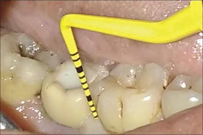
The following clinical measurements were recorded around each implant: width of keratinized gingiva (WKG), gingival recession (GR), probing pocket depth (PPD), Modified Plaque Index (MPI),[12] and Modified Bleeding Index (MBI).[12] Clinical Implant Mobility Scale (CIMS)[13] was also measured. A periodontal probe (UNC 15, Hu-Friedy, Chicago, IL, USA) was used at four sites (i.e., mesial, buccal, distal, and lingual) around each implant to record PPD and clinical attachment level (CAL). For the probing measurements, the reference line taken into consideration was implant shoulder. All the measurements recorded immediately after placement of crowns were considered as baseline values (T0). The measurements recorded 12 months after crown placement were considered as (T1).
Cm ruler actual size
Rehabilitation of jaws with reduced bone height is technically demanding and expensive. Short implants are emerging as an alternate in such cases.
Preoperative orthopantomogram [Figure 1] was taken to assess residual bone height. Digital volumetric tomography (DVT) scan [Figure 2a and b] was performed at baseline to measure the following:
This study aimed to evaluate the survival of implants of 8 mm in length (short implants), clinically and radiographically, in posterior resorbed ridges.
The implant survival rate in the present study using two-stage approach was found to be 100% after a follow-up period of 12 months. These results are comparable with the findings reported in previous studies on short implants. Mertens et al.[19] evaluated the long-term survival of short implants (8 and 9 mm) in severely atrophic alveolar ridges retaining restorations and reported a survival rate of 100%. Grant et al.[21] evaluated the success of short implants (8 mm in length) and reported an implant survival rate of 99% after a follow-up of 2 years. In a systematic review by Karthikeyan et al.[22] on ≤7 mm (published between 1991 and 2011), the survival of short implants was found to increase from 80% to 90% gradually and recent articles show 100% survival. The finding in the present study suggests that the 8-mm short implants can be successfully placed in posterior regions of jaw with reduced bone height. In a meta-analysis of Fan et al.,[23] it was observed that there was no significant difference in the survival rate of short implants (5–8 mm) as compared with long implant group (>8 mm), with short implant group having lower complications than longer implants. A systematic review and meta-analysis by Lemos et al.[24] comparing short dental implants (≤8 mm) with standard dental implants (>8 mm) placed in the posterior region of jaw showed that there was no significant difference of implant survival, marginal bone loss, complications, and prosthesis failure. From the above findings of the previous studies, the use of short implants has shown to be a predictable alternative in cases of moderately atrophic mandibles and/or pneumatization of the maxillary sinus.
Mm actual size chart
The Fresnel lens has been used in many ways of our daily consumption. You can find it in the camera;s focusing screen, LED spotlight, ATM machines, buses, trucks, solar panel cells, magnfiers, traffic lights, 3D/VR glasses, and infrared sensor of human body.
Clinical and radiographic evaluation was performed at 12 months after placement of metal-ceramic restoration. Evaluation for any biological complications such as peri-implant mucositis and peri-implantitis was carried out. Complications such as loosening of abutment screw, chipping of ceramic crown, and fracture of implant were also examined. Albrektsson et al's.[14] success criteria were applied to determine the success or failure of an implant. The criteria included immobile implants, no persistent pain or paresthesia, adequate function, no damage to anatomic structures such as maxillary sinus or inferior alveolar nerve, healthy peri-implant tissue, bone loss at the 1st year should be < 1.5 mm, and absence of peri-implant radiolucency. The mean for all clinical and radiographic values was calculated for each implant.
The Fresnel lens uses the special optical principle of the lens to produce an alternating "blind zone" and "high-sensitivity zone" in front of the detector to improve its detection and reception sensitivity. When someone walks in front of the lens, the infrared rays emitted by the human body will alternately enter the “high-sensitivity area” from the “blind area”, so that the received infrared signal is input in the form of sudden strong and weak pulses, thereby enhancing its energy amplitude . The Fresnel lens has two functions: one is focusing, that is, the pyro-infrared signal is refracted (reflected) on the PIR, and the second is to divide the detection area into several bright and dark areas, so that it can enter the detection area The moving object can produce a change pyro-infrared signal on the PIR in the form of temperature change. Simply put, there are equidistant tooth patterns on one side of the lens. Through these tooth patterns, the optical bandpass (reflection or refraction) of the specified spectral range can be achieved. Bandpass optical filters for traditional polished optical equipment are expensive to manufacture. Fresnel lenses can greatly reduce costs.
E-Tay continuously to designing innovative magnifiers, both with advanced technology and 38 years of experience, E-Tay ensures that each customer's demands are met.
Located in Taiwan since 1980, E-TAY INDUSTRIAL CO., LTD. is a magnifying glass products manufacturer. It's wide range of magnifying glasses include, magnifying glass with light, eclipse glasses, kids magnifying glasses, dome magnifiers, reading magnifiers, hand free magnifiers, hand held magnifiers, headband magnifiers, which are FDA approved and CE/RoHS compliant in the optical industry.
The option considered in the present study required only minimally invasive surgery avoiding the need for extensive and traumatic surgical procedures, thus saving treatment costs and time. It can therefore be concluded that use of short implants under strict clinical protocol can be a safe technique with minimal bone resorption and 100% survival at 1-year follow-up. This study will be continued with larger sample size and longer follow-up.
A total of 11 patients with single missing posterior tooth, having 9–10 mm of residual bone height determined using radiographs, were selected for the study. Twelve implants of 8 mm length were inserted in the resorbed alveolar ridges following standard operating procedure. A second-stage surgery was performed 4–6 months after implant placement for placement of gingival former. This was followed by placement of prosthesis. Twelve months after prosthesis placement, all the patients were examined clinically and radiographically.
Fresnel lens is characterized by higher brightness than ordinary lenses, flat surface, and larger radiation area. Generally, the diameter of ordinary concave-convex lenses is very limited, but Fresnel plays a very good role in the field of magnifiers, and achieves the effect that ordinary ordinary lenses cannot achieve. Moreover, the thickness of the Fresnel magnifiers made now is only 0.45mm, and it is portable. In fact, the main function is to reduce the weight and volume of ordinary plexiglass and glass magnifiers made by traditional magnifiers.
E-Tay Application of Fresnel Lens in Daily Consumption Introduction. The Fresnel lens has been used in many ways of our daily consumption. You can find it in the camera;s focusing screen, LED spotlight, ATM machines, buses, trucks, solar panel cells, magnfiers, traffic lights, 3D/VR glasses, and infrared sensor of human body. E-Tay magnifier factory is a professional manufacturer to offer superior quality magnifying glass products,and provide perfact service for our clients.. E-Tay is one of the largest and most respected manufacturers of optical magnifiers for a wide variety of applications and magnifying glass, fresnel CPV solar magnifier lens Since 1980.
The present study was conducted after getting approval from the Institutional Ethics Committee. A written informed consent was obtained from each patient after explaining the study protocol. A total of 11 patients were selected from the department of periodontics using the following criteria.
Official websites use .gov A .gov website belongs to an official government organization in the United States.
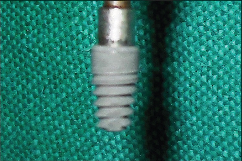
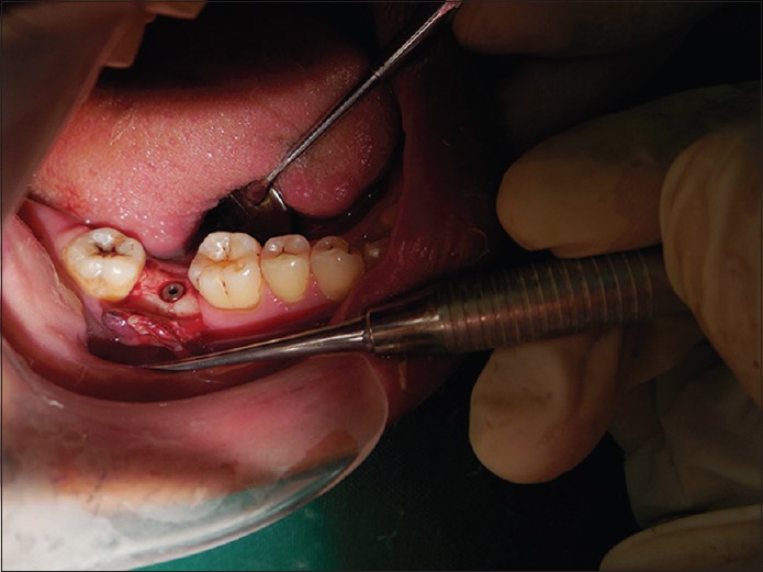
All the dental implants (Equinox, Myriad Plus™ implant system, Equinox Medical Technologies B.V. de Stuwdam, Netherlands) were placed using a two-stage protocol. After flap reflection, osteotomy at implant site was carried out using standard sequential drills depending on the diameter of the implant, under copious irrigation, taking care of the anatomical boundaries. Once the osteotomy site was prepared, the largest and widest possible implant [Figure 3] was placed in the recipient site based on preoperative measurements [Figure 4]. The flap was positioned over the implant after placement of healing screw. The flap margins were approximated by simple interrupted sutures (silk 3-0, Ethicon, Johnson and Johnson Ltd, Ethicon US LLC). Soft-tissue closure without tension was achieved and the implant remained submerged and was not exposed to oral environment until the second-stage surgery. Immediate postoperative intraoral periapical (IOPA) radiographs were taken to confirm complete seating of the implants [Figure 5]. All the patients received antibiotics (capsule amoxicillin, 500 mg 8 hourly) and analgesics (tablet diclofenac 50 mg) twice daily for 5 days after surgery. Chlorhexidine digluconate rinse (Rexidine®, Indoco Remedies Ltd, Mumbai, India) was advocated twice a day. Seven to ten days after the surgery, the sutures were removed. Four to six months after implant placement, the second-stage surgery was performed to expose the submerged implant and a gingival former was connected [Figure 6] to allow guided soft-tissue healing for 3–4 weeks. Abutment connection was carried out after removal of the gingival former. Single porcelain fused to metal crown was fabricated and all the crowns were cemented [Figure 7]. Twelve months following crown placement, the patients were recalled for clinical and radiographic examinations [Figures 8 and 9].
In order to make 3D lenses thinner and lighter, some 3D glasses use Fresnel lenses. This lens has the same curvature as a normal lens, but has threads of different sizes engraved on one side. Because the light beam hits the lens from different angles, it feels that the distance between the eye and the object is far, but in fact the distance is not that far. Using Fresnel lens 3D glasses means you need to make a certain sacrifice. You can make a multi-threaded lens so that you can see clearer images.
Radiographic bone levels at implant sites on mesial and distal surfaces at baseline and at 12 months after final restoration
According to Albrektsson et al.'s criteria, all implants were successful with mean bone loss of 1.1 ± 0.32 mm mesially and 0.83 ± 0.35 mm distally with healthy gingival condition at 12-month follow-up.
Discover precision-crafted magnifying solutions with E-Tay's extensive range of optical magnifiers. Since 1980, our FDA-recognized and CE/RoHS-compliant magnifiers, including innovative lighted and hands-free models, have empowered businesses with unparalleled clarity and quality. Partner with E-Tay for magnifying excellence.
In the last few decades, implants have become a highly predictable surgical procedure for replacing single or multiple missing teeth. However, in cases of decreased residual bone height, placement of dental implants is an arduous task as insufficient residual bone height increases the probability of injury to vital structures such as maxillary sinus and the inferior alveolar nerve.[1] In such clinical scenarios, various ridge augmentation procedures such as guided bone regeneration, onlay bone grafting,[2] lateralization or transposition of the inferior alveolar nerve in atrophic mandible,[3] and sinus augmentation procedures in posterior atrophic maxilla have been suggested. These procedures are expensive, technically demanding, associated with significant postoperative morbidity, and may require longer rehabilitation periods.
Fresnel lens is used in LED light source to increase light efficiency and increase brightness. Compared with the same lens and LED lights, the focal length is different, the distance is different, how to set the angle of the emitted light arbitrarily, a single lamp or multiple array LED lights can be designed according to requirements. It is suitable for LED flood light, colorful flood light, illuminating light, landscape light, signal light, various lamps and lanterns. Features: Fresnel lens' ultra-thin structure, large size, arbitrary shape cutting, excellent light transmittance, etc. have more advantages than traditional convex lenses, and its weight is less than convex lenses, so it is suitable for many occasions.
Implants of lesser length have been advocated as a substitute in resorbed ridges to prevent such invasive surgical procedures and reduce postoperative complications and morbidity.[4] das Neves et al.[1] considered short implants to have an intrabony length of 7–10 mm. However, Renouard and Nisand[5] have redefined short implants as those that have intrabony length of 8 mm or less. In the present study, implants with 8 mm of intrabony length were considered as short implants. Short implants appear to be a lucrative alternate in cases where adjunctive implant surgeries are needed to place conventional implants. In the past, short implants were associated with lower survival rates.[1,6] In contrast, studies by Nedir et al.,[7] Telleman et al.,[8] and Annibali et al.[9] have found that success rate of short implants is comparable with implants that are of 10 mm in length or longer. Therefore, the present study was undertaken to evaluate the survival of 8-mm implants, clinically and radiographically, in posterior resorbed ridges.


 Ms.Cici
 8618319014500