
HANDI-VAC Vacuum Sucking Pen, ESD Safe ... - handi vac
Take a look around the space you are in. Can you find an example of the Fresnel Effect? Look at shiny floors and plastic surfaces. Crouch down as I did and see how the intensity of the reflection changes.
Excellent explanation, making simple the (apparently) complex. Indeed, your description demonstrates that “complexity is n the eye of the beholder.’
A demonstration on how internal lens elements in a high numerical aperture dry objective may be correctly adjusted with these varied cover glass thickness and dispersion fluctuations is featured in this tutorial.
What isaberration
Explore the relationship between astigmatism and comatic aberrations, and how astigmatism aberrations are manifested by the off-axis image of a specimen point appearing as a line or ellipse instead of a point.
Thank you Kevin! I wanted to use as few technical terms as possible in order to keep the article short and easy to understand. The surface normal is a very useful concept and I like your way of using it to describe the principle behind the Fresnel Effect.
Here is another example: reflections change across distance – because the angle of incidence changes. As you look down to the ground close to your feet, the angle of incidence is very steep. If you look at a point on the ground that’s further away from you, the angle gets more shallow – and the reflection becomes more visible.
Difference between chromatic and monochromaticaberration
And here is what we see from the viewpoint of the person. Use the arrows to the left and right of the image below to switch between versions with Fresnel Effect and without Fresnel Effect.
I was blind to the Fresnel Effect until someone pointed it out to me — now I can see that it is everywhere! If you’re looking for it, you’ll find it.
Opticalaberrations of the eye
The angle they actually care about when calculating the amount of reflected light (and possibly refracted light, for transparent objects) is between the vector (or ray , if you prefer) created from the viewer to the spot you’re looking at on the object, and the surface normal of the object.
Explore the two most prevalent types of distortion, positive and negative, and how they can often be present in very sharp images that are otherwise corrected for spherical, chromatic, comatic, and astigmatic aberrations.
Curvature of field in optical microscopy is an aberration that is familiar to most experienced microscopists. This artifact is the natural result of using lenses that have curved surfaces.
Opticalaberrations pdf
I had a hard time understanding this concept till I found this explanation, very simple and easy to grasp, thanks for keeping things simple.
Learn about comatic aberrations and how they are mainly encountered with off-axis light fluxes and are most severe when the microscope is out of alignment, as well as the result of when these aberrations occur.

Types ofaberrationin optics
Objectives are made with differing degrees of optical correction for both monochromatic (spherical, astigmatism, coma, distortion) and polychromatic aberrations, field size and flatness, transmission wavelengths, freedom from fluorescence, birefringence and other factors contributing to background noise. Depending upon the degree of correction, objectives are generally classified as achromats, fluorites, and apochromats, with a plan designation added to lenses with low curvature of field and distortion. This section addresses some of the more common optical aberrations that are commonly found (and often corrected) in microscope objectives.
Hey John! The two are related. Highlights are “specular reflections” and the fresnel effect describes how the intensity of specular reflection changes depending on your angle of view. This video might clarify it: https://www.dorian-iten.com/see-more/
Monochromaticaberration
Aberrations are divided into two main categories: errors that occur when polychromatic light (white light) is passed through a lens, and errors that are present when only a single wavelength (monochromatic) of light is utilized. The selected references listed in this section contain information about the cause and correction of the most common optical aberrations encountered with microscope and other lens systems. Bear in mind that the optical designer must correct for both polychromatic and monochromatic aberrations simultaneously in the production of well-corrected microscope objectives.
For microscope objectives having apertures, the optical properties and thickness of the medium lying between the front lens element and the specimen affect the calculations necessary to correct for image aberrations.
Learn more about chromatic aberrations and how when white light passes through a simple or complex lens system, the component wavelengths are refracted according to their frequency.
Curvature of field in optical microscopy and how it is a common and annoying aberration that is familiar to most experienced microscopists is explained in this featured interactive tutorial.
An ideal microscope objective produces a symmetrical diffraction limited image of an Airy pattern from an infinitely small object point. The image plane is generally located at a fixed distance from the objective front lens in a medium of defined refractive index. Microscope objectives offered by the leading manufacturers have remarkably low degrees of aberrations and other imperfections, provided the appropriate objective is selected for the task and the objective is utilized properly in accordance with the manufacturer's recommendations. It should be emphasized that objective lenses are not made to be perfect from every standpoint, but are designed to meet certain specifications depending on their intended use, constraints on physical dimensions, and price ranges.
Optical aberrationexamples
Departures in lens action from the ideal conditions of optics are known as aberrations. Optical trains typically suffer from as many as five common aberrations: spherical, chromatic, curvature of field, comatic, and astigmatic.
Astigmatismaberration
Learn about the most serious of the monochromatic defects that occurs with microscope objectives, the spherical aberration, which causes the specimen image to appear hazy or blurred and slightly out of focus.
Brian O. Flynn, John C. Long, Matthew Parry-Hill, and Michael W. Davidson - National High Magnetic Field Laboratory, 1800 East Paul Dirac Dr., The Florida State University, Tallahassee, Florida, 32310.

To understand the Fresnel Effect, you have to understand the basics of reflections.We’ll keep this minimal – the key is the Angle of Incidence.The Angle of Incidence is the angle between your line of sight and the surface of the object you are looking at.
PS. If you find this interesting, you might enjoy learning about the 11 Modeling Factors – light effects that create the sensation of form.
Hi Vaughn! Yes, usually the surface normal is used to explain the Fresnel effect. My goal with this article was to make the concept of the Fresnel effect as simple to grasp as possible. Adding the concept of surface normals would have increased complexity and cognitive load unnecessarily. I included a link to the Wikipedia entry for those readers that want to get the full technical explanation. 🙂
In other words, if you look directly down at the table, the angle is close to zero, and as you get closer to eye level with the table, the angle increases. This makes it easier to explain that “as the angle increases, the amount of reflection increases.”
Discover an aberration-free meridional section of a point source of light located at a depth in the specimen layer having a refractive index and imaged with a virtual microscope objective.
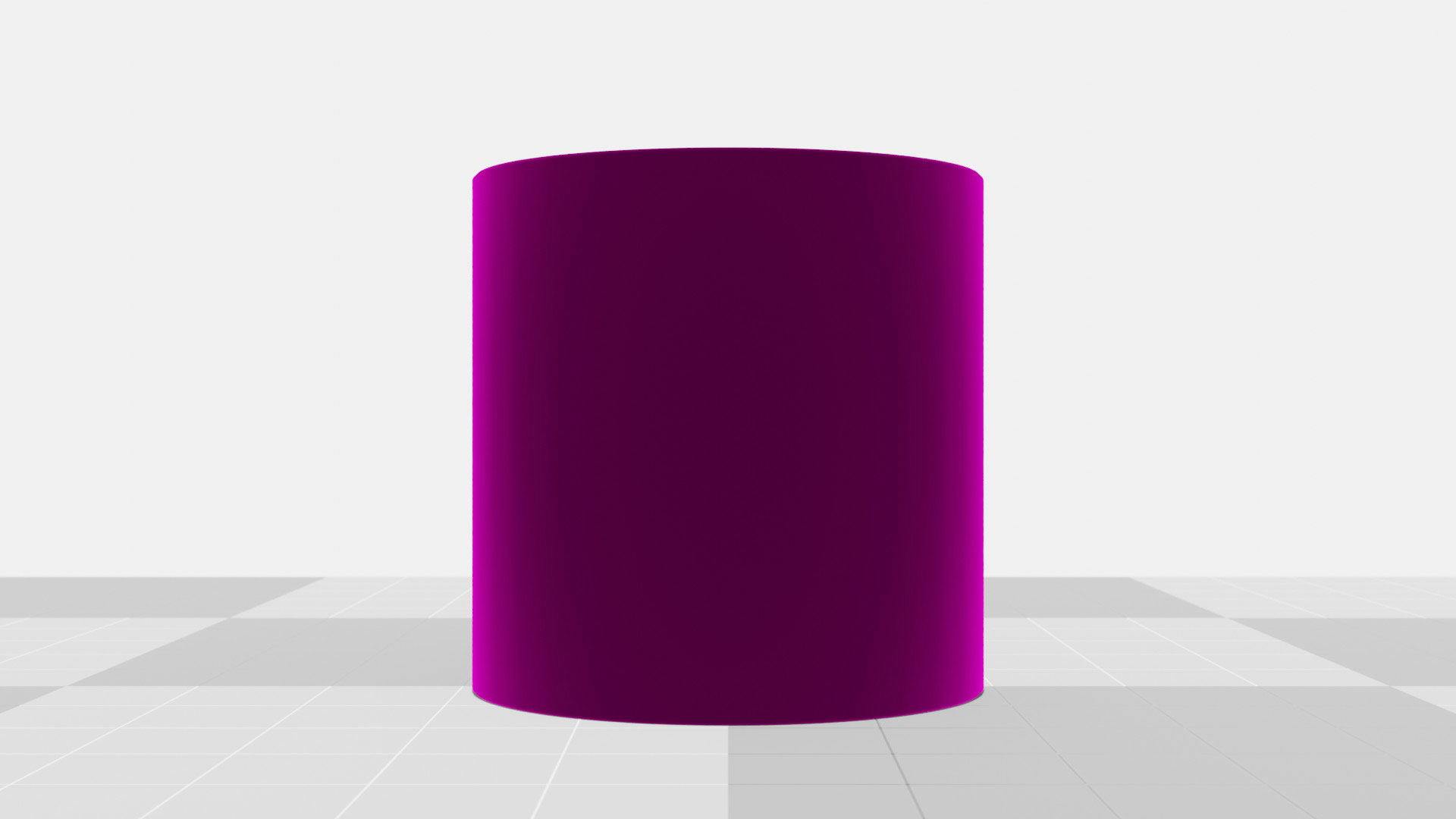
On curved surfaces, the angle of incidence gets steeper towards the edges of the form. On a cylinder, the fresnel effect leads to the specular reflections being most visible in the red areas:
I thought the angle of incidence is taken from the normal to the surface, why is yours taken from parallel to the surface?


 Ms.Cici
 8618319014500