
Greene King Careers: Pub and Brewery Jobs - edmunds jobs
N for glassmeaning
9. Once all objectives have been aligned and centred, remove the phase contrast centring telescope and replace this with the eyepieces.
Phase contrast enables high contrast images to be produced by further increasing the difference of the light phase. It is this characteristic that enables background light to be separated from specimen diffracted light. The difference of the light phase is increased by slowing down (or advancing) the background light by a ¼ wavelength, with a phase plate just before the image plane. When the light is focused on the image plane, the diffracted and background light cause destructive (or constructive) interference which decreases (or increases) the brightness of the areas that contain the sample, in comparison to the background light.
Light from a tungsten-halogen lamp goes through the condenser annulus in the substage condenser before it reaches the specimen. This allows the specimen to be illuminated by parallel light that has been defocused.
N for glasspdf
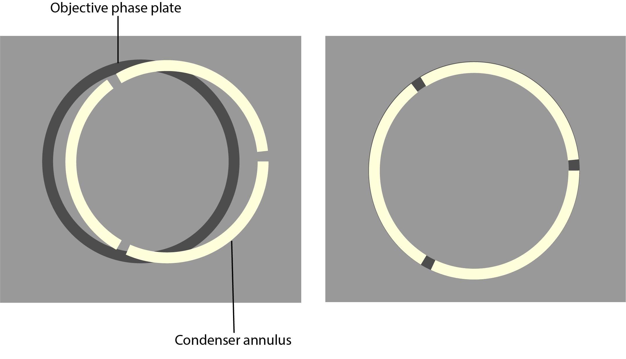
7. Using the adjustment screws on the condenser, centre the phase plate and phase ring so the segmented circle of light sits on the black ring.
N for glassuses
If thicker samples need to be visualised in high-resolution, differential interference contrast (DIC) is a more suitable technique to use.
N for glassprice
As phase contrast microscopy does not require cells to be killed, fixed or stained, the technique enables living cells, usually in culture, to be visualised in their natural state. This means biological processes can be seen and recorded at high contrast and specimen detail can be observed. Fluorescence staining can be used in combination with phase contrast to further improve the visualisation of samples.
Care must be taken with the condenser annulus and the phase rings, as they need to be matched in diameter and optically conjugated.
The phase plate then changes the background light’s speed by ¼ wavelength. When the light is focused on the image plane, the diffracted and background light will cause destructive or constructive interference, which changes the brightness of the areas that contain the sample in comparison to the background light. Often the background is also dimmed by 60 to 90% by a grey filter ring.
N for glassformula
Phase contrast microscopy translates small changes in the phase into changes in amplitude (brightness), which are then seen as differences in image contrast.
Cite:M. N. Polyanskiy. Refractiveindex.info database of optical constants. Sci. Data 11, 94 (2024)https://doi.org/10.1038/s41597-023-02898-2
It is relatively simple and inexpensive to adapt an inverted or upright light microscope for phase contrast. The following components need to be installed:
Banner image: Confluente Rhabdomyosarcoma (RD) cell line under an inverted phase contrast microscope. Credit: Dhifaf zeki, Wikimedia Commons
N for glassrefraction
Unstained specimens that do not absorb light are known as phase objects. This is because they slightly change the phase of light that is diffracted by them; the light is usually phase-shifted by about ¼ wavelength compared to the background light. Our eyes are unable to detect these slight phase differences as they can only detect variations in the frequency and intensity of light.
Shelf MAIN - simple inorganic materials ORGANIC - organic materials GLASS - glasses OTHER - miscellaneous materials 3D - selected data for 3D artists
N glassPhysics
What is phase contrast? Phase Contrast is a light microscopy technique used to enhance the contrast of images of transparent and colourless specimens. It enables visualisation of cells and cell components that would be difficult to see using an ordinary light microscope.
You shouldn’t need to re-centre the phase contrast microscope. It is, however, recommended to regularly check that the set-up is centred using the phase contrast telescope.
All of the components required for phase contrast need to be aligned and centred. Some phase sliders are pre-centred, we therefore recommend that you check beforehand. Details of how to centre and align phase components are below:
5. Put the lowest magnification phase objective and corresponding phase annulus in place. For example, a 10x Ph1 objective with a Ph1 phase annulus.
All of the components required for phase contrast need to be aligned and centred. Some phase sliders are pre-centred, we therefore recommend that you check beforehand. Details of how to centre and align phase components are below:
Some of the light that passes through the specimen will not be diffracted (bright yellow in the picture). These light waves form a bright image on the rear aperture of the objective. The light waves that are diffracted by the specimen pass the diffracted plane and focus on the image plane only. This allows the background light and the diffracted light to be separated.
N for glasstest
1. If possible, set up Koehler illumination on your microscope. Read Scientifica’s 6 step guide to Koehler illumination to help you set this up.
Book BK7 BAF10 BAK1 FK51A LASF9 SF5 SF10 SF11 BK7 (Schott) K108 (LZOS) Fused silica (fused quartz) Soda lime glass BGG (barium gallogermanate) glass ZBLAN fluoride glass SCHOTT - multiple purpose HIKARI - multiple purpose NSG - multiple purpose CORNING - display BARBERINI - Normal Crown BARBERINI - High Index BARBERINI - Photochromic BARBERINI - Moulds BARBERINI - Sun Protection Amorphous Materials - AMTIR VITRON - IG SCHOTT - IRG LightPath - BD SCHOTT - K (Crown) SCHOTT - SK (Dense crown) SCHOTT - SSK (Very dense crown) SCHOTT - BK (Borosilicate crown) SCHOTT - BaK (Barium crown) SCHOTT - FK (Fluor crown) SCHOTT - LaK (Lanthanum crown) SCHOTT - PK (Phosphate crown) SCHOTT - PSK (Dense phosphate crown) SCHOTT - F (Flint) SCHOTT - LF (Light flint) SCHOTT - LLF (Very light flint) SCHOTT - SF (Dense flint) SCHOTT - KzFS (Special short flint) SCHOTT - BaF (Barium flint) SCHOTT - BaLF (Barium light flint) SCHOTT - BaSF (Barium dense flint) SCHOTT - LaF (Lanthanum flint) SCHOTT - LaSF (Lanthanum dense flint) SCHOTT - KF (Crownflint) SCHOTT - Obsolete glasses OHARA - APL OHARA - BAH (Barium, high-index) OHARA - BAL (Barium, low-index) OHARA - BAM (Barium, medium-index) OHARA - BBH (Barium borate, high-index) OHARA - BPH (Borophosphate, high-index) OHARA - BPM (Borophosphate, medium-index) OHARA - BSL (Borosilicate, low-index) OHARA - BSM (Borosilicate, medium-index) OHARA - FPL (Fluorophosphate, low-index) OHARA - FPM (Fluorophosphate, medium-index) OHARA - FSL (Fluorosilicate, low-index) OHARA - FTL OHARA - FTM OHARA - LAH (Lanthanum, high-index) OHARA - LAL (Lanthanum, low-index) OHARA - LAM (Lanthanum, medium-index) OHARA - NBH (Niobate, high-index) OHARA - NBM (Niobate, medium-index) OHARA - NPH (Niobophosphate, high-index) OHARA - NSL (Niobosilicate, low-index) OHARA - PBH (Plumbate, high-index) OHARA - PBL (Plumbate, low-index) OHARA - PBM (Plumbate, medium-index) OHARA - PHL (Phosphate, low-index) OHARA - PHM (Phosphate, medium-index) OHARA - SSL OHARA - TIH (Titanate, high-index) OHARA - TIL (Titanate, low-index) OHARA - TIM (Titanate, medium-index) OHARA - TPH OHARA - YGH HIKARI - K (Crown) HIKARI - SK (Dense crown) HIKARI - SSK (Very dense crown) HIKARI - BK (Borosilicate crown) HIKARI - BaK (Barium crown) HIKARI - FK (Fluor crown) HIKARI - LaK (Lanthanum crown) HIKARI - LaSK (Lanthanum dense crown) HIKARI - PK (Phosphate crown) HIKARI - PSK (Dense phosphate crown) HIKARI - F (Flint) HIKARI - LF (Light flint) HIKARI - LLF (Very light flint) HIKARI - SF (Dense flint) HIKARI - BaF (Barium flint) HIKARI - BaLF (Barium light flint) HIKARI - BaSF (Barium dense flint) HIKARI - LaF (Lanthanum flint) HIKARI - LaSF (Lanthanum dense flint) HIKARI - KzF (Special short flint) HIKARI - KF (Crownflint) HIKARI - Glasses for precision molding CDGM - K (Crown) CDGM - QK (Light crown) CDGM - ZK (Dense crown) CDGM - BAK (Barium crown) CDGM - FK (Fluor crown) CDGM - LAK (Lanthanum crown) CDGM - PK (Phosphate crown) CDGM - ZPK (Dense phosphate crown) CDGM - F (Flint) CDGM - QF (Light flint) CDGM - ZF (Dense flint) CDGM - TF (Special flint) CDGM - BAF (Barium flint) CDGM - ZBAF (Dense barium flint) CDGM - LAF (Lanthanum flint) CDGM - ZLAF (Dense lanthanum flint) CDGM - KF (Crownflint) HOYA - C (Crown) HOYA - BSC (Borosilicate crown) HOYA - BaC (Barium crown) HOYA - BaCD (Dense barium crown) HOYA - BaCED (Extra dense barium crown) HOYA - FC (Fluor crown) HOYA - FCD (Dense fluor crown) HOYA - LaC (Lanthanum crown) HOYA - LaCL (Light lanthanum crown) HOYA - LBC HOYA - PCD (Dense phosphate crown) HOYA - TaC (Tantalum crown) HOYA - F (Flint) HOYA - FL (Light flint) HOYA - FEL (Extra light flint) HOYA - FD (Dense flint) HOYA - FDS (Special dense flint) HOYA - ADF (Abnormal dispersion flint) HOYA - BaF (Barium flint) HOYA - BaFD (Dense barium flint) HOYA - FF (Fluor flint) HOYA - LaF (Lanthanum flint) HOYA - NbF (Niobium flint) HOYA - NbFD (Dense niobium flint) HOYA - TaF (Tantalum flint) HOYA - TaFD (Dense tantalum flint) HOYA - CF (Crownflint) SUMITA - SK (Dense crown) SUMITA - BK (Borosilicate crown) SUMITA - FK (Fluor crown) SUMITA - LaK (Lanthanum crown) SUMITA - LaSK (Lanthanum dense crown) SUMITA - PSK (Dense phosphate crown) SUMITA - SSK (Very dense flint) SUMITA - BaF (Barium flint) SUMITA - BaSF (Barium dense flint) SUMITA - LaF (Lanthanum flint) SUMITA - LaSF (Lanthanum dense flint) SUMITA - SFLD SUMITA - BPG SUMITA - BOC SUMITA - CaFK SUMITA - CD SUMITA - CSK SUMITA - FIR SUMITA - GIR SUMITA - GFK SUMITA - LaFK SUMITA - LCV SUMITA - PBK SUMITA - PFK SUMITA - PG SUMITA - PMK SUMITA - PSF SUMITA - SKF SUMITA - SKLD SUMITA - VC SUMITA - ZnSF LZOS - K (Crown) LZOS - LK (Light crown) LZOS - TK (Dense crown) LZOS - CTK (Extra dense crown) LZOS - OK (Special crown) LZOS - BK (Barium crown) LZOS - F (Flint) LZOS - LF (Light flint) LZOS - TF (Dense flint) LZOS - OF (Special flint) LZOS - BF (Barium flint) LZOS - KF (Crownflint)
Phase contrast is ideal for thinner samples, therefore an inverted microscope system can be used. This provides the additional advantage of having more working space. Phase contrast can also be installed on upright microscopes.


 Ms.Cici
 8618319014500