
DC Micro (M12) Connection Systems | Allen-Bradley | US - m12 cable color code
Brightfieldmicroscope
Now the light source is huge. Shadows no longer exist as the light is big enough to shine all around the object or model.
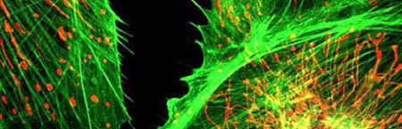
The physical principle of fluorescence is used to selectively visualize and localize defined fluorescent structures, while non-fluorescent structures remain dark in order to obtain a high image contrast. For this purpose, fluorescent dyes (fluorochromes) are used with specific excitation and emission filters installed in the optical path of the microscope. A wide range of fluorescent dyes with different colors are available, which are used in molecular biological, biomedical and clinical research. For example, in immunohistochemistry, fluorescence-in-situ-hybridization and for visualization of cells or cellular components in living/fixed specimens.
JavaScript seems to be disabled in your browser. For the best experience on our site, be sure to turn on Javascript in your browser.
Bright field lighting
Get more information for All Electronics Repair in Charlotte, NC. See reviews, map, get the address, and find directions.
Vision inspection systems (sometimes referred to as machine vision systems) provide image-based inspection automated for your convenience for a variety of ...
Polarization microscopy is used for the analysis of optically anisotropic samples. The primary objective of polarization microscopy is not magnification of an object, but rather the analysis of optical properties such as refractive index or birefringence for sample analysis. The method is mainly used in mineralogy and in industry for testing plastics or mineral building materials in order to gain insights into their composition.
Bright fieldvsdark fieldvsphase contrast
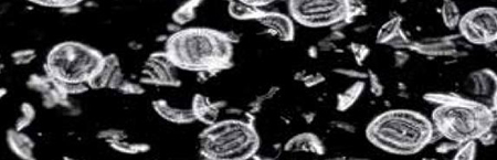
A dark field microscope produces a contrast-enhanced image by indirect illumination of the specimen, thereby also unstained specimens can be displayed with high contrast. Using this technique, direct light is bypassing the objective, only light scattered by the specimen enters the objective. As a result, the background appears dark or black, only the specimen is illuminated and even small structures can be visualized with high contrast. Dark field microscopy in biology and medicine is especially used for transparent and low contrast specimens, for example, studying blood, small animals or micro-particles in material science.
Integro Technologies is a principal source for machine vision industrial applications for a wide array of industries. As an AIA Certified Systems Integrator, ...
Dark field lighting photography
Phase contrast microscopy is a contrast-enhancing technique to visualize structures difficult to detect with bright-field microscopy due to a lack of contrast, without the need of a staining. When penetrating a medium, light propagates with different speeds depending on the refractive index of the medium. This leads to phase differences, which are converted to differences in brightness by the microscope using phase rings. Areas of application of phase contrast microscopes are mainly the observation of living biological samples in order to resolve fine structures with high contrast.
Our Vehicle Bulb Finder is a quick and easy way to locate LED bulbs for your . Whether you're searching for replacements for burned-out or dim incandescent ...
An inverted microscope is an upside-down standard light microscope. This type of microscope is characterized by locating the objective underneath the stage and pointing upwards to the specimen. Inverted microscopes are mainly used for live cell analysis of cell cultures growing in culture medium. Culture dishes are not only available with standard coverslip bottoms (thickness 0.17mm) but also in various material and bottom thicknesses. Therefore, for some models special objectives are available that can correct for different bottom glass thicknesses. In addition, inverse microscopes are also used for studies of thicker specimens. Inverted microscopes are also available as dark field, polarization or fluorescence microscopes.
Choose the focal lengths you want to compare · According to your choice of focal lengths, you can now choose the available aperture · Check whether you own or ...
What are the three basic categories of light? Looking at the hardness (or softness) of light is certainly the most simple and easy way to classify it. However, we can never call a certain light shaper hard or soft (with the exception of a point light source that is always hard). Depending on the size and the distance between the object and the light, the same light shaper can once be hard, soft or even diffused. Let’s have a closer look at these three categories:
Darkfield vs brightfieldmicroscopy
Die Verwendung einer LED-Beleuchtung hat zahlreiche Vorteile, darunter: Niedriger Energieverbrauch; LED-Leuchten haben eine Lebensdauer von 10.000 bis 30.000 ...
Looking at the light of a point light source, we will see very clearly defined shadows. On a background or underground there is either light or shadow, but nothing in between, no gradations. Even the finest details provoke a clear shadow. The structure of any object (e.g., textile, skin) is pointed out very clearly. A very hard light source is the only one that does not change its characteristics if we vary the distance to the object (but according to the inverse square law it does change the power). The shadows remain the same: very sharp. Hard lights may increase the contrast of the object. The areas directly lit may be burnt while the shadows remain very dark.The hardness of the light finally has an influence on the color saturation. Small and hard lights increase the saturation of the picture while soft, and especially diffused lights reduce it.
We use cookies to give you the best experience on our website. If you continue, we assume that you are happy to receive all cookies on this website. For more information on cookies, please refer to our cookies page.
2022726 — Coherent light is electromagnetic radiation that has a certain wavelength. So it is always a single -colour light, since the wavelength ...
A metallurgical microscope is a special version of a standard light microscope for the study of materials, such as Metals, plastics, ceramics and others. Since materials are usually not transparent solid structures, metallurgical microscopes often have an upright light unit. Moreover, this type of microscope is characterized by extensive magnification, e.g. for detailed investigations of surface structures. Application areas of metallurgical microscopy are industry, material science and research.
Darkfield vs brightfieldreddit

A good example to illustrate the difference between hard and soft shadows: Through a very narrow opening of curtains, daylight is falling into this hotel room. Horizontally, the opening is only a few centimeters wide – the corresponding shadows are very hard. The vertical shadows, however, are very soft because the curtains let some light in from the ceiling to the floor. In the studio, this effect can be simulated with a Striplite 60, a litestick and with narrow softboxes like the 30x120 or the 30x180 cm softbox, especially when these are equipped with the additional strip mask.
20161230 — The EMVA 1288 standard characterizes image sensor quality (but not full camera systems, which include lenses). Imatest implemented a subset of ...
Bright field and dark field microscopy PDF
These cookies are used to provide you with a more personalized experience on our website and to remember the choices you make when you use our website. For example, we may use functionality cookies to remember your language preferences, direct you to your local distributor, or remember your login details.
Difference between dark field and phase contrast microscopy
Average soft light sources have about the same sizes as the objects or set-ups they illuminate: Let’s say a 50 by 50 cm softbox for a narrow-cropped portrait or an 80 by 140cm softbox for a full-body shot. The shadows on the under- and backgrounds are still clearly visible, even when they are not sharply defined anymore. Big parts of these shadows show gradations, and a small core shadows still exist. Small and fine details, however, do not appear in the shadow. The texture of our object is now shown in lower contrast and is therefore not as clear as in hard light. Soft light still increases the contrast of the object a little, but less than a hard one. The color saturation finally is somewhere in between the one derived from a hard light (high) and a diffused light (low). Being soft, our light source got a certain size (it is not a point anymore) and the distance from it becomes very important: The closer we get, the bigger the light source becomes (seen from the perspective of the object or model). This means that our light becomes softer when we get closer, and harder, when we use it over larger distances. A light of about 100 by 100cm placed at 4 meters from the model has the same hardness as a source of half the size (50 by 50 cm) at half the distance (2 meters). Due to the inverse square law, we expect other effects (higher contrast when placing the light closer to the object or model). When we bring the 100 by 100 cm softbox to half the distance (we will have to reduce the power by about 2 f-stops) the light will be a lot softer.The following light shapers can be used as soft lights:
MicroBrite White and Color LED Lights | TC Pool Equipment Co. Best Brands For Less!
Standard bright-field microscopes are used for daily laboratory routine in research and diagnostics for simple, standard applications that require no special equipment. Therefore simple optical systems and lenses are applied.
Sony E-Mount 16-50mm f3.5-5.6 II Power Zoom Lens ... The Sony E-Mount 16-50mm f3.5-5.6 II Power Zoom Lens is a versatile lens designed for Sony APS-C cameras. It ...
These cookies are used to collect information to analyze the traffic to our website and how visitors are using our website. For example, these cookies may track things such as how long you spend on the website or the pages you visit which helps us to understand how we can improve our website site for you. The information collected through these tracking and performance cookies do not identify any individual visitor.
The light does not show any direction anymore and the only contrast remaining in the photograph is the contrast of the object itself. The structure of the object’s surface is as flat as possible, almost invisible and the color saturation is heavily reduced.The following light shapers can be used as diffused lights:


 Ms.Cici
 8618319014500