
Fused Silica Capillary Tubing 10m x 0.375mm x 0.005mm - 0.375 mm
Axial Resolution: point-to-point resolving power in the plane parallel to the optical axis. It is usually defined at the shortest distance between two longitudinal points on the specimen plane that can still be distinguished as separate entities.
Electric polarization
Lateral Resolution: point-to-point resolving power in the plane perpendicular to the optical axis. It is usually defined as the shortest distance between two lateral points on the specimen plane that can still be distinguished as separate entities.
Polarizedlight and Optical systems PDF
Stereo microscopes have low magnifications that can range from 2 to 100x depending on the microscope, and are designed for viewing whole objects like rocks, plants, flowers, and invertebrate organisms by reflecting light off the specimen, producing a 3-dimensional image. Sometimes there is a light located in the base of the microscope that will allow transmitted light.
A circularly polarized laser beam can be constructed by coherently combining two linearly-polarized beams satisfying three conditions: The two beams should have perpendicular polarizations, a 90-degree phase difference, and the same amplitude. These conditions can be met by defining two orthogonal aperture-grating structures on the device facet. By tailoring the separations between the aperture and the nearest groove in the two aperture-grating structures, we can control the respective phase and amplitude of the surface waves propagating along the left and the right grating, and therefore control the scattered light from the left and from the right grating to achieve circularly-polarized light in the far-field.
Conventionally, manipulation of the polarization state of a light output is conducted externally using bulky and expensive optical components such as beam-splitting polarizers and wave plates. Our integrated plasmonic polarizer provides a compact solution allowing the development of polarization-controllable active and passive devices over a wide frequency range, from the visible to the far-infrared. The design involves the integration of an aperture-grating plasmonic structure on the emission surface of a light source.
Note: The microscope is now set to maximize resolution of the specimen. If you adjust the condenser height to gain contrast or adjust light intensity you will sacrifice the resolution capability. Use the aperture diaphragm and /or the illumination intensity to adjust contrast.
Depth of Field: is determined by the distance from the nearest specimen plane in focus to that of the farthest plane also simultaneously in focus. The thickness of the optical section along the optical axis within which objects in the specimen plane are in focus. High-magnification objectives have a decreased depth of field. The reverse is true of low-magnification objectives Field of View: the visible area seen through the microscope when the specimen is in focus. The greater the magnification the smaller the view. Focus: a specimen is in focus at the desired magnification when the image seen through the ocular lens is sharp and clear.
We used mid-infrared quantum cascade lasers (QCLs) as a model system to demonstrate two types of polarization control: to filter the linearly-polarized output of a QCL to produce linearly-polarized emission along other directions and to transform a linearly-polarized QCL into a circularly-polarized device.
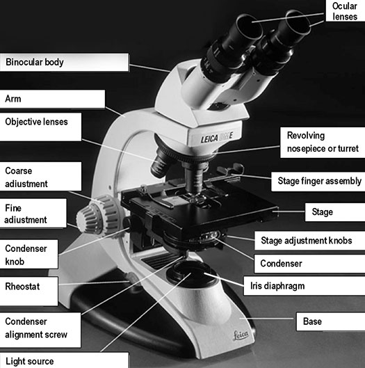
Illuminator or light source: the light source can be built into the base of the microscope, transmitting light through the specimen and/or the light source may be above the specimen as incident light. The lights can be turned on using rheostat (light) control knob on the front of the base.
Ocular lens or eyepiece: the secondary optical system that you look through. The ocular lens further magnifies (10x) the image and brings the light rays to a focal point. A binocular microscope has two ocular lenses and a monocular microscope has one ocular lens that sit on the adjustable binocular body. Binocular lenses can be adjusted to fit the distance between your eyes by gently pulling the oculars apart or by pushing them closer together.
Focusing knob: the knob that allows you to focus on the object at each magnification by moving the stereo head up or down.
Light sources with a desirable polarization state are of high interests for many applications. For example, satellite communications use two orthogonal polarizations to double the capacity of the services; circularly-polarized light sources are of great importance in chemistry and biology for detecting molecules exhibiting circular dichroism; laser sources with a variety of polarization states are used for quantum cryptography. However, semiconductor lasers are mostly linearly-polarized along a fixed direction (determined by the optical selection rules of the gain medium). Other state of polarizations proved difficult to achieve.
Polarization is defined as the orientation of the oscillation of the electric field in the plane perpendicular to the electromagnetic wave's direction of travel. If the tip of the electric field vector traces out a single straight line, it is called linear polarization; if the electric field vector traces out a circle in the plane, it is called circular polarization. Other situations are called elliptical polarization.
Linear polarization
Objective lenses: the primary optical system which produces a magnified image of the specimen. There are typically four objective lenses attached to the nosepiece with the magnification of each objective is engraved on its side.
Microscopes must be calibrated so accurate measurements can be made. To calibrate a microscope both an ocular and a stage micrometer are used.
A compound microscope is a high power microscope that uses a compound lens system. Higher magnification is achieved by using two lenses rather than just a single magnifying lens. While the eyepieces and the objective lenses create high magnification, a condenser beneath the stage focuses the light directly into the sample. A compound microscope has multiple lenses: the objective lens (typically 4x, 10x, 40x or 100x) is compounded (multiplied) by the eyepiece lens (typically 10x) to obtain a high magnification of 40x, 100x, 400x and 1000x. The objective lenses of a compound microscope causes the orientation of the image of the specimen to be inverted compared to the orientation of the actual specimen which means that a specimen viewed through a compound microscope will look upside down and backwards compared to how the specimen is mounted on the slide.
P-polarized light
The 100X objective lens is called an oil immersion lens because oil is placed between the lens and the microscope slide to increase resolution (i.e., the level of detail that can be observed in an image). Light bends when it passes from the glass slide to air because of differing refractive indices. A drop of immersion oil between the slide and lens eliminates this problem because the oil has the same refractive index as the glass slide. Never use the 100X objective lens without oil and do not get oil on the 4X, 10X, or 40X lenses.
To achieve the results, the researchers sculpted a metallic structure, dubbed a plasmonic polarizer directly on the facet of a quantum cascade (QC) laser. The QC laser emitted at a wavelength of ten microns (in the invisible part of the spectrum known as the mid-infrared where the atmosphere is transparent). The team was able to control the state of polarization by generating both linearly polarized light along an arbitrary direction and circularly polarized light.
Coarse adjustment or coarse focusing knob: the large knob towards the back of the instrument that is used to significantly raise or lower the stage, when you first focus on a specimen at low power. It is never used when high power objectives are in place.
Condenser: the lens located below the stage, which focuses light (from the illuminator) through the specimen being observed. Most microscopes have a movable condenser allowing its distance from the specimen to be adjusted using the condenser knob and condenser alignment screws.
Stage: the flat surface upon which the slide with your specimen is placed. Most microscopes have a stage finger assembly to hold the slide on the stage. The entire mechanism including the slide moves horizontally across the stationary stage (left/right and forward/back) using two stage adjustment knobs situated under the stage (variably on the left or right side, in front of the focusing knobs).
The research was partially supported by Air Force Office of Scientific Research. The authors also acknowledge the support of two Harvard-based centers, the NSF-funded Nanoscale Science and Engineering Center (NSEC) and the Center for Nanoscale Systems (CNS), a member of the National Nanotechnology Infrastructure Network (NNIN).
The resolving power of a microscope is dependent on the numerical apertures of the optical lenses and the wavelength of light used to examine the specimen. It is the smallest distance between two points (measured in microns) that can be seen with the microscope. If two small objects close together can be seen clearly as two distinct objects, a microscope is said to have high resolving power.
S-polarization
CAMBRIDGE, Mass. – April 13, 2009 – Applied scientists at the Harvard School of Engineering and Applied Sciences (SEAS) in collaboration with researchers from Hamamatsu Photonics in Hamamatsu City, Japan, have demonstrated, for the first time, lasers in which the direction of oscillation of the emitted radiation, known as polarization, can be designed and controlled at will.
Illuminator or light source: the light source is usually built into the base of the microscope, and directs light through the condenser to the specimen.Alternatively, the light source may be separate, and be directed toward the condenser with a mirror. The intensity of the light can be adjusted using the rheostat (light) control knob. The microscope you are using has a rheostat on the front of the base and a switch on the left of the base.
Elliptical polarization
Light sources with a desirable polarization state are useful for a wide variety of applications. For example, satellite communications use two orthogonal polarizations to double the capacity of the channel; circularly-polarized light sources are necessary to detect certain biomolecules; and laser sources with a variety of polarization states have relevance for quantum cryptography.
To project the linear polarization of a QCL into other directions we used a one-dimensional aperture-grating structure patterned on the metal-coated laser facet. The slit aperture and the straight grating grooves are oriented perpendicular to the direction that we want to project the laser polarization into. Only the component of the laser polarization perpendicular to the slit aperture/grating grooves couples to surface electromagnetic waves propagating along the grating. These surface waves are scattered into far-field by the grating grooves, and the scattered waves coherently combine in the far field to produce linearly-polarized light with the desired orientation.
Iris diaphragm: a unit below the condenser that controls the amount of light directed to the specimen. The diameter of the diaphragm can be adjusted by turning it to increase or decrease the size of the hole that light passes through.
Circularlypolarizedlight
Spearheaded by graduate student Nanfang Yu (above right) and Federico Capasso (above left), Robert L. Wallace Professor of Applied Physics and Vinton Hayes Senior Research Fellow in Electrical Engineering, both of SEAS, and by a team at Hamamatsu Photonics headed by Dr. Hirofumi Kan, General Manager of the Laser Group, the findings will be published as a cover feature of the April 13 issue of Applied Physics Letters.
Polarization
The innovation opens the door to a wide range of applications in photonics and communications. Harvard University has filed a broad patent on the invention.
Base: the bottom of the microscope, which supports the entire instrument. The stage plate is located directly on the base surface upon which a specimen is placed. The stage can have a removable black or white tile (that can be removed and cleaned) or it will have a light that will transmit light through the specimen.
Microscope are used by the students in many lab exercises. Instructors also need to learn to use the instructor microscope with the Leica camera and required LAS EZ & Leica AirLab Icon Guide software which will allow them to project the microscope images in real time.
Fine adjustment or fine focusing knob: the smaller knob towards the back of the instrument that is used to make small adjustments in the height of the stage for final focusing on a specimen. It is the only focusing knob used with high power objectives.
Diopter: compensates for focusing differences between your eyes, it is very important this is set correctly, in order to prevent eye strain.
Köhler illumination is the alignment of the image-forming light path and the illumination light path of the microscope. In this process the con-denser is centered and focused, thereby providing an evenly illuminated field of view and more importantly maximum resolution of the specimen
"Polarization is one of the key features defining a laser beam. Controlling it represents an important new step towards beam engineering of lasers with unprecedented flexibility, tailored for specific applications," explains Capasso. "The novelty of our approach is that instead of being conducted externally, which requires bulky and expensive optical components, manipulation of the beam polarization is achieved by directly integrating the polarizer on the laser facet. This compact solution is applicable to semiconductor lasers and other solid-state lasers, all the way from communication wavelengths to the mid-infrared and Terahertz spectrum."
Magnification is the process of enlarging the apparent size, not physical size, of something. In microscopy, it is the ratio between the size of an image produced by the microscope and its actual size. Microscopes magnify thin specimens mounted on microscope slides. They are ideal for observing unicellular or very small organisms, cells, and cell structures. We will use the compound and dissecting microscopes many times over the course of the semester. It is important to familiarize yourself with microscope use.
The team's co-authors are postdoctoral researcher Qijie Wang and research associates Christian Pflügl and Laurent Diehl (all from Harvard) and researchers Tadataka Edamura, Sninichi Furuta, and Masamichi Yamanishi (both from Hamamatsu Photonics).


 Ms.Cici
 8618319014500