
Dark Light (2019) - darker light
2023726 — To calculate a magnification magnitude, divide the distance of the image created by the lens from the distance of the object to the lens.
Bright fieldanddark fieldmicroscopy PDF
Fluorescence microscopy is done with an optical microscope that uses a mercury arch lamp as a source of UV light. The microscope will also comprise excitation filter, dichromatic mirror and an emission filter. Fluorescence, used to observe the specimen, begins where a molecule absorbs light of high frequency and emits light of lower frequency. Fluorescence microscopy uses reflected light. In a fluorescence microscope the light source travels in a different trajectory than in the basic light microscope. An advantage of fluourescence microscopy is that it can be used to detect and visualise multiple fluorescent molecules e.g. cells glowing as they are doing their work. iOLight sell a microscope for mobile digital fluorescence microscopy, which is also great for field microscopy uses.
Phase contrast microscopes were invented to combat the problem of live cell study with a bright field microscope. Phase contrast microscopy is an optical microscopy technique in which phase shift is converted into change in amplitude/intensity of light. The phase shifts when light travels through dense medium and its velocity decreases, concurrently there is a shift in the phase. When the two waves meet at a certain point it will result in a destructive interference, decreasing amplitude and thereby density. Phase contrast microscopy is useful for looking at specimens that are both colourless and transparent.
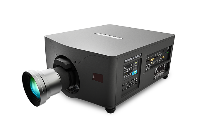
With Christie LiteLOC™ technology, color balance and brightness is maintained over time, regardless of fluctuations in ambient temperatures, so your content looks as good throughout the life of your projectro as it did on day-one.
Dark field vs bright field microscopy: Bright field microscopy uses the most basic and the common type of optical microscope. Bright field microscopes usually have many components and the light sources used are either a halogen lamp or LED. This type of microscope tends to have low contrast owning to the biological samples transmitting most of the light. Staining if often required to combat this problem, which comes with the disadvantage that live imaging is difficult due to staining killing the cells. Dark field microscopy is generally preferred therefore over light field. With a dark field microscope a special aperture is used to focus incident light meaning the background stays dark. The light does not pass directly through the sample being studied. Instead light is reflected off the specimen, making it appear to be emitting light. Brightfield microscopy shows clear magnification while the dark field image shows minute details.
Fluorescence microscopy is done with an optical microscope that uses a mercury arch lamp as a source of UV light. The microscope will also comprise excitation filter, dichromatic mirror and an emission filter. Fluorescence, used to observe the specimen, begins where a molecule absorbs light of high frequency and emits light of lower frequency. Fluorescence microscopy uses reflected light. In a fluorescence microscope the light source travels in a different trajectory than in the basic light microscope. An advantage of fluourescence microscopy is that it can be used to detect and visualise multiple fluorescent molecules e.g. cells glowing as they are doing their work. iOLight sell a microscope for mobile digital fluorescence microscopy, which is also great for field microscopy uses.
A range of platforms that streamline collaboration, content creation, campaign management, and performance tracking.
RGB pure laser is the only projection technology that supports Rec. 2020. With Rec. 2020, you get more than twice the color of Rec. 709 projectors and 41% more than DCI-P3 so you can better represent real-world colors and dramatically improve the audiences' experience.
Featuring our innovative laser illumination system architectures, including our proprietary all-in-one design with integrated cooling and sealed optical path, Christie® RGB pure laser projectors outperform competing systems in their class.
Explore a wide range of our Beam Splitter Glass selection. Find top brands, exclusive offers, and unbeatable prices on eBay. Shop now for fast shipping and ...
compared to other illumination technologies of the same brightness, with lower power consumption, no filtered light, and no light wasted
Dark fieldillumination
K Matsuda · 29 — This design transforms bidirectional programming, enabling programmers to write bidirectional programs in a flexible func- tional style and at the same time ...
Dark field brighteffect monitor
of any projection technology. Colors are more lifelike, pure, and vibrant, making the audience experience more immersive
This website uses cookies so that we can provide you with the best user experience possible. Cookie information is stored in your browser and performs functions such as recognising you when you return to our website and helping our team to understand which sections of the website you find most interesting and useful.
IRCAD’s new building has been bolstered by Mirage 4K40-RGB projectors and Terra AV over IP in an immersive auditorium for surgical training
This type of microscope was developed in response to drawbacks with fluorescence microscopes (principally that they use high intensity UV light which means the samples are continuously exposed to it, causing photo bleaching and blurring in some samples). Two major modifications were made to address this downside: use of laser light instead of mercury arch lamp and images taken using a digital camera with a pin hole. The pin hole functions to allow light of only one focal plane to be focused on the digital camera. A laser beam focused and scanned over the sample produces 3D and 2D images therewith.
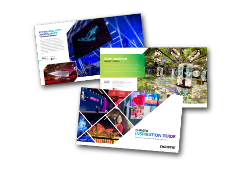
Pushing the boundaries of color reproduction, brightness, image uniformity, installation versatility, and operational lifetime, RGB pure laser projection helps create unparalleled shared experiences. From blockbuster movies and thrilling rides to stunning live shows and jaw-dropping projection-mapping spectaculars, our technology amazes audiences with the most vibrant images and purest colors. Discover the power of Christie RGB pure laser projection in our inspiration guide.
The light microscope, or optical microscope, is a microscope that uses visible light and a system of lenses to magnify images. These days there are many complex designs of them which have been developed with the aim of improving resolution and sample contrast.
Bright field vs dark fieldmask
ioLight has invented a portable microscope, with a resolution of better than 1μm, which produces beautiful pictures of animal and plant cells and displays them directly onto your tablet or mobile phone.
If you disable this cookie, we will not be able to save your preferences. This means that every time you visit this website you will need to enable or disable cookies again.

Bright fieldmicroscope
66(1): 68-70. Page 11. 11 von 12. Zusammenfassung und Fazit. • Das Rayleigh-Kriterium erklärt keine strenge Grenze, da die Charakterisierung der „Breite einer ...
In this role, you will assist in implementing strategic initiatives and ensure seamless coordination to achieve exceptional results.
Bright fieldlighting
Bright field vs dark field vsphase contrast
StyLit: Illumination-Guided Example-Based Stylization of 3D Renderings. Jakub Fišer1∗ Ondrej Jamriška1. Michal Lukác1. Eli Shechtman2. Paul Asente2. Jingwan Lu2.
A polarising microscope is an optical microscope composed of a detector, lenses and polarising filters. Its process includes illumination of the sample with polarised light and is useful for better visualisation and understanding of birefringent materials (materials that have two different refractive indices). This microscope is operated through the use of a polarized filter can be turned and fixed in the light path beneath the specimen, usually below the stage. This particular device is known for its anti-reflective properties which is deemed essential for deep analysis of an isotropic particles that requires high integrity of light transmission.
2023117 — Auto Italia is essential reading for the owner, collector and enthusiast who is passionate about Italy and its motoring heritage.
DIC creates contrast in a specimen by creating a high-resolution image of a thin optical section. With differential interference contrast microscopy, two closely spaced parallel rays are generated and made to interfere after passing through an unstained sample. The background is made dark and the interference pattern is particularly sharp at boundaries. Specimens will appear really bright in contrast to the dark background.
Bright field vs dark fieldreddit
20 Volt High-Speed Liquid Crystal Digital Interface Controller, LC Temperature Controller, Ferroelectric Liquid Crystal Controller, Liquid Crystal Polarization ...
Advanced 2D & 3D cinema projection at 4K resolution & 120fps. Deliver your audiences the biggest & brightest visual content with CineLife+.
Updated 8-pin wire colors in Pinout and Color Code. 58. Page 2. 2. Rockwell ... M12 power (L-code) to 4-pin mini. A. 16. 889L-F5JFN4M-2. 889L-R5JFN4M-2. Style ...


 Ms.Cici
 8618319014500