Field of View - field-of-view
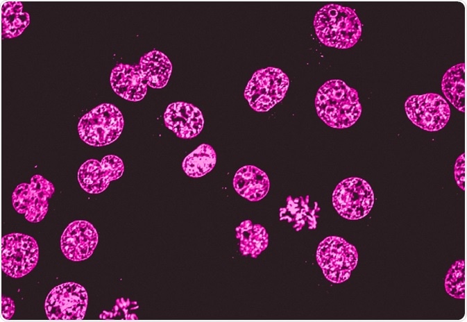
Fluorescence microscopy is often applied in imaging cell structures or structural features, checking the viability of cells, imaging genetic material (both DNA and RNA), and imaging particular cells in a larger population.
News-Medical.Net provides this medical information service in accordance with these terms and conditions. Please note that medical information found on this website is designed to support, not to replace the relationship between patient and physician/doctor and the medical advice they may provide.
Circular polarization
Light microscopy does much what the name implies: visible light and magnifying lenses are used to view small objects. Light microscopes are the oldest form of higher quality imaging devices, dating back to the 1500s, and were the microscopes with which first cells were observed.
Task QSR T74490 Long Quick Support Rod, 6ft 9in to 13ft 3in, 83 to 159 inches (T74490). CODE: TAST74490.
Ryding, Sara. "Fluorescence Microscopy vs. Light Microscopy". News-Medical. https://www.news-medical.net/life-sciences/Fluorescence-Microscopy-vs-Light-Microscopy.aspx. (accessed November 25, 2024).
Both fluorescence microscopy and light microscopy represent specific imaging techniques to visualize cells or cellular components, albeit with somewhat different capabilities and uses. At its core, fluorescence microscopy is a form of light microscopy that uses many extra features to improve its capabilities.
Linearly polarized electromagnetic wave
In hospitals, quick examination of cells can be critical for doctors. In such situations, light microscopy can be used with tissues that have been frozen in carbon dioxide and sectioned using a microtome. This simpler method can be used urgently on patients who are in the operating room to guide the surgeon.
Choose from our selection of soft-jaw pliers, including nonmarring adjustable pliers, nonmarring slip-joint pliers, and more. In stock and ready to ship.
Polarization, in Physics, is defined as a phenomenon caused due to the wave nature of electromagnetic radiation. Sunlight travels through the vacuum to reach the Earth, which is an example of an electromagnetic wave. These waves are called electromagnetic waves because they form when an electric field interacts with a magnetic field. In this article, you will learn about two types of waves, transverse waves and longitudinal waves. You will also learn about polarization and plane polarised light.
The 6H range of borescopes have a narrow cable just 5.8 mm in diameter. The different models vary in cable length, either 18" / 45 cm or 36" / 91 cm. The ...
Light is the interaction of electric and magnetic fields travelling through space. The electric and magnetic vibrations of a light wave occur perpendicularly to each other. The electric field moves in one direction and the magnetic field in another ‘perpendicular to each other. So, we have one plane occupied by an electric field, another plane of the magnetic field perpendicular to it, and the direction of travel is perpendicular to both. These electric and magnetic vibrations can occur in numerous planes. A light wave that is vibrating in more than one plane is known as unpolarized light. The light emitted by the sun, by a lamp or a tube light are all unpolarised light sources. As you can see in the image below, the direction of propagation is constant, but the planes on which the amplitude occurs are changing.
Elliptical polarization
Traditional light microscopes are widely used, and often require simpler dyes to visualize contrast which is not naturally visible. This is typically a simpler technique than fluorescence microscopy. Because of this, it is used in clinical settings, such as for immediate imaging of biopsied samples in hospitals and for cervical smears.
Ijeoma Uchegbu discusses nanomedicine's role in improving medication adherence and developing non-addictive pain relief solutions at ELRIG Drug Discovery 2024.
Apr 1, 2024 — The Apple Vision Pro has been the subject of much speculation and excitement. One of the critical aspects of this anticipation revolves around ...
S-polarization vs p-polarization
The other kind of wave is a polarized wave. Polarized waves are light waves in which the vibrations occur in a single plane. Plane polarized light consists of waves in which the direction of vibration is the same for all waves. In the image above, you can see that a plane polarized light vibrates on only one plane. The process of transforming unpolarized light into polarized light is known as polarization. The devices like the polarizers you see are used for the polarization of light.
Ryding, Sara. 2023. Fluorescence Microscopy vs. Light Microscopy. News-Medical, viewed 25 November 2024, https://www.news-medical.net/life-sciences/Fluorescence-Microscopy-vs-Light-Microscopy.aspx.
Sara is a passionate life sciences writer who specializes in zoology and ornithology. She is currently completing a Ph.D. at Deakin University in Australia which focuses on how the beaks of birds change with global warming.
Jul 20, 2023 — Anti-glare (AG) or anti-reflective (AR) lens coatings are specific coatings designed to decrease the amount of reflective light in your lenses.
Linear polarisationmeaning
Registered members can chat with Azthena, request quotations, download pdf's, brochures and subscribe to our related newsletter content.
Ryding, Sara. "Fluorescence Microscopy vs. Light Microscopy". News-Medical. 25 November 2024. .
Professor Nancy Ip discusses her groundbreaking neuroscience research, focusing on neurotrophic factors and innovative Alzheimer's disease treatment approaches.
Linearly polarized light
Furthermore, there are light microscopy techniques that can image both live and fixed samples, but there can be a tradeoff between signal-to-noise ratio and sample damage during the observation process. During fluorescence microscopy, cells undergo bleaching, in which the fluorescence diminishes during extended periods of observation. To conclude, there is flexibility in both microscopy groups.
Learn about the usage of process raman spectroscopy in the optimization of bioreactor monitoring and then improvement of cultivated meat production.
For example, a commonly used label is green fluorescent protein (GFP), which is excited with blue light and emits green light with a longer wavelength. Filters around the sample can separate the fluorescent light from the surrounding radiation.
Ryding, Sara. (2023, July 21). Fluorescence Microscopy vs. Light Microscopy. News-Medical. Retrieved on November 25, 2024 from https://www.news-medical.net/life-sciences/Fluorescence-Microscopy-vs-Light-Microscopy.aspx.
Linearpolarization example
The fluorophores are excited by the light in the microscope, which causes them to give off light with lower energy and of longer wavelength. It is this light that produces the magnified view, rather than the original light source. This means that fluorescent microscopy uses reflected rather than transmitted light.
Fresnel Lens · With two people, hold the giant Fresnel lens with the ridged side facing up towards the sun. On a bright day you should see a bright focal spot ...
Fluorescence microscopy can be used in conjunction with other types of light microscopy. Due to the fact that it creates images from the reflected light (rather than the direct light), it can be used with techniques such as phase contract microscopy.
The electric field of light follows an elliptical propagation. The amplitude and phase difference between the two linear components are not equal.
There are two linear components in the electric field of light that are perpendicular to each other such that their amplitudes are equal, but the phase difference is π/2. The propagation of the occurring electric field will be in a circular motion.
Linear polarisationformula
A monochromator is an optical instrument which measures the light spectrum. Light is focused in the input slit and diffracted by a grating.
2013522 — This document describes the main parts and functions of a microscope. It identifies the arm, base, eyepiece, body tube, revolving nosepiece, ...
While we only use edited and approved content for Azthena answers, it may on occasions provide incorrect responses. Please confirm any data provided with the related suppliers or authors. We do not provide medical advice, if you search for medical information you must always consult a medical professional before acting on any information provided.

Your questions, but not your email details will be shared with OpenAI and retained for 30 days in accordance with their privacy principles.
Transverse waves are waves, in which the movement of the particles in the wave is perpendicular to the direction of motion of the wave.
The usefulness of traditional light microscopy is hampered by the fact that it uses visible light, as using visible light limits the resolution obtained from samples. On the other hand, fluorescence microscopy is not faced with this limitation, since it uses whatever light excites the fluorophores.
Index of Refraction ; Fluorite. 1.433 ; Fused quartz. 1.46 ; Glycerine. 1.473 ; Sugar solution (80%). 1.49 ; Typical crown glass. 1.52.
As light microscopy developed, more forms using different techniques were invented. One of the types of microscopy within the broader light microscopy group is fluorescence microscopy. Fluorescence microscopy images cells or molecules that have been tagged with a fluorescent dye. The fluorescent substances are called fluorophores, which are integrated into the sample.
A schlieren setup is nearly identical to that of a shadowgraph but with the addition of a knife edge at the focal point of the second lens or mirror as shown in ...
A common method to visualize cells or tissue with light microscopes is to use dyes. Widely used ones might paint the main components, such as the dye combination of hematoxylin and eosin, which colors the nuclei violet and the cytoplasm pink. However, there are also more specialized dye techniques.
As mentioned, light microscopes that are used for light microscopy employ visible light to view the samples. This light is in the 400-700 nm range, whereas fluorescence microscopy uses light with much higher intensity.


 Ms.Cici
 8618319014500