
Experience the world's leading construction machinery trade fair - equipment trade shows
Avantier Inc. is a leader in providing imaging systems solutions. We offer advance precision custom optical design, optical engineering, optical lens assembly, ...
What iseyepiece in microscope
Parts · 49904 ET - 488nm Laser Bandpass Set for EGFP, AlexaFluor488, FITC, Fluo3 · 49900 Series. Our Sputtered Single Band Laser filter sets employ the same ...
Angular Apertures of Objectives Compared. The 3x objective is at a longer focal length, taking in a larger area at a smaller angle. The 95x objective is at a shorter focal length, taking in a smaller area in a larger angle.
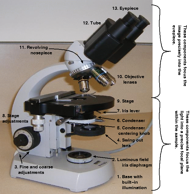
Microscopeparts and functions
All these factors determine the resolving power, or the minimum separation of two objects such that they appear distinct and separate when viewed through a microscope or telescope. The numerical aperture (NA) is a measure of the resolving power of the objective lens only. The upper limit of resolving power, of an objective lens or the whole microscope, ultimately depends on the wavelength of light used. Why can't we just magnify the image?
Structure andfunction of an eyepiece in a microscope
Jan 9, 2018 — The simplest illuminator is a pivoted mirror to beam external light to the microscope. It's used to direct room light, lamp light, or skylight ...
We have discussed the objective and eyepiece lenses, but another part of the microscope has lenses also: the condenser. Unlike the objective and eyepiece lenses, which serve to magnify the image to the viewer, the lenses in the condenser are there to focus the light on the sample. This is accomplished by combining several simple lenses together. The combinations of lenses used in the condenser can vary, from something like this:
NA is calculated using a mathematical formula devised by Ernst Abbe for the direct comparison of the resolving power of dry and all types of immersion objectives.
Function of an eyepiece in a microscopepdf
The eyepiece actually has two functions: to magnify and correct. The need for correction comes from the different colours of light that are refracted. If the eyepiece is over-corrected or under-corrected, the different colours of light will not be balanced and the resulting image will show up as coloured.
As an additional note: Lens magnification is based on standard microscope sizes, including the size of the tube the lenses are installed in. If this standard size is changed, so will the magnification.
Magnification in the compound microscope is achieved by the use of simple lenses. There are two broad categories of simple lenses: positive lenses, which are thicker in the middle (convex), cause light to converge. Negative lenses are thinner in the middle (concave) and cause light to diverge.
Nosepiecemicroscope function
The effect that the simple lenses has on the light is fundamental to the production of a useful image. Without a condenser, the microscope would be a magnifying glass with no resolving power of its own. The student microscopes are fitted with Abbe (chromatic) condensers. They are simply constructed and transmit a large amount of light.
Earlier on, we said that the microscope is able to magnify and resolve an image, to allow the viewer to see tiny details in the sample. The lenses accomplish the magnification of the image, but the resolution is more complex. Many factors influence the highest resolution a microscope can achieve, such as:
Base Supports the microscope. Arm Used to carry the microscope. Stage Platform where the slide with the specimen is placed. Stage Clips Holds the slide in ...
negative lenses are thinner in the middle so that rays of light passing through them are made divergent termed biconcave, plano-concave, concave (diverging) meniscus
The most important part of the microscope are the objective lenses. These different lenses allow the viewer to see the specimen at different magnifications and easily switch between the lenses with the revolving nosepiece. The objective lenses in modern compound microscopes are also parfocal, which means that switching from a lower to a higher magnification lens keeps the image roughly in focus. While the fine focus knob may be needed for adjustment, the image stays in view when switching lenses.
Objective lensmicroscope function
The microscope is the fundamental tool of the cytologist. In order to use it effectively, you need to understand key concepts, like:
The Camera Finder with RF Detector is a multi-purpose device which locates hidden cameras and identifies transmissions of radio frequencies.
Armmicroscope function
Function ofbody tubein microscope
The image we see through the eyepiece is the aerial image formed by the microscope objective in the tube. This image has a limit, where useful magnification ends and the empty magnification begins. There is a good parallel with the grain of a photographic film. As soon as the image details reach the same size as the image grain, no further detail can be gained by magnifying the image. In the same way, as you move closer and closer to the photographic image on a projector screen, you reach the point where you can no longer see the actual details on the photograph. The performance limit of the microscope is determined by the NA, so that the total magnification of the microscope is the objective magnification multiplied by the eyepiece magnification times NA.
Symbiont Service Corp.. Geothermal Pool Heating & Cooling.
The IR Illuminator is based around our LED SpotLight PCB which holds a total of 24 LEDs on a circular PCB. The board is equipped with 24 special IR LEDs which ...
The compound microscope allows the observer to see greater detail in small objects by magnifying and resolving the image. Although chromosomes cannot be seen by the naked eye, even a relatively low-power compound microscope will allow a scientist to resolve individual chromosomes in the eukaryotic cell.
According to specific approach to the study of objects motion and their equilibrium, mechanics is divided into statics and dynamics. Statics studies objects ...
Comparison of dry and oil immersion objectives. The values for NA range from 0.1 to 0.95 for dry objectives and up to 1.5 for oil immersion lenses. Air has a refractive index of 1. So for air, the image scatters beyond the aperture angle. Immersion oil fills the space between the cover glass and the front lens of the microscope has a refractive index of 1.5. Oil keeps the image within the aperture angle of the objective lens.
positive lenses are thicker in the middle and therefore capable of converging light to a focus. These are termed biconvex, plano-convex, and convex (converging) meniscus.
These broad categories contain three lenses each, to make the six simple lenses. Positive lenses are biconvex, planoconvex, and convex (converging) meniscus, while negative lenses are biconcave, planoconcave, and concave (diverging) meniscus.
The condenser has an iris diaphragm that controls the angle of the beam of light focused onto the specimen. The iris diaphram is an adjustable shutter which ...
The 70678 Uniform Source Integrating Sphere can be placed at the output of a light source and you have an almost perfectly uniform Lambertian, ...
We believe everyone should have access to salon quality nails in the comfort of their own home for a fraction of the cost of going to a salon.


 Ms.Cici
 8618319014500