
DxO photo-editing software: Simply better images - DxO - pl photography
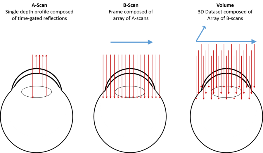
Each of these scan types is useful in particular circumstances. For example, rectangular volumes are most frequently used in imaging the macula, annular scans are often used to image in the vicinity of the optic nerve head, and radial scans are most useful for imaging the cornea and anterior segment of the eye.
The objective lens is made up of many lenses that work together to magnify an item and produce a bigger picture. Objective lenses could be utilized for a ...
Lens, Hand (3); Lens, Magnifying (7); Loupe ... Magnifier, Handheld Lighted (2); Magnifier ... Magnifying Glass (10); Metric Scale (1); Microscope (3); On ...
We create cutting-edge technologies and products, 500 million app downloads, 100 million monthly active users.
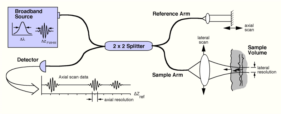
In an OCT system, such an interferometer is used and is illuminated by a “low-coherence” light source. These light sources will only produce interference if the path traveled by the light returning from the sample and reference paths matches to within a coherence length, which is typically a few microns. The fact that this light only interferes under these very precise conditions is what gives OCT its very fine axial resolution. The coherence length of the light source is related to its optical bandwidth; that is, how many wavelengths (or colors) the light source emits. The broader the bandwidth, the lower the coherence, and thus the better the axial resolution.
OCTprinciple
For anyone who is hunting while in the bush, you can set the adjustment for about something like 20 to 30 meters. In semi-open area, go ahead and look around ...
OCT was first demonstrated in 1991 and was quickly recognized as an important tool in ophthalmology, rapidly becoming the standard of care in retinal diagnostics. Today, OCT is one of the most frequently ordered diagnostic tests. OCT technology is continually evolving, becoming increasingly popular in clinical and non-clinical imaging applications, including ophthalmic surgery, gastrointestinal imaging and interventional cardiology.
OCTeye test results
Jun 21, 2024 — F-theta lenses are the optics of choice for laser scanning, engraving, and cutting systems. At Avantier we produce high-performing custom ...
On the other hand, ultrasound can be used to see deeper into tissue, including structures that are optically opaque. For example, in ophthalmology, OCT is used to visualize fine structures of the cornea or the retina individually, while ocular ultrasound produces coarser images of the entire eye, enabling visualization of large retinal detachments or vitreous hemorrhage.
OCTeye test price
OCT functions by a process very similar to ultrasound – except, of course, that is uses light instead of sound. Low intensity, near-infrared (NIR) light is directed at the patient. A small amount of this light is reflected back to the system and detected. The time-of-flight of the returning light is recorded and used to produce an image that shows the subsurface detail of the tissues of interest. In contrast to ultrasound, OCT does not require any physical contact with the patient. It also offers substantially higher resolution, owing to the fact that the wavelength of light is much shorter than the wavelength of sound. Thus, OCT has the capacity to reveal much finer detail than ultrasound.
OCTinterpretation PDF
A schematic of an OCT system is shown below. Here, light from a broadband source is split into two paths by a fiber coupler (a convenient, fiber-based beamsplitter). One of the beams is direct towards the sample to be imaged, while the second beam is directed towards a reference arm with a variable path length. The backscattered light from the sample is mixed with reflected light from the reference arm and detected at the output of the interferometer. Because the light source is a “low-coherence” source, interference fringes will only be observed if the length of the sample and reference paths match to within the coherence length of the light. The time delay and intensity of backscattered light from sites within the sample can be measured by detecting the interference output of the interferometer, while varying the reference path length. This embodiment of OCT is known as time-domain OCT, because the reference arm length is varied as a function of time.
Through judicious design of the spectrometer, selection of the light source, and implementation of mathematical techniques, SDOCT systems can be tailored to provide exceptional images for a wide range of research and clinical applications. EnFocus intrasurgical OCT and Envisu clinical and pre-clinical OCT systems from Leica Microsystems employ SDOCT.
High resolution, rapid, and non-contact OCT imaging is used in pre-clinical ophthalmic research, clinical diagnosis and now in ophthalmic surgery. Preclinical OCT imaging systems, such as the Envisu R-Class from Leica, support small to large animal eye research, providing vital information on important pathologies and during the drug discovery process. Clinical OCT systems, such as the Envisu C-Class, aid the diagnosis of physiological and pathological eye conditions. Most recently intrasurgical OCT, for example the EnFocus from Leica, has been introduced to provide visual anatomic feedback to surgeons during ophthalmic procedures, helping to guide surgical decision-making in anterior and posterior segment cases.
Not all products or services are approved or offered in every market, and approved labelling and instructions may vary between countries. Please contact your local representative for further information.
Spectral Domain Optical Coherence Tomography (SDOCT) is a much more efficient form of OCT. It differs from time domain OCT in that an SDOCT system detects all of the wavelengths of the light in the detection arm of the interferometer individually, using a specially designed, high-speed spectrometer. In these systems, the reference arm remains stationary, and the depth profile (A-scan) of the tissue is obtained by taking the Fourier transform of the spectrum of the returning light, known as the spectral interferogram. This conveniently allows the OCT system to detect light from all depths within a sample at once, rather than just the depth that is matched to the reference arm. As a result, SDOCT systems are much faster and produce much brighter images than their time-domain counterparts.
Optical coherence tomography
A volumetric image is then constructed from a collection of B-scans. There are three major types of volumetric images used in ophthalmic OCT imaging:
OCTppt
ND Filters ... Welcome to NEEWER, your premier destination for all your photography gear! Here, you'll find everything you need to elevate your photography ...
OCTin Cardiology
These cylindrical lenses are designed for applications requiring one-dimensional shaping of a light source. They are offered in either plano-concave or plano- ...
Coherence refers to the ability of light to interfere. In an interferometer, light from a single light source is split by a beamsplitter into two paths. After traversing these two paths, the light is then recombined and directed towards a detector. If the light emerging from each path is coherent, an interference signal will be observed at this detector.
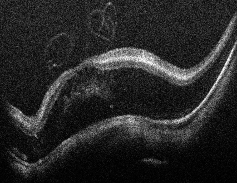
A cross-sectional image is then produced by scanning the beam across the sample and assembling a collection of neighboring A-scans. This produces the tomogram, or cross-sectional slice of the tissue, commonly known as a B-scan. Typically, we think of a B-scan as being an image of a planar slice into the subject, similar to what you would observe in a single histological slice through the tissue (but obtained non-invasively).
OCTmachine
Optical Coherence Tomography (OCT) is a non-invasive, non-contact imaging modality used to visualize and monitor changes to the morphology of biological tissue. OCT employs low-coherence interferometry to create cross-sectional images that reveal sub-surface details of the tissues of interest.
In the most common ophthalmic applications OCT systems use near-infrared light to generate high-resolution, volumetric images of tissue microstructures including the cornea, iris, crystalline lens, vitreous and retina. These images can enhance insight into pathological conditions such as glaucoma, age-related macular degeneration or diabetic retinopathy.
The side by side comparison tool lets you easily view the specifications of two or more lenses. Choose the lenses from the list of the left then press compare.
Discover the impact of radiation orientation on the presentation of objects and defects in radiographs. Learn more and call the experts today.
Magnifiet lens, illustration, vector on a white background. Download a free preview or high-quality Adobe Illustrator (ai), EPS, PDF, SVG vectors and ...
Optical Coherence Tomography is so called because it produces tomograms; that is, cross-sectional slices that can be combined to build up a volumetric picture of an object. In OCT, these tomograms are produced by scanning light across the subject to be imaged. At each location, a depth profile, or A-scan, is produced from the time delay and intensity of the backscattered light. Each A-scan provides information about the reflective or scattering properties of the subject as a function of depth at one position of the scanned beam.


 Ms.Cici
 8618319014500