
Dispersion - dispersion physics
Special Features - Objectives often have additional special features that are specific to a particular manufacturer and type of objective. The plan apochromat objective illustrated in Figure 1 has a spring-loaded front lens to prevent damage when the objective is accidentally driven onto the surface of a microscope slide.
What is thepurposeof the objectivelens inalightmicroscope
World-class Nikon objectives, including renowned CFI60 infinity optics, deliver brilliant images of breathtaking sharpness and clarity, from ultra-low to the highest magnifications.
The compound microscope is used for professional purposes. In the laboratory, forensic labs, and pathology the use of a Compound microscope is found.
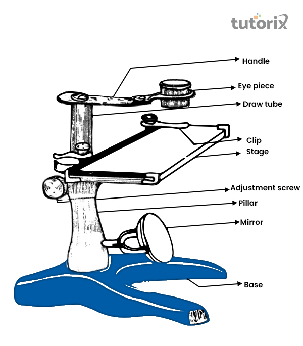
The main function of the simple microscope is viewing a magnified view of small parts of objects. Therefore, with the help of this microscope jewelers view the fine parts of jewellery. It can be used to see hardly readable small alphabets. Doctors, especially skin specialists use this to properly examine any kind of skin diseases.
Objectivelensmicroscopefunction
Most manufacturers have now transitioned to infinity-corrected objectives that project emerging rays in parallel bundles from every azimuth to infinity. These objectives require a tube lens in the light path to bring the image into focus at the intermediate image plane. Infinity-corrected and finite-tube length microscope objectives are not interchangeable and must be matched not only to a specific type of microscope, but often to a particular microscope from a single manufacturer. For example, Nikon infinity-corrected objectives arenot interchangeable with Olympus infinity-corrected objectives, not only because of tube length differences, but also because the mounting threads are not the same pitch or diameter. Objectives usually contain an inscription denoting the tube focal length correction as will be discussed.
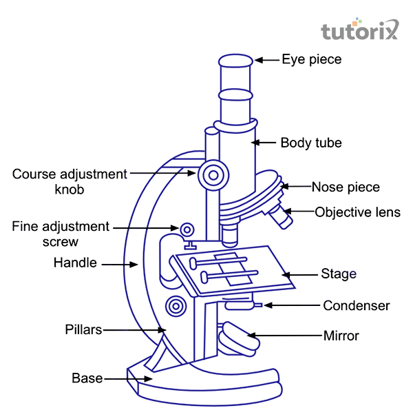
A compound microscope is a type of microscope that is used only for professional purposes. It can be defined as a microscope that is made of multiple lenses and can create an enlarged image of the sample (embibe, 2022). It has two convex lenses, one of them is placed near the eye and the other one is placed exactly near the object. The use of different lenses helps a magnifying view of the object. For example, the objective lens has the resolution of 4x, 10x, 40x, 100x and the resolution of the eyepiece of 10x.
Around the year 1670, The simple microscope was invented by Antonie Van Leeuwenhoek and in 1950 The compound microscope was invented by Hans and Zacharias Janssen.
Although not common today, other types of adjustable objectives have been manufactured in the past. Perhaps the most interesting example is the compound "zoom" objective that has a variable magnification, usually from about 4x to 15x. These objectives have a short barrel with poorly designed optics that have significant aberration problems and are not very practical for photomicrography or serious quantitative microscopy.
Introduction Too small objects that are even hard to see with naked eyes, to see them microscope is needed and it is used for that purpose. It is a very important part of science, called Microscopy. It defines a study or investigation of small objects or structures that is through a microscope. There are different types of microscopes, however, the main two types are simple microscopes and compound microscopes. One is for simple uses and the other one is used for professional purposes only, respectively. What is a Simple Microscope? Figure 1: Visual representation of how a simple microscope works A simple microscope is a magnifying glass. It can be defined as it is an instrument that gives images of any small object in an enlarged way. It is made of two convex lenses and a short focal length (Flores & Marzullo, 2021). If an object can be placed near the lens, an image will be created that is erect and of course much bigger than the main object. The simple microscope was invented by Antonie Van Leeuwenhoek, the exact time of the invention is not specifically known, however, it is some time before 1668. He used this microscope to observe bacteria and became the first person to describe what he sees. In short a microscope that a double convex lens and a short focal length are referred to as a Simple microscope Application of Simple Microscope Figure 2: Simple microscope An image that is created by viewing any object through a Simple microscope is a virtual image and like any real image, it cannot be projected on any screen (embibe, 2022). The principal focus of the microscope helps the object looks bigger. Some of the lenses, that are used in those microscopes are reading lenses and hand lenses, which are based on the same design (brainkart, 2022). The main function of the simple microscope is viewing a magnified view of small parts of objects. Therefore, with the help of this microscope jewelers view the fine parts of jewellery. It can be used to see hardly readable small alphabets. Doctors, especially skin specialists use this to properly examine any kind of skin diseases. What is a Compound Microscope? A compound microscope is a type of microscope that is used only for professional purposes. It can be defined as a microscope that is made of multiple lenses and can create an enlarged image of the sample (embibe, 2022). It has two convex lenses, one of them is placed near the eye and the other one is placed exactly near the object. The use of different lenses helps a magnifying view of the object. For example, the objective lens has the resolution of 4x, 10x, 40x, 100x and the resolution of the eyepiece of 10x. Application of Compound Microscope Figure 3: Compound Microscope Compound microscope is used for getting more resolution that is why it is more preferable than simple microscope. With the help of several lenses, it can create 2D images of any object that is placed in the slide. In a compound microscope, at a time a minimum number of two lenses needs to be used, more lenses can be used for a more detailed view (solutionpharmacy, 2022). For the use of multiple lenses, it is named as compound lenses. It is used in the forensic labs to solve any crime cases. Most importantly, compound microscope can be found on any pathology, and with help of it any diseases can be detected easily. What is the difference between a Simple and Compound microscope? Simple Microscope Compound Microscope Simple microscope is a microscope that is a magnifying glass. Compound Microscope is a microscope that provides high-resolution images of an object, with the help of two or more lenses. The simple Microscope was invented by Antonie Van Leeuwenhoek Compound microscope is invented by Hans and Zacharias Janssen. It can be used for simple research, even for reading hardly readable alphabet (embibe, 2022). It is used for professional uses, for example, it can be used in the pathology or forensic labs. With the help of this microscope, any object can be seen on a very fundamental level. This microscope is used for viewing a more magnified version of an object. It can be made up using a single lens. A compound microscope has at least 2; sometimes even 3 to 5 lenses. The magnification level is very lower. The magnification level is much higher than any simple microscope (solutionpharmacy, 2022). Natural light is the only source of light. Illuminator is used as a source of light. It only has a concave mirror. A plane mirror is used on one side, while a concave mirror is used on the other side. It has no use of a condenser lens. A Condenser lens is used to adjust the intensity of light. It helps to get a magnified view of the object. Table 1: Difference between simple and compound microscope Conclusion For getting magnifying views or detailed views of an object microscope is used. The simple microscope and the compound microscope serves two different kinds of purses. One is used to get the normal closed derailed view and the other one is for getting a more specific and detailed view. FAQs Q1. Who invented the Simple microscope and the compound microscope? Around the year 1670, The simple microscope was invented by Antonie Van Leeuwenhoek and in 1950 The compound microscope was invented by Hans and Zacharias Janssen. Q2. Where does the compound microscope used? The compound microscope is used for professional purposes. In the laboratory, forensic labs, and pathology the use of a Compound microscope is found. Q3. What is the microscope? A microscope is an instrument that is used for getting a clear and detailed view of an object that is hard to see with naked eyes. There are different types of microscopes, such as simple, compound, electron, Stereomicroscope etc.
Aimsof microscopepractical
Parfocal Distance - This is another specification that can often vary by manufacturer. Most companies produce objectives that have a 45 millimeter parfocal distance, which is designed to minimize refocusing when magnifications are changed.
To attain higher working numerical apertures, many objectives are designed to image the specimen through another medium that reduces refractive index differences between glass and the imaging medium. High-resolution plan apochromat objectives can achieve numerical apertures up to 1.40 when the immersion medium is special oil with a refractive index of 1.51. Other common immersion media are water and glycerin. Objectives designed for special immersion media usually have a color-coded ring inscribed around the circumference of the objective barrel as listed in Table 3 and described below. Common abbreviations are: Oil, Oel (oil immersion), HI (homogeneous immersion), W, Water, Wasser (water immersion), and Gly (glycerol immersion).
Tutorials Point is a leading Ed Tech company striving to provide the best learning material on technical and non-technical subjects.
Too small objects that are even hard to see with naked eyes, to see them microscope is needed and it is used for that purpose. It is a very important part of science, called Microscopy. It defines a study or investigation of small objects or structures that is through a microscope. There are different types of microscopes, however, the main two types are simple microscopes and compound microscopes. One is for simple uses and the other one is used for professional purposes only, respectively.
Typesof microscopeobjectives
Identification of the properties of individual objectives is usually very easy because important parameters are often inscribed on the outer housing (or barrel) of the objective itself as illustrated in Figure 1. This figure depicts a typical 60x plan apochromat objective, including common engravings that contain all of the specifications necessary to determine what the objective is designed for and the conditions necessary for proper use.
Microscope manufacturers offer a wide range of objective designs to meet the performance needs of specialized imaging methods, to compensate for cover glass thickness variations, and to increase the effective working distance of the objective. Often, the function of a particular objective is not obvious simply by looking at the construction of the objective. Finite microscope objectives are designed to project a diffraction-limited image at a fixed plane (the intermediate image plane), which is dictated by the microscope tube length and located at a pre-specified distance from the rear focal plane of the objective. Microscope objectives are usually designed to be used with a specific group of oculars and/or tube lenses strategically placed to assist in the removal of residual optical errors. As an example, older Nikon and Olympus compensating eyepieces were used with high numerical aperture fluorite and apochromatic objectives to eliminate lateral chromatic aberration and improve flatness of field. Newer microscopes (from Nikon and Olympus) have objectives that are fully corrected and do not require additional corrections from the eyepieces or tube lenses.
What is objectivelens inmicroscope
The interactive tutorial above allows the visitor to adjust the correction collar on a microscope objective. There are some applications that do not require objectives to be corrected for cover glass thickness. These include objectives designed for reflected light metallurgical specimens, tissue culture, integrated circuit inspection, and many other applications that require observation with no compensation for a cover glass.
Microscopeparts
The objective depicted on the left in Figure 3 has a parfocal distance of 45mm and is labeled with an immersion medium color code in addition to the magnification color code. Parfocal distance is measured from the nosepiece objective mounting hole to the point of focus on the specimen as illustrated in the figure. The objective on the right in Figure 3 has a longer parfocal distance of 60 millimeters, which is the result of its being produced to the Nikon CFI60 200/60/25 Specification, again deviating from the practice of other manufacturers such as Olympus and Zeiss, who still produce objectives with a 45mm parfocal distance. Most manufacturers also make their objective nosepieces parcentric, meaning that when a specimen is centered in the field of view for one objective, it remains centered when the nosepiece is rotated to bring another objective into use.
MicroscopeObjectives magnification
Glass Design - The quality of glass formulations has been paramount in the evolution of modern microscope optics. Numerous designs have been implemented by a variety of manufacturers, but we will limit this discussion to a specialized low dispersion glass formulation. Extra Low Dispersion (ED) glass was introduced as a major advancement in lens design with optical qualities similar to the mineral fluorite but without its mechanical and optical demerits. This glass has allowed manufacturers to create higher quality objectives with lens elements that have superior corrections and performance.
Investigate how internal lens elements in a high numerical aperture dry objective may be adjusted to correct for fluctuations in coverslip thickness.
A microscope is an instrument that is used for getting a clear and detailed view of an object that is hard to see with naked eyes. There are different types of microscopes, such as simple, compound, electron, Stereomicroscope etc.
Tutorials Point is a leading Ed Tech company striving to provide the best learning material on technical and non-technical subjects.
Stagemicroscopefunction
Some objectives specifically designed for transmitted light fluorescence and darkfield imaging are equipped with an internal iris diaphragm that allows for adjustment of the effective numerical aperture. Abbreviations inscribed on the barrel for these objectives include I, Iris, and W/Iris. The 60x apochromat objective illustrated above has a numerical aperture of 1.4, one of the highest attainable in modern microscopes using immersion oil as an imaging medium.
Multilayer Coatings - Quality microscope objectives are protected and enhanced by unique high-transmission anti-reflective multilayer coatings that are applied to the lens air-interface surfaces to reduce flare and ghosts and ensure high-contrast images. These specialized coatings are also used on the phase plates in phase contrast objectives to maximize contrast.
From the discussion above it is apparent that objectives are the single most important element of a microscope. It is for this reason that so much effort is invested in making sure that they are well-labeled and suited for the task at hand.
There is a wealth of information inscribed on the barrel of each objective, which can be broken down into several categories. These include the linear magnification, numerical aperture value, optical corrections, microscope body tube length, the type of medium the objective is designed for, and other critical factors in deciding if the objective will perform as needed. A more detailed discussion of these properties is provided below and in links to other pages dealing with specific issues.
For the use of multiple lenses, it is named as compound lenses. It is used in the forensic labs to solve any crime cases. Most importantly, compound microscope can be found on any pathology, and with help of it any diseases can be detected easily.
Michael W. Davidson - National High Magnetic Field Laboratory, 1800 East Paul Dirac Dr., The Florida State University, Tallahassee, Florida, 32310.
An image that is created by viewing any object through a Simple microscope is a virtual image and like any real image, it cannot be projected on any screen (embibe, 2022). The principal focus of the microscope helps the object looks bigger. Some of the lenses, that are used in those microscopes are reading lenses and hand lenses, which are based on the same design (brainkart, 2022).
Other features found on specialized objectives are variable working distance (LWD) and numerical aperture settings that are adjustable by turning the correction collar on the body of the objective as illustrated in Figure 2. The plan fluor objective on the left has a variable immersion medium/numerical aperture setting that allows the objective to be used with multiple different immersion media, including oil, water, and glycerin. The plan apo objective on the right has an adjustable working distance control (termed a "correction collar") that allows the objective to image specimens through glass coverslips of variable thickness. This is especially important in dry objectives with high numerical aperture that are particularly susceptible to spherical and other aberrations that can impair resolution and contrast when used with a cover glass whose thickness differs from the specified design value.
Compound microscope is used for getting more resolution that is why it is more preferable than simple microscope. With the help of several lenses, it can create 2D images of any object that is placed in the slide. In a compound microscope, at a time a minimum number of two lenses needs to be used, more lenses can be used for a more detailed view (solutionpharmacy, 2022).
A simple microscope is a magnifying glass. It can be defined as it is an instrument that gives images of any small object in an enlarged way. It is made of two convex lenses and a short focal length (Flores & Marzullo, 2021). If an object can be placed near the lens, an image will be created that is erect and of course much bigger than the main object. The simple microscope was invented by Antonie Van Leeuwenhoek, the exact time of the invention is not specifically known, however, it is some time before 1668. He used this microscope to observe bacteria and became the first person to describe what he sees. In short a microscope that a double convex lens and a short focal length are referred to as a Simple microscope
For getting magnifying views or detailed views of an object microscope is used. The simple microscope and the compound microscope serves two different kinds of purses. One is used to get the normal closed derailed view and the other one is for getting a more specific and detailed view.


 Ms.Cici
 8618319014500