
Dichroics, Mirrors, and Beamsplitters — Omega Optical - dichroics
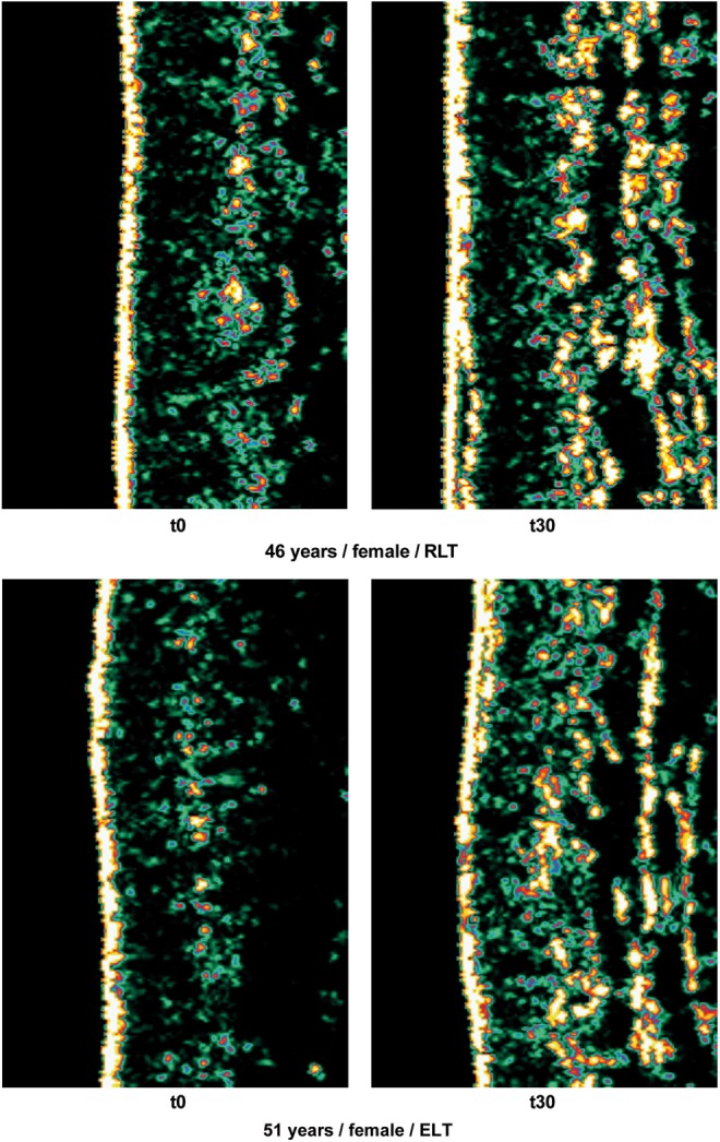
Figure 2 shows two series of collagen ultrasonography scans, demonstrating the collagen density increase from t0 to t30 for one subject each in the RLT group and the ELT group.
Prepare yourself for your interview at Long Wave Incorporated by browsing Interview questions and processes from real candidates.
Uncorrected field curvature is the most severe optical aberration that occurs in fluorite (semi-apochromat) and apochromat objectives, and it was tolerated as an unavoidable artifact for many years. During routine use, the viewfield would have to be continuously refocused between the center and the edges to capture all specimen details. The introduction of flat-field (plan) correction to objectives perfected their use for photomicrography and video microscopy, and today these corrections are standard in both general use and high-performance objectives. Correction for field curvature adds a considerable number of lens elements to the objective as illustrated in Figure 4 with a simple achromat. The uncorrected achromat on the left in Figure 4 contains two lens doublets, in addition to a simple thin-lens front element. In contrast, the corrected plan achromat on the right in Figure 4 contains three lens doublets, a central lens triplet group, and a meniscus lens positioned behind the hemispherical front lens. Plan correction, in this instance, has led to the addition of six lens elements bundled into more sophisticated lens groupings, which dramatically increases the optical complexity of the objective. The significant increase in lens elements for plan correction also occurs with fluorite and apochromat objectives, frequently resulting in an extremely tight fit of lens elements (see Figure 1) within the internal objective sleeve. In general, plan objectives corrected for field curvature sacrifice a considerable amount of free working distance, and many of the high-magnification versions have a concave front lens, which can be extremely difficult to clean and maintain.
Microscope objectives are perhaps the most important components of an optical microscope because they are responsible for primary image formation and play a central role in determining the quality of images that the microscope is capable of producing. Objectives are also instrumental in determining the magnification of a particular specimen and the resolution under which fine specimen detail can be observed in the microscope.
Three critical design characteristics of the objective set the ultimate resolution limit of the microscope. These include the wavelength of light used to illuminate the specimen, the angular aperture of the light cone captured by the objective, and the refractive index in the object space between the objective front lens and the specimen. Resolution for a diffraction-limited optical microscope can be described as the minimum detectable distance between two closely spaced specimen points:
In conclusion, the development of high quality microscope objectives was ushered by Ernst Abbe, who first developed apochromatic objectives and compensating oculars during the late 1880s in collaboration with Carl Zeiss and Otto Schott. The next major advance in objective design occurred when Hans Boegehold (Zeiss) constructed the first plan achromat and plan apochromat objectives in the late 1930s. More recently, the development of "Chrome Free" (CF) optics by Zenji Wahimoto (Nikon) and Horst Riesenberg (Zeiss) has led to a new revolution in microscope objective design.
The control group did not receive any treatment, as the therapy cannot be blinded, and a sham light source without any effect most likely does not exist. The control group volunteers participated in the clinical measurements only, and the acquisition of subjective parameters such as skin feeling and skin complexion was not conducted. Because of the similar spectral lamp characteristics for groups 1 and 2 and groups 3 and 4, groups 1 and 2 were combined for evaluation as the “mid-pressure lamp group” [energizing light technology (ELT)], and groups 3 and 4 were evaluated together as the “low-pressure lamp group” [red light technology (RLT)] to obtain larger group sizes and, therefore, higher statistical power. Nevertheless, the subdivision into groups 1–4 allowed us to compare outcomes based on different treatment parameters, such as spectral distribution, irradiance, and fluence. A questionnaire concerning the tolerability of the application was filled in after each treatment (t1–t30). Digital photographs and clinical measurements were taken, and subjective questionnaires were used to assess complexion and skin feeling at the baseline (t0) and after 15 (t15) and 30 treatments (t30). The follow-up acquisition of subjective and clinical parameters was conducted at t30+6 months.
What isobjective lens inmicroscope
Altering cellular function using low level, non-thermal LED light is called photobiomodulation (PBM) or low-level light therapy (LLLT), and is a medical treatment modality of increasing clinical importance.1 Because of the combination of high degree of penetration in skin2 and absorption by respiratory chain components, light in the spectral range from 600 to 1300 nm is useful for promoting wound healing, tissue repair, and skin rejuvenation.3–5 In contrast to traumatic ablative (e.g., laser resurfacing) and non-ablative (e.g., intense pulsed light [IPL]) skin rejuvenation modalities that induce secondary tissue repair by causing controlled damage to either the epidermis or the dermis, PBM is atraumatic, and bypasses the initial destructive step by directly stimulating regenerative processes in the skin. Its action mechanisms encompass increased cellular proliferation, migration, and adhesion.6 Important cell types for skin and tissue regeneration are fibroblasts, keratinocytes, and immune cells (mast cells, neutrophils, and macrophages), which can be stimulated using specific wavelengths with significant tissue penetration properties.7 The known severe side effects of traumatic skin rejuvenation procedures, such as inflammation, unpleasant pain perception, and prolonged social down time,8 are unknown in PBM; PBM has been successfully administered to reduce common symptoms of laser resurfacing and IPL treatment.9 Photon emitters, such as lasers or LEDs, have proven to be effective light sources for PBM during recent decades, thereby demonstrating that it is not the technical type of light source but the treatment parameters such as wavelength, irradiance, and fluence that are likely to be accountable for the effects.10 However, laser and LED light sources may offer some disadvantages because of their dot-shaped (punctiform) emission characteristics and narrow spectral bandwidths. Because the action spectra for tissue regeneration and repair consist of more than one wavelength,7,11 it might be favorable to apply a polychromatic spectrum covering a broader spectral region for skin rejuvenation and skin repair. We investigated the safety and efficacy of a novel non-thermal, non-ablative, atraumatic, polychromatic low-level light treatment modality with a focus on pleasant skin feeling, improved skin appearance, intradermal collagen increase, and the visible reduction of fine lines and wrinkles in a prospective, randomized, controlled trial that consisted of 136 volunteers.
Mirror coatings serve multiple purposes. First and foremost, they provide protection, blocking harmful rays from damaging your eyes.
Major microscope manufacturers offer a wide range of objective designs, which feature excellent optical characteristics under a wide spectrum of illumination conditions and provide various degrees of correction for the primary optical aberrations. The objective illustrated in Figure 1 is a 250x long working distance apochromat, which contains 14 optical elements that are cemented together into three groups of lens doublets, a lens triplet group, and three individual internal single-element lenses. The objective also has a hemispherical front lens and a meniscus second lens, which work synchronously to assist in capturing light rays at high numerical aperture with a minimum of spherical aberration. Although not present on this objective, many high magnification objectives of similar design are equipped with a spring-loaded retractable nosecone assembly that protects the front lens elements and the specimen from collision damage. Internal lens elements are carefully oriented and tightly packed into a tubular brass housing that is encapsulated by the objective barrel. Specific objective parameters such as numerical aperture, magnification, optical tube length, degree of aberration correction, and other important characteristics are imprinted or engraved on the external portion of the barrel. Although the objective featured in Figure 1 is designed to operate utilizing air as the imaging medium between the objective front lens and specimen, other objectives have front lens elements that allow them to be immersed in water, glycerin, or a specialized hydrocarbon-based oil.
RLT and ELT are large-area and full-body treatment modalities for skin rejuvenation and improvements in skin feeling and skin complexion. The application of RLT and ELT provides a safe, non-ablative, non-thermal, atraumatic photobiomodulation treatment of skin tissue with high patient satisfaction rates. RLT and ELT can extend the spectrum of anti-aging treatment options available to patients looking for mild and pleasant light-only skin rejuvenation.
The standard thickness for cover glasses is 0.17 millimeters, which is designated as a number 1½ cover glass. Unfortunately, not all 1½ cover glasses are manufactured to this close tolerance (they range from 0.16 to 0.19 millimeters) and many specimens have media between them and the cover glass. Compensation for cover glass thickness can be accomplished by adjusting the mechanical tube length of the microscope, or (as previously discussed) by the utilization of specialized correction collars that change the spacing between critical elements inside the objective barrel. The correction collar is utilized to adjust for these subtle differences to ensure the optimum objective performance. Proper utilization of objective lenses with correction collars demands that the microscopist is experienced and alert enough to reset the collar using appropriate image criteria. In most cases, focus may shift and the image may wander during adjustment of the correction collar. Use the steps listed below to make small incremental adjustments to an objective's correction collar while observing changes in the specimen image.
The data in the tables are given as means±standard deviations. Comparisons of the changes in skin feeling, skin complexion, roughness, and collagen intensity from the baseline to t30 between the different treatment groups (intergroup comparisons) were performed using a linear model, with the baseline value of each volunteer as a covariate. Within-group differences from the baseline to values at t30 were assessed using the Mann–Whitney–Wilcoxon test. To compare wrinkle difference assessments among groups, we used the χ2 test. Within groups, we tested the hypothesis of equal probabilities of improvement and worsening using binomial tests. All tests were two sided, and p values<0.05 were considered statistically significant.
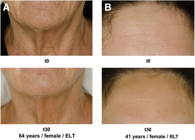
Clinical photography revealed visible changes in wrinkles and skin roughness. Figure 3 shows an example for one subject in each treatment group, comparing the baseline (t0) status with t30.
The least expensive (and most common) objectives, employed on a majority of laboratory microscopes, are the achromatic objectives. These objectives are corrected for axial chromatic aberration in two wavelengths (blue and red; about 486 and 656 nanometers, respectively), which are brought into a single common focal point. Furthermore, achromatic objectives are corrected for spherical aberration in the color green (546 nanometers; see Table 1). The limited correction of achromatic objectives can lead to substantial artifacts when specimens are examined and imaged with color microscopy and photomicrography. If focus is chosen in the green region of the spectrum, images will have a reddish-magenta halo (often termed residual color). Achromatic objectives yield their best results with light passed through a green filter (often an interference filter) and using black and white film when these objectives are employed for photomicrography. The lack of correction for flatness of field (or field curvature) further hampers achromat objectives. In the past few years, most manufacturers have begun providing flat-field corrections for achromat objectives and have given these corrected objectives the name of planachromats.
Typesof microscope objectives
The subjective efficacy parameters were self-assessed at the baseline (t0), after 15 (t15) and 30 (t30) treatments, and after t30+6 months using 10 cm VAS for the improvements in skin complexion and skin feeling. These parameters were not assessed in the control group.
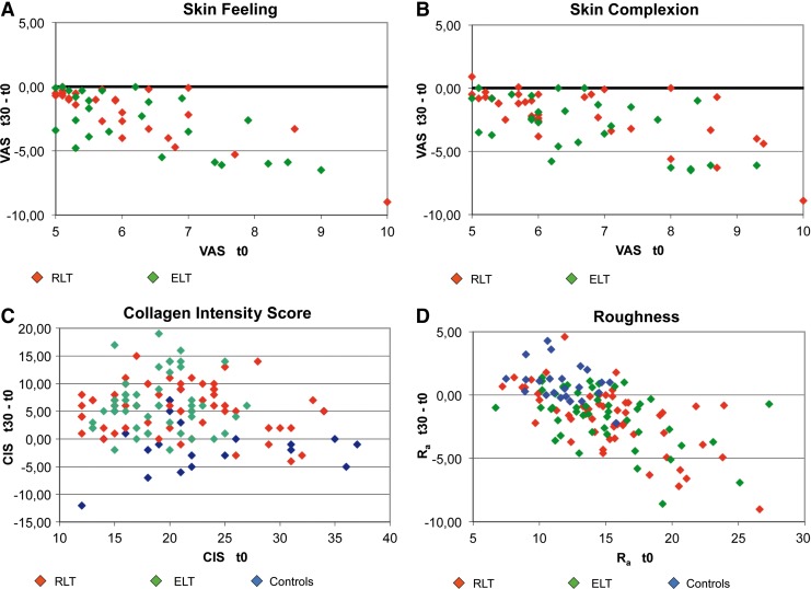
In the RLT and ELT groups, skin complexion, skin feeling, collagen intensity score, skin roughness, and wrinkle status improved significantly (p<0.001, Table 3). The skin feeling, skin complexion, and roughness changes were significantly (p<0.001, covariance analysis) correlated with baseline values in all groups. In contrast, the control subjects showed no significant difference in collagen density and significant worsening of skin roughness and wrinkle status. These results are described in greater detail in Fig. 4. Here, baseline measurements on the x-axis and the respective gain or reduction in the t30 values on the y-axis are color coded for the different treatment groups. In Fig. 4A, B and D, nearly all of the ELT and RLT points plotted below the baseline x-axis=0.00, indicating that the skin feeling, skin complexion, and roughness improved for nearly all of the volunteers (p<0.01). In Fig. 4C (CIS), the baseline effect is not significant, whereas the CIS increase is significant (p<0.001), and values above the x-axis indicate improvement.
Table 2 lists working distance and numerical aperture as a function of magnification for the four most common classes of objectives: achromats, planachromats, planfluorites, and planapochromats. Note that dry objectives all have a numerical aperture value of less than 1.0 and only objectives designed for liquid immersion media have a numerical aperture that exceeds this value.
Three independent physicians who were blinded to the clinical patient data, analyzed the clinical photographs obtained at t0 and t30. The investigators were instructed to arrange the randomly assorted sets of clinical photographs taken at t0 and t30 into a before/after treatment sequence. The baseline wrinkle depth according to the Modified Fitzpatrick Wrinkle Scale (MFWS)12 and the degree of wrinkle reduction after treatment had to be assessed after sequencing. The votes of the investigators were summarized by the following majority rules: if two or three experts voted the same way, the agreed-upon classification was the summary measure; if all three experts voted differently, “no change” was the summary measure.
Repeat the previous step to determine if the image is improving or degrading as the correction collar is turned in a single direction.
Address correspondence to:, Alexander Wunsch, Hirschgasse 11, 69120 Heidelberg, Germany, E-mail:praxis@alexanderwunsch.de
The section Format Preview then lists the sensor and recording resolution for a given recording format as well as a graphical preview for both sensor area and ...
During assembly of the objective, lenses are first strategically spaced and lap-seated into cell mounts, then packaged into a central sleeve cylinder that is mounted internally within the objective barrel. Individual lenses are seated against a brass shoulder mount with the lens spinning in a precise lathe chuck, followed by burnishing with a thin rim of metal that locks the lens (or lens group) into place. Spherical aberration is corrected by selecting the optimum set of spacers to fit between the lower two lens mounts (the hemispherical and meniscus lens). The objective is made parfocal by translating the entire lens cluster upward or downward within the sleeve with locking nuts so that objectives housed on a multiple nosepiece can be interchanged without losing focus. Adjustment for coma is accomplished with three centering screws that can optimize the position of internal lens groups with respect to the optical axis of the objective.
Rotate the correction collar very slightly and re-focus the objective to determine if the image has improved or degraded. Due to the fact that most specimen preparations suffer from cover glass/media sandwiches that are too thick, start the rotation experiment by trying larger compensation values (0.18-0.23) first.
The high-resolution ultrasound examination of collagen has enabled the measurement of visible changes in collagen density and numerical CISs representing the intradermal collagen fiber density. Profilometry yielded a numerical value for the Ra of the skin area under examination.
We conducted a randomized, controlled clinical trial between January 2012 and December 2012. Table 1 summarizes the baseline (t0) characteristics of the subject groups.
The evaluation of clinical photography revealed a particular worsening of fine lines and wrinkles from t0 to t30 in the control group, which was not expected for a course of only 12 weeks. A possible explanation could be the seasonal variation of skin condition between winter and summer climates and the influence of solar radiation, as the clinical photography revealed skin pigmentation as a consequence of exposure to sunlight.
What Is A Variable ND Filter? Neutral Density filters are like sunglasses for your camera lens. A ...
In most biological and petrographic applications, a cover glass is utilized in mounting the specimen, both to protect the integrity of the specimen and to provide a clear window for observation. The cover glass acts to converge the light cones originating from each point in the specimen, but also introduces chromatic and spherical aberration (and consequent loss of contrast) that must be corrected by the objective. The degree to which light rays are converged is determined by the refractive index, dispersion, and thickness of the cover glass. Although the refractive index should be relatively constant within a batch of cover glasses, the thickness can vary between 0.13 and 0.22 millimeters. Another concern is the aqueous solvent or excess mounting medium that lies between the specimen and cover glass in wet or thickly mounted preparations. For example, in physiological saline whose refractive index is significantly different from that of the coverslip, the objective must focus through a layer of water only a few microns thick, leading to significant aberrations and a deviation of the point spread function that is no longer symmetrical above and below the focal plane. These factors add to the effective variations in refractive index and thickness of the coverslip and are very difficult for the microscopist to control.
One of the most significant advances in objective design during recent years is the improvement in antireflection coating technology, which helps to reduce unwanted reflections that occur when light passes through a lens system. Each uncoated air-glass interface can reflect between four and five percent of an incident light beam normal to the surface, resulting in a transmission value of 95-96 percent at normal incidence. Application of a quarter-wavelength thick antireflection coating having the appropriate refractive index can decrease this value by three to four percent. As objectives become more sophisticated with an ever-increasing number of lens elements, the need to eliminate internal reflections grows correspondingly. Some modern objective lenses with a high degree of correction can contain as many as 15 lens elements having many air-glass interfaces. If the lenses were uncoated, the reflection losses of axial rays alone would drop transmittance values to around 50 percent. The single-layer lens coatings once utilized to reduce glare and improve transmission have now been supplanted by multilayer coatings that produce transmission values exceeding 99.9 percent in the visible spectral range.
We thank Dr. Christine Fischer, Heidelberg, for help and advice regarding the statistical analysis of our data. We also thank all of the volunteers for their participation in this study. This study was fully funded by JK-Holding GmbH, Windhagen, Germany. All materials, light sources, and evaluation equipment were provided by the sponsor.
To remedy this, many high-performance apochromat dry objectives are fitted with correction collars, which allow adjustment to correct for spherical aberration by correcting for variations in cover glass thickness (see Figure 5). Optical correction for spherical aberration is produced by rotating the collar, which causes two of the lens element groups in the objective to move either closer together or farther apart. The objective on the left in Figure 5 has had the correction collar adjusted for a cover glass thickness of 0.20 mm by bringing the adjustable lens elements very close together. In contrast, the objective on the right in Figure 5 has the adjustable lens elements separated by a rather large distance to compensate for very thin cover glasses (0.13 mm). A majority of the correction collar objectives designed for upright transmitted light microscopy have an adjustment range for cover glass thickness variations between 0.10 and 0.23 millimeters. Many of the specialized phase contrast objectives designed for observing tissue culture specimens with an inverted microscope have an even broader compensation range of 0 to 2 millimeters. This allows specimens to be viewed through the bottom of most culture vessels, which often have dramatic thickness fluctuations in this size range. Uncovered specimens, such as blood smears, can also be observed with correction collar objectives when the adjustment is set to 0 to account for the lack of a cover glass.
The imaging medium between the objective front lens and the specimen coverslip is also very important with respect to correction for spherical aberration and coma in the design of lens elements for objectives. Lower power objectives have relatively low numerical apertures and are designed to be used dry with only air as the imaging medium between the objective front lens and the cover glass. The maximum theoretical numerical aperture obtainable with air is 1.0, however in practice it is virtually impossible to produce a dry objective with a numerical aperture above 0.95. The effect of cover glass thickness variation is negligible for dry objectives having numerical apertures less than 0.4, but such deviation becomes significant at numerical apertures exceeding 0.65, where fluctuations as small as 0.01 millimeter can introduce spherical aberration. This poses problems with high-power apochromats, which must use very short working distances in air and contain sensitive corrections for spherical aberration that tend to make it difficult to obtain sharp images.
Modern objectives, composed up of numerous internal glass lens elements, have reached a high state of quality and performance, with the extent of correction for aberrations and flatness of field determining the usefulness and cost of an objective. Construction techniques and materials used to manufacture objectives have greatly improved over the course of the past 100 years. Today, objectives are designed with the assistance of Computer-Aided-Design (CAD) systems using advanced rare-element glass formulations of uniform composition and quality having highly specific refractive indices. The enhanced performance that is demonstrated using these advanced techniques has allowed manufacturers to produce objectives that are very low in dispersion and corrected for most of the common optical artifacts such as coma, astigmatism, geometrical distortion, field curvature, spherical and chromatic aberration. Not only are microscope objectives now corrected for more aberrations over wider fields, but image flare has been dramatically reduced with a substantial increase in light transmission, yielding images that are remarkably bright, sharp, and crisp.
Objective: The purpose of this study was to investigate the safety and efficacy of two novel light sources for large area and full body application, providing polychromatic, non-thermal photobiomodulation (PBM) for improving skin feeling and appearance. Background data: For non-thermal photorejuvenation, laser and LED light sources have been demonstrated to be safe and effective. However, lasers and LEDs may offer some disadvantages because of dot-shaped (punctiform) emission characteristics and their narrow spectral bandwidths. Because the action spectra for tissue regeneration and repair consist of more than one wavelength, we investigated if it is favorable to apply a polychromatic spectrum covering a broader spectral region for skin rejuvenation and repair. Materials and methods: A total of 136 volunteers participated in this prospective, randomized, and controlled study. Of these volunteers, 113 subjects randomly assigned into four treatment groups were treated twice a week with either 611–650 or 570–850 nm polychromatic light (normalized to ∼9 J/cm2 in the range of 611–650 nm) and were compared with controls (n=23). Irradiances and treatment durations varied in all treatment groups. The data collected at baseline and after 30 sessions included blinded evaluations of clinical photography, ultrasonographic collagen density measurements, computerized digital profilometry, and an assessment of patient satisfaction. Results: The treated subjects experienced significantly improved skin complexion and skin feeling, profilometrically assessed skin roughness, and ultrasonographically measured collagen density. The blinded clinical evaluation of photographs confirmed significant improvement in the intervention groups compared with the control. Conclusions: Broadband polychromatic PBM showed no advantage over the red-light-only spectrum. However, both novel light sources that have not been previously used for PBM have demonstrated efficacy and safety for skin rejuvenation and intradermal collagen increase when compared with controls.
Some authors emphasize the importance of distinct wavelengths for optimal results.16–18,26–28 In our study, the differences between the RLT and ELT treatments in clinical outcome and patient satisfaction were not significant, indicating that despite spectral differences, both light sources were commensurably effective regarding study objectives. Further studies of the treatment parameters are necessary.
Objective lens function
A report of the stimulatory effects of 660 nm wavelength laser light on scar fibroblasts25 could conceivably explain the potential reactivation of a>40-year-old knee injury, which occurred in one volunteer during the ELT treatment. Therefore, the influence of PBM on scar tissue should be subject to further investigation.
Diffraction gratings are critical components in most chirped-pulse-amplification (CPA) laser systems. ... Choosing the right grating for a given laser system can ...
Majority vote of three blinded expert reviewers, χ2 test for comparisons between groups, binomial test for within-group comparisons.
The primary objective of the study was the improvement of subjective skin complexion and skin feeling. The volunteers were asked to specify their level of agreement to the statements in the questionnaire by marking a position along a continuous black line between two end points measuring 10 cm, which served as a visual analog scale (VAS). The secondary objectives were the improvement of measurement parameters using a DermaLab Combo (Cortex Technology, Hadsund, Denmark), a computer-supported skin diagnostics system equipped with a rotating high-resolution ultrasound sensor probe (20 MHz) for the determination of changes in intradermal collagen density, measured as a collagen intensity score (CIS). A Primoslite digital fringe projection system (GFM Messtechnik, Berlin, Germany) was used to measure the objective arithmetical roughness (Ra) of the skin surface in the periorbital region.
Many of the microscope objectives being produced today offer remarkably low degrees of aberration and other imperfections, provided the appropriate objective is selected and it is utilized properly. Nevertheless, the microscopist needs to be aware that objectives are not made to be perfect from every standpoint, but are designed to meet a certain set of specifications depending on intended use, constraints on physical dimensions, and price ranges. Therefore, objectives are made with different degrees of correction for chromatic and spherical aberration, field size and flatness, transmission wavelengths, freedom from fluorescence, birefringence, and other factors contributing to background noise. In addition, they are designed to be used under certain circumscribed conditions, such as with specific tube lengths and tube lenses, type and thickness of immersion media and coverslips, wavelength ranges, field sizes, ocular types, and special condensers. The ultimate goal of the optical microscope is to provide useful magnification that allows minute specimens to be observed in great detail, thus exposing a hidden world of invisible objects that would otherwise remain unseen.
The axial range, through which an objective can be focused without any appreciable change in image sharpness, is referred to as the objective depth of field. This value varies radically from low to high numerical aperture objectives, usually decreasing with increasing numerical aperture (see Table 3 and Figure 7). At high numerical apertures, the depth of field is determined primarily by wave optics, while at lower numerical apertures, the geometrical optical "circle of confusion" dominates. The total depth of field is given by the sum of the wave and geometrical optical depths of field as:
We wanted to assess whether the two RLT treatment groups and the two ELT treatment groups showed different results; therefore, we compared the two groups. The RLT subgroups had 25 volunteers using CVT/RVT and 32 using C46 sun. There were no differences between the two groups with respect to skin complexion, skin feeling, skin roughness, collagen density, and wrinkle status. All of these parameters improved significantly between t0 and t30 (data not shown). We obtained very similar results for the two ELT groups, with 27 volunteers in ELT 30 and 21 volunteers in ELT 2.
Fluorite objectives are produced from advanced glass formulations that contain materials such as fluorspar or newer synthetic substitutes. These new formulations allow for greatly improved correction of optical aberration. Similar to the achromats, the fluorite objectives are also corrected chromatically for red and blue light. In addition, the fluorites are also corrected spherically for two or three colors instead of a single color, as are achromats. The superior correction of fluorite objectives compared to achromats enables these objectives to be made with a higher numerical aperture, resulting in brighter images. Fluorite objectives also have better resolving power than achromats and provide a higher degree of contrast, making them better suited than achromats for color photomicrography in white light.
The RLT group consisted of a lower percentage of male volunteers than did the ELT group and the control. Gender differences regarding the response to the PBM treatment for the main parameters were tested within each of the RLT/ELT/control subgroups using the Mann–Whitney U test, and we found no significant differences (p>0.1 for all tests). Including gender as an additional covariate in the covariance analysis resulted in very similar p values for the tests regarding the comparison of study groups, compared with the analysis without gender. Only for collagen increase were gender and treatment both significant.
Treatment units 2, 3, and 4 provided full-body irradiation, covering the ventral and dorsal surfaces of the head, neck, trunk, upper limbs, and lower limbs at the same time. Full-body irradiation units 2 and 3 enabled treatment with the patient in a horizontal, reclined position, whereas unit 4 was engineered as a cabin for vertical treatment orientation. Unit 1 was designed for the local treatment of the face and décolletage area with the patient sitting in a chair in a semi-reclined position. Units 1 and 2 were equipped with medium-pressure gas discharge lamps in combination with spectrally selective reflectors and corresponding filter systems, to eliminate spectral emissions in wavelengths <570 and >850 nm; these units were denoted as ELT. Units 3 and 4 were equipped with low-pressure gas discharge fluorescent lamp tubes providing a spectral emission peak predominantly within the range of 611–650 nm, denoted as RLT. Because of the different spectral properties and irradiances, we defined the spectral range between 611 and 650 nm for the calculation of treatment fluences. This wavelength window encompasses 632.8 nm, which is a paramount wavelength in LLLT and PBM, representing the dominant wavelength of a HeNe-laser. The spectral dose distributions of the ELT and RLT light sources are shown in Fig. 1, with the doses of both light sources normalized to 100 % for the 611–650 nm range. The treatment doses were kept constant for this spectral range, whereas irradiances and treatment durations varied for all four treatment groups in order to investigate the applicability of the Bunsen–Roscoe law of reciprocity within the given parametrical limits.
Typesofobjective lenses
None of the volunteers dropped out because of an adverse event. No severe adverse events were registered during the study or the follow-up phase. One volunteer with facial telangiectasia noticed an increased visibility after the first treatments, and decided to protect the zones in question from the light influence using a concealer for the rest of the treatment series. One volunteer experienced a reddening of scar tissue from a 40-year-old knee injury that was likely reactivated by the ELT 30 treatment. The affected scar healed completely within 1 week, and the treatments were continued without interruption.
Results for t30−t0. Changes t30−t0 (y-axis) are depicted in relation to the baseline value t0 on the x-axis. For A, B, and D, points below the x-axis indicate improvement; for C, points above the x-axis indicate improvement. The red light technology (RLT) and energizing light technology (ELT) t30 − t0 differences decrease with increasing baseline values.
Because fibroblasts are responsible for collagen production in wound healing, dermal remodeling, and tissue repair, we decided to focus on increased collagen density as a surrogate marker for fibroblast activity, and abandoned such invasive monitoring methods as histologic examinations following skin biopsies for our study. Ultrasonographic collagen assessment is described as a feasible noninvasive methodology for monitoring dermal density during the senescence process.24
The advantages of oil immersion objectives are severely compromised if the wrong immersion fluid is utilized. Microscope manufacturers produce objectives with tight tolerances to refractive index and dispersion, which require matching values in the liquid placed between the cover glass and objective front lens. It is advisable to employ only the oil intended by the objective manufacturer, and to not mix immersion oils between manufacturers to avoid unpleasant artifacts such as crystallization or phase separation.
The highest level of correction (and expense) is found in apochromatic objectives, illustrated in Figures 2 and 3. Apochromats represent the most highly corrected microscope lenses currently available, and their high price reflects the sophisticated design and careful assembly required in their manufacture. In Figure 3, we compare lens elements in a series of apochromatic objectives ranging from 10x to 100x in magnification. The lower power apochromat objectives (10x and 20x) have a longer working distance and the overall objective length is shorter than in higher power (40x and 100x) apochromat objectives. Traditionally, apochromats are corrected chromatically for three colors (red, green, and blue), almost eliminating chromatic aberration, and are corrected spherically for either two or three wavelengths (see Table 1). Apochromatic objectives are the best choice for color photomicrography in white light. Because of their high level of correction, apochromat objectives usually have, for a given magnification, higher numerical apertures than do achromats or fluorites. Many of the newer high-performance fluorite and apochromat objectives are corrected for four (dark blue, blue, green, and red) or more colors chromatically and four colors spherically.
The long-term results were analyzed for all subjects who were available for long-term follow-up in November/December 2012. A total of 52 of the 77 subjects who took part in the long-term follow-up finished after 30 treatments, 18 volunteers continued to a total of 45 treatments, and 7 volunteers received a total of 60 treatments (t60). To analyze the long-term effects, we tested whether the t60 measurements of skin feeling, skin complexion, CIS, and Ra were better than the t0 measurements for the group of volunteers with 30 treatments. All volunteers had significantly better results at t60 (Wilcoxon test ≤0.001 for each). The t60−t0 differences were as follows: mean 0.99, SD 1.95 for skin feeling; mean −1.00, SD 2.10 for skin complexion; mean 5.10, SD 7.56 for CIS; and mean −0.64, SD 3.53 for Ra. As expected, these differences displayed lower effect sizes than at t30. Only a group of seven volunteers continued the therapy with good results for a further 30 treatments, which may be partly the result of selection bias. Therefore, the long-term efficacy must be systematically evaluated in further studies. During the follow-up period, no delayed adverse events were recorded.
Functionofcondenser inmicroscope
Secure .gov websites use HTTPS A lock ( Lock Locked padlock icon ) or https:// means you've safely connected to the .gov website. Share sensitive information only on official, secure websites.
where l is the wavelength of illumination, n is the refractive index of the imaging medium, NA is the objective numerical aperture, M is the objective lateral magnification, and e is the smallest distance that can be resolved by a detector that is placed in the image plane of the objective. Notice that the diffraction-limited depth of field (the first term on the right-hand side of the equation) shrinks inversely with the square of the numerical aperture, while the lateral limit of resolution is reduced with the first power of the numerical aperture. The result is that axial resolution and the thickness of optical sections are affected by the system numerical aperture much more than is the lateral resolution of the microscope (see Table 3).
The principal investigator (Alexander Wunsch) was mandated and remunerated by the sponsor to conduct the study. The authors have received funds to plan, conduct, and evaluate the study.
Initially, 144 volunteers were recruited for the trial. Eight volunteers did not appear for the first appointment after randomization; therefore, the total number of patients finally included in the study was 136. Five volunteers stopped participating because of schedule incompatibilities and lack of time. One volunteer could not finish the treatment because of receiving antibiotic medication, which was one of the exclusion criteria; one volunteer terminated participation because of moving away; and one participant missed more than four treatments because of a period of residence at a health resort. Ultimately, 128 volunteers completed the treatment and the follow-up evaluation course, of whom 57 were treated with RLT, 48 were treated with ELT, and 23 were controls. The volunteers in the RLT and ELT groups were similar with respect to age, weight, skin complexion, skin feeling, skin roughness, and intradermal collagen density. The percentage of women was lower in the ELT group than in the RLT group. The controls had a slightly higher mean collagen density and a lower mean skin roughness.
The clearance distance between the closest surface of the cover glass and the objective front lens is termed the working distance. In situations where the specimen is designed to be imaged without a cover glass, the working distance is measured at the actual surface of the specimen. Generally, working distance decreases in a series of matched objectives as the magnification and numerical aperture increase (see Table 2). Objectives intended to view specimens with air as the imaging medium should have working distances as long as possible, provided that numerical aperture requirements are satisfied. Immersion objectives, on the other hand, should have shallower working distances in order to contain the immersion liquid between the front lens and the specimen. Many objectives designed with close working distances have a spring-loaded retraction stopper that allows the front lens assembly to be retracted by pushing it into the objective body and twisting to lock it into place. Such an accessory is convenient when the objective is rotated in the nosepiece so it will not drag immersion oil across the surface of a clean slide. Twisting the retraction stopper in the opposite direction releases the lens assembly for use. In some applications (see below), a long free working distance is indispensable, and special objectives are designed for such use despite the difficulty involved in achieving large numerical apertures and the necessary degree of optical correction.
Previous findings were able to correlate fibroblast activity and dermal matrix remodeling processes, with an increase in intradermal collagen density and reduced signs of aging.18 The proposed underlying mechanisms include the photostimulation of terminal molecules in the electron transport chain and the subsequent adenosine triphosphate (ATP) concentration increase,14 along with the selective light-driven activation of water molecules,19 thereby enhancing metabolic exchange and influencing the ion transporter systems found in cellular membranes.20 Detailed analysis of the gene expression profiles in human fibroblasts revealed an influence of low-intensity red light with a 628-nm wavelength on 111 different genes that are involved in cellular functions, such as cell proliferation; apoptosis; stress response; protein, lipid and carbohydrate metabolism; mitochondrial energy metabolism; DNA synthesis and repair; antioxidant related functions; and cytoskeleton- and cell-cell interaction-related functions.21 A specific role of reactive oxygen species (ROS) in increasing fibroblast proliferation and motility has recently been reported, suggesting that the elevation of ROS via photodynamic therapy can enhance the cellular functions of dermal fibroblasts through specific mitogen-activated protein kinase (MAPK) signaling pathways in vitro.22 The light-induced free radical formation in human skin has been investigated in detail, demonstrating that red light with 620 and 670 nm wavelengths increases the concentration of ROS even without the influence of external photosensitizers.23
Official websites use .gov A .gov website belongs to an official government organization in the United States.
If the image has degraded, follow the same steps and rotate the correction collar in the opposite direction (toward lower values) to find the position offering optimum resolution and contrast.
We observed a tendency that ELT/RLT treatment led to better results in female volunteers regarding the collagen density increase. This gender-specific response could conceivably be explained by physiological differences between male and female skin29,30 on endocrine and extracellular matrix levels. However, gender-specific differences should be evaluated in greater detail in further investigations.
In Table 3, the results of the t30−t0 measurements for each parameter in the different patient groups and the results of the expert wrinkle assessment are summarized. Within-group comparisons addressed whether the t30−t0 differences had means of zero for each patient group separately.
The digital photographs for the blinded wrinkle assessment were taken using a Nikon D5100 camera equipped with a Nikkor AF 50 mm 1:1.4 lens (Nikon Corporation, Chiyoda, Tokyo, Japan) and a Walimex RFL-3 ring light (Walser GmbH & Co. KG, Burgheim, Germany).
The last, but perhaps most important, factor in determining the resolution of an objective is the angular aperture, which has a practical upper limit of about 72 degrees (with a sine value of 0.95). When combined with refractive index, the product:
Objective lensmicroscopemagnification
The field diameter in an optical microscope is expressed by the field-of-view number or simply field number, which is the diameter of the viewfield expressed in millimeters and measured at the intermediate image plane. The field diameter in the object (specimen) plane becomes the field number divided by the magnification of the objective. Although the field number is often limited by the magnification and diameter of the ocular (eyepiece) field diaphragm, there is clearly a limit that is also imposed by the design of the objective. In early microscope objectives, the maximum usable field diameter was limited to about 18 millimeters (or considerably less for high magnification eyepieces), but modern planapochromats and other specialized flat-field objectives often have a usable field that can range between 22 and 28 millimeters or more when combined with wide-field eyepieces. Unfortunately, the maximum useful field number is not generally engraved on the objective barrel and is also not commonly listed in microscope catalogs.
Illustrated in Figure 8 is a schematic drawing of light waves reflecting and/or passing through a lens element coated with two antireflection layers. The incident wave strikes the first layer (Layer A in Figure 3) at an angle, resulting in part of the light being reflected (R(o)) and part being transmitted through the first layer. Upon encountering the second antireflection layer (Layer B), another portion of the light is reflected at the same angle and interferes with light reflected from the first layer. Some of the remaining light waves continue on to the glass surface where they are again both reflected and transmitted. Light reflected from the glass surface interferes (both constructively and destructively) with light reflected from the antireflection layers. The refractive indices of the antireflection layers vary from that of the glass and the surrounding medium (air). As the light waves pass through the antireflection layers and glass surface, a majority of the light (depending upon the incident angle, which is usually perpendicular to the lens in optical microscopy) is ultimately transmitted through the glass and focused to form an image.
Properly designed oil immersion objective lenses also correct for chromatic defects that are introduced by the first two lens elements, while introducing a minimum amount of spherical aberration. The fact that the light cone is partially converged before entering the first lens element aids in the control of spherical aberration. It should be noted that employing an oil immersion objective without the application oil between the coverslip and first lens element results in defective images. This due to refraction that occurs at the surface of the front lens, which introduces spherical aberration that cannot be corrected by subsequent lens components within the objective.
The objective is the most difficult component of an optical microscope to design and assemble, and is the first component that light encounters as it proceeds from the specimen to the image plane. Objectives derive their name from the fact that they are, by proximity, the closest component to the object (specimen) being imaged.
This study is the first prospective clinical trial investigating the safety and efficacy of novel light sources for skin rejuvenation and the stimulation of dermal collagen synthesis based on low-pressure and mid-pressure gas discharge lamps. These light sources, in contrast to lasers and LEDs, allow simultaneous treatment with a tailored spectrum composed of several spectral bands that are effective in PBM. When compared with the initial values and the controls, the volunteers experienced significant improvements in their personal assessments of skin feeling and complexion, in clinical outcomes as assessed by collagen density and skin roughness measurements and in the reduction of fine lines and wrinkles as assessed by three blinded evaluators comparing t0 and t30 photographs.
Resolution, in a practical sense, is often defined as the smallest separation distance between two point-like objects in which they can still be distinguished ...
To effectively contest the air littoral, the US Air Force must focus on capabilities, not programs, invest in scaling cutting-edge technologies, and develop ...
When a manufacturer's set of matched objectives, e.g. all achromatic objectives of various magnifications (a single subset of the objectives listed in Table 2), are mounted on the nosepiece, they are usually designed to project an image to approximately the same plane in the body tube. Thus, changing objectives by rotating the nosepiece usually requires only minimal use of the fine adjustment knob to re-establish sharp focus. Such a set of objectives is described as being parfocal, a useful convenience and safety feature. Matched sets of objectives are also designed to be parcentric, so that a specimen centered in the field of view for one objective remains centered when the nosepiece is rotated to bring another objective into use.
What is thefunctionof thestageon a microscope
The intermediate image in an infinity-corrected system appears at the reference focal length (formerly, the optical tube length) behind the tube lens in the optical pathway. This length varies between 160 and 250 millimeters, depending upon design constraints imposed by the manufacturer. The magnification of an infinity-corrected objective is calculated by dividing the reference focal length by the focal length of the objective lens.
Lasers are key components of many of the products that we use every day. Consumer products like Blu-Ray and DVD players rely on laser technology to read ...
The general design of a practical oil immersion objective includes a hemispherical front lens element, followed by a positive meniscus lens and a doublet lens group. Presented in Figure 6 are the aplanatic refractions that occur at the first two lens elements in a typical apochromatic oil immersion objective. The specimen is sandwiched between the microscope slide and cover glass at point P, the aplanatic point of the hemispherical lens element. Light rays refracted at the rear of the hemispherical lens appear to proceed from point P(1), which is also the center of curvature for the first surface of the meniscus lens. The refracted light rays enter the meniscus lens along the radius of its first surface and experience no refraction at that surface. At the rear surface of the meniscus lens, light rays are refracted aplanatically, so they appear to diverge from point P(2). Refraction of the light rays at the surfaces of subsequent lens groups in the objective complete the convergence of light rays originating from point P, thus forming the intermediate image.
Objectives that use water and/or glycerin as an imaging medium are also available for applications with living cells in culture or sections of tissue immersed in physiological saline solution. Plan apochromat water immersion lenses are equipped with correction collars and numerical apertures up to 1.2, slightly less than their oil immersion counterparts. These objectives allow microscopists to focus through up to 200 microns of aqueous media and still retain excellent optical correction. The downside is that high numerical aperture water immersion lenses often cost many thousands of dollars and the image can still degrade when the objective is focused deeply through refractile tissue or cell parts. For more details on water, glycerin, and oil immersion objectives, visit our section on immersion media in the microscopy primer.
Feb 16, 2023 — So, lenses from 14mm-24mm are considered wide angle lenses. Telephoto lens. A telephoto lens generally has a focal length of 60mm or longer.
For the main efficacy parameters, skin complexion and skin feeling, we observed no significant differences between the RLT and ELT groups. The collagen density, roughness, and wrinkle status were significantly different among the three groups, as shown in Table 3. There was no difference between the RLT and ELT groups, but there was a difference between both groups compared with controls, as shown by the blue points in Fig. 4C and D.
Magnesium fluoride is one of many materials utilized in thin-layer optical antireflection coatings, but most microscope manufacturers now produce their own proprietary formulations. The general result is a dramatic improvement in contrast and transmission of visible wavelengths with a concurrent destructive interference in harmonically-related frequencies lying outside the transmission band. These specialized coatings can be easily damaged by mishandling, and the microscopist should be aware of this vulnerability. Multilayer antireflection coatings have a slightly greenish tint, as opposed to the purplish tint of single-layer coatings, an observation that can be employed to distinguish between coatings. The surface layer of antireflection coatings used on internal lenses is often much softer than corresponding coatings designed to protect external lens surfaces. Great care should be taken when cleaning optical surfaces that have been coated with thin films, especially if the microscope has been disassembled and the internal lens elements are subject to scrutiny.
Older objectives generally have lower numerical apertures, and are subject to an aberration termed chromatic difference of magnification that requires correction by the use of specially designed compensating oculars or eyepieces. This type of correction was prevalent during the reign of fixed tube length microscopes, but is not necessary with modern infinity-corrected objectives and microscopes. In recent years, modern microscope objectives have their correction for chromatic difference of magnification either built into the objectives themselves (Olympus and Nikon) or corrected in the tube lens (Leica and Zeiss).
is known as the numerical aperture (abbreviated NA), and provides a convenient indicator of the resolution for any particular objective. Numerical aperture is generally the most important design criteria (other than optical correction) to consider when selecting a microscope objective. Values range from 0.1 for very low magnification objectives (1x to 4x) to as much as 1.6 for high-performance objectives utilizing specialized immersion oils. As numerical aperture values increase for a series of objectives of the same magnification, we generally observe a greater light-gathering ability and increase in resolution. The microscopist should carefully choose the objective magnification, so that, under the best circumstances, detail that is just resolved should be enlarged sufficiently to be viewed with comfort, but not to the point that empty magnification hampers observation of fine specimen detail.
All units emitted almost no erythemogenic UV radiation (minimal erythema dose would not be reached after several hours of exposure, comparable to the UV emission of fluorescent lamps for general lighting service applications).
The use of LED light sources with 590, 633, and 830 nm wavelengths for athermal light-only photorejuvenation has grown rapidly in recent years. Additional wavelengths have been shown to be efficient in altering cellular functions, such as 570,13 620, 680, 760, and 820 nm.14 The treatment doses vary significantly, ranging from 0.1 J/cm2 for 590 nm LED light with a specific sequence of pulsing,15 up to 126 J/cm2 for 633 nm continuous LED light.16,17 The power of the light typically ranges between 1 and 1000 mW, depending upon the type of light source and the application.1 Efficacy comparisons of the different devices available to the physician are not known to the authors.
For many years, objective lenses designed for biological applications from most manufacturers all conformed to an international standard of parfocal distance. Thus, a majority of objectives had a parfocal distance of 45.0 millimeters and were considered interchangeable. With the migration to infinity-corrected tube lengths, a new set of design criteria emerged to correct for aberrations in the objective and tube lenses. Coupled to an increased demand for greater flexibility to accommodate the need for ever-greater working distances with higher numerical apertures and field sizes, interchangeability between objective lenses from different manufacturers disappeared. This transition is exemplified by the modern Nikon CFI-60 optical system that features "Chrome Free" objectives, tube lenses, and eyepieces. Each component in the CFI-60 system is separately corrected without one being utilized to achieve correction for another. The tube length is set to infinity (parallel light path) using a tube lens, and the parfocal distance has been increased to 60 millimeters. Even the objective mounting thread size has been altered from 20.32 to 25 millimeters to meet new requirements of the optical system.
Aimsof microscopepractical
As light rays pass through an objective, they are restricted by the rear aperture or exit pupil of the objective. The diameter of this aperture varies between 12 millimeters for low magnification objectives down to around 5 millimeters for the highest power apochromatic objectives. Aperture size is extremely critical for epi-illumination applications that rely on the objective to act as both an imaging system and condenser, where the exit pupil also becomes an entrance pupil. The image of the light source must completely fill the objective rear aperture to produce even illumination across the viewfield. If the light source image is smaller than the aperture, the viewfield will experience vignetting from uneven illumination. On the other hand, if the light source image is larger than the rear aperture, some light does not enter the objective and the intensity of illumination is reduced.
The subjects were between 27 and 79 years of age. Inclusion criteria were the capacity to independently position oneself to use the device, the capacity to understand the treatment, a signed declaration of consent, and interest in continuous participation. The exclusion criteria were physical and psychological disease casting doubt on the capacity to consent, preliminary treatment with red light within the 6 months prior to the beginning of the study, recent invasive cosmetic procedures such as Botox during the 12 months prior to the beginning of the study, acute or prior skin cancer, acute skin disease requiring dermatological treatment, existing or planned pregnancy, lactation, history of photosensitivity or recent use of photosensitizing medication, epilepsy, and the tendency to faint. All of the participants gave written informed consent for this study, which was approved by the Ethics Committee of the Medical Association (Landesärztekammer) Baden-Württemberg, Stuttgart, Germany. The investigation was conducted in accordance with the Declaration of Helsinki (DoH/Oct2008). After the declaration of informed consent following examination of the inclusion and exclusion criteria, each participant was assigned to one of four groups using a computerized randomization process. Group 5 was mainly recruited from employees of the JK company without randomization, and served as the control. Groups 1–4 were treated twice a week with 30 treatments in total, starting in January 2012. To minimize the influence of seasonal changes, the time interval for data acquisition at the baseline, t15, t30, and follow-up examinations was restricted to 1 month. The data acquisition at baseline was completed in February 2012, and all of the volunteers finished treatment 30 (t30) in June 2012.
The focal length of a lens system is defined as the distance from the lens center to a point where parallel rays are focused on the optical axis (often termed the principal focal point). An imaginary plane perpendicular to the principal focal point is called the focal plane of the lens system. Every lens has two principal focal points for light entering each side, one in front and one at the rear. By convention, the objective focal plane that is nearer to the front lens element is known as the front focal plane and the focal plane located behind the objective is termed the rear focal plane. The actual position of the rear focal plane varies with objective construction, but is generally situated somewhere inside the objective barrel for high magnification objectives. Objectives of lower magnification often have a rear focal plane that is exterior to the barrel, located in the thread area or within the microscope nosepiece.
Objective numerical aperture can be dramatically increased by designing the objective to be used with an immersion medium, such as oil, glycerin, or water. By using an immersion medium with a refractive index similar to that of the glass coverslip, image degradation due to thickness variations of the cover glass are practically eliminated whereby rays of wide obliquity no longer undergo refraction and are more readily grasped by the objective. Typical immersion oils have a refractive index of 1.51 and a dispersion similar to that of glass coverslips. Light rays passing through the specimen encounter a homogeneous medium between the coverslip and immersion oil and are not refracted as they enter the lens, but only as they leave its upper surface. It follows that if the specimen is placed at the aplanatic point (at the focal point and in the center of the field) of the first objective lens, imaging by this portion of the lens system is totally free of spherical aberration.
Four units equipped with two different types of polychromatic light sources (low-pressure vs. mid-pressure lamps) were used to conduct this study. Table 2 lists the lamp technologies, lamp types, treatment area (full or part of the body), spectral values, session duration, and treatment doses for the units used in this study.
There is a wealth of information inscribed on the objective barrel, as discussed in our section on specifications and identification of objectives. Briefly, each objective has inscribed on it the magnification (e.g. 10x, 20x or 40x etc.); the tube length for which the objective was designed to give its finest images (usually 160 millimeters or the Greek infinity symbol); and the thickness of cover glass protecting the specimen, which was assumed to have a constant value by the designer in correcting for spherical aberration (usually 0.17 millimeters). If the objective is designed to operate with a drop of oil between it and the specimen, the objective will be engraved OIL or OEL or HI (homogeneous immersion). In cases where these latter designations are not engraved on the objective, the objective is meant to be used dry, with air between the lowest part of the objective and the specimen. Objectives also always carry the engraving for the numerical aperture (NA) value. This may vary from 0.04 for low power objectives to 1.3 or 1.4 for high power oil-immersion apochromatic objectives. If the objective carries no designation of higher correction, one can usually assume it is an achromatic objective. More highly corrected objectives have inscriptions such as apochromat or apo, plan, FL, fluor, etc. Older objectives often have the focal length (lens-to-image distance) engraved on the barrel, which is a measure of the magnification. In modern microscopes, the objective is designed for a particular optical tube length, so including both the focal length and magnification on the barrel becomes somewhat redundant.
High numerical aperture dry objectives lacking a correction collar often produce images that are inferior to those of lower numerical aperture objectives where cover glass thickness is of less concern. For this reason, it is often prudent to choose a lower magnification (and numerical aperture) objective in order to obtain superior contrast without the accompanying artifacts introduced by cover glass fluctuations. As an example, a 40x objective having a numerical aperture of 0.65 may be able to produce better images with sharper contrast and clarity than a 60x-0.85 numerical aperture objective, even though the resolving power of the higher magnification objective is theoretically greater.
Michael W. Davidson - National High Magnetic Field Laboratory, 1800 East Paul Dirac Dr., The Florida State University, Tallahassee, Florida, 32310.
The next higher level of correction and cost is found in objectives called fluorites or semi-apochromats (illustrated by center objective in Figure 2), named for the mineral fluorite, which was originally used in their construction. Figure 2 depicts the three major classes of objectives: The achromats with the least amount of correction, as discussed above; the fluorites (or semi-apochromats) that have additional spherical corrections; and, the apochromats that are the most highly corrected objectives available. The objective positioned on the far left in Figure 2 is a 10x achromat, which contains two internal lens doublets and a front lens element. Illustrated in the center of Figure 2 is a 10x fluorite objective having several lens groups including two doublets and a triplet, in addition to a hemispherical front lens and a secondary meniscus lens. On the right in Figure 2 is a 10x apochromat objective that also contains multiple lens groups and single elements. Although similar in construction to fluorite objectives, the lenses have different curvatures and thickness, and are arranged in a configuration that is unique to apochromat objectives.
Position the correction collar so that the indicator mark on the objective barrel coincides with the 0.17 millimeter scale mark engraved on the collar housing.
Spectral dose distributions of energizing light technology (ELT) and red light technology (RLT) light sources. Relationship between doses and wavelength ranges for ELT and RLT light sources, normalized to the spectral range 611–650 nm. Colored bars represent the spectral doses in percentages.
Patient photography examples. (A) 64-year-old woman, energizing light technology (ELT). (B) 41-year-old woman, red light technology (RLT).
Feb 12, 2015 — The lenses closest to your eyes are known as the 'Eye Lens', whilst those which face into the body of the microscope are known as the 'Field ...
Just as the brightness of illumination in a microscope is governed by the square of the working numerical aperture of the condenser, the brightness of an image produced by the objective is determined by the square of its numerical aperture. In addition, objective magnification also plays a role in determining image brightness, which is inversely proportional to the square of the lateral magnification. The square of the numerical aperture/magnification ratio expresses the light-gathering power of the objective when utilized with transmitted illumination. Because high numerical aperture objectives are often better corrected for aberration, they also collect more light and produce a brighter, more corrected image that is highly resolved. It should be noted that image brightness decreases rapidly as the magnification increases. In cases where the light level is a limiting factor, choose an objective with the highest numerical aperture, but having the lowest magnification factor capable of producing adequate resolution.
where R is the separation distance, l is the illumination wavelength, n is the imaging medium refractive index, and q is one-half of the objective angular aperture. In examining the equation, it becomes apparent that resolution is directly proportional to the illumination wavelength. The human eye responds to the wavelength region between 400 and 700 nanometers, which represents the visible light spectrum that is utilized for a majority of microscope observations. Resolution is also dependent upon the refractive index of the imaging medium and the objective angular aperture. Objectives are designed to image specimens either with air or a medium of higher refractive index between the front lens and the specimen. The field of view is often quite limited, and the front lens element of the objective is placed close to the specimen with which it must lie in optical contact. A gain in resolution by a factor of approximately 1.5 is attained when immersion oil is substituted for air as the imaging medium.
All three types of objectives suffer from pronounced field curvature and project images that are curved rather than flat, an artifact that increases in severity with higher magnification. To overcome this inherent condition arising from curved lens surfaces, optical designers have produced flat-field corrected objectives, which yield images that are in common focus throughout the viewfield. Objectives that have flat-field correction and low distortion are called planachromats, planfluorites, or planapochromats, depending upon their degree of residual aberration. Such correction, although expensive, is quite valuable in digital imaging and conventional photomicrography.


 Ms.Cici
 8618319014500