
Dichroic Mirrors Explained: A Comprehensive Guide - dichroic mirror
Since ancient times, measurement has been necessary in various aspects of human society, including pyramids. In manufacturing, "what cannot be measured correctly cannot be processed correctly," and "measurement" is essential to evaluate the quality of products. A micrometer is one of the indispensable tools for precision measurement.
Microscope objectives come in a range of designs, including apochromat, semi-apochromat, and achromat, among others. Our expansive collection of microscope objectives suits a wide variety of life science applications and observation methods. Explore our selection below to find a microscope objective that meets your needs. You can also use our Objective Finder tool to compare options and locate the ideal microscope objective for your application.
Many microscopes have several objective lenses that you can rotate the nosepiece to view the specimen at varying magnification powers. Usually, you will find multiple objective lenses on a microscope, consisting of 1.25X to 150X.
The ocular lens is located at the top of the eyepiece tube where you position your eye during observation, while the objective lens is located closer to the sample. The ocular lens generally has a low magnification but works in combination with the objective lens to achieve greater magnification power. It magnifies the magnified image already captured by the objective lens. While the ocular lens focuses purely on magnification, the objective lens performs other functions, such as controlling the overall quality and clarity of the microscope image.
These extended apochromat objectives offer high NA, wide homogenous image flatness, 400 nm to 1000 nm chromatic aberration compensation, and the ability to observe phase contrast. Use them to observe transparent and colorless specimens such as live cells, biological tissues, and microorganisms.
These semi-apochromat objectives enable phase contrast observation while providing a high level of resolution, contrast, and flatness for unstained specimens.
For example, more than 3,000 micrometers are actually used at production sites of aircraft-related companies, playing an important role in air safety. *The above is a description of the outer micrometers.
Objective lensesmicroscope function
Micrometers are indispensable precision measuring tools in the manufacturing field. This section provides basic explanations of the measurement methods and features of micrometers, as well as correct usage and precautions.
Optimized for multiphoton excitation imaging, these objectives achieve high-resolution 3D imaging through fluorescence detection at a focal point of a large field of view. They enable high-precision imaging of biological specimens to a depth of up to 8 mm for in vivo and transparent samples.
For relief contrast observation of living cells, including oocytes, in plastic vessels, our universal semi-apochromat objectives feature a long working distance. These also provide high image flatness and high transmission up to the near-infrared region.
Here we explain the main usage and precautions, using a standard outside micrometer as an example. Micrometers can measure from 0 mm to 2000 mm. Confirm the measuring range and select a type suitable for the shape of the object to be measured and the purpose of use.
Designed for clinical research and routine examination in labs using phase contrast illumination, these achromat objectives offer excellent field flatness.
Unsure of what microscope objective is right for you? Use our guide on selecting the right microscope objective to weigh your options.
Aug 1, 2024 — Knowledge of the MTF curves of the lenses and camera sensors involved in an optical system is used for optimization of the optical system ...
A micrometer is a simple-looking tool, but the standard model can read down to 1 μm(micrometer). 1 μm is 1/1000 of 1 mm, which is about 90 μm for copy paper, 70 to 80 μmfor a hair, and 10 μm for food film. This unit is very fine, showcasing how precise a tool such as the micrometer is.
This semi-apochromat objective series provides flat images and high transmission up to the near-infrared region of the spectrum. Acquiring sharp, clear images without color shift, they offer the desired quality and performance for fluorescence, brightfield, and Nomarksi DIC observations.
Designed for phase contrast observation of cell cultures in transmitted light, these achromat objectives combine field flatness and easy focusing with cost efficiency. They are well suited for routine microscopy demands.
Objective lenses are responsible for primary image formation, determining the quality of the image produced and controlling the total magnification and resolution. They can vary greatly in design and quality.
For relief contrast observation of living cells, including oocytes, in plastic vessels using transmitted light, these achromat objectives provide excellent field flatness.
For clinical research requiring polarized light microscopy and pathology training, these achromat objectives enable transmitted polarized light observation at an affordable cost.
Ocular lens
Browse Getty Images' premium collection of high-quality, authentic Cyan Square stock photos, royalty-free images, and pictures. Cyan Square stock photos are ...
Optimized for polarized light microscopy, these semi-apochromat objectives provide flat images with high transmission up to the near-infrared region of the spectrum. They are designed to minimize internal strain to meet the requirements of polarization, Nomarski DIC, brightfield, and fluorescence applications.
Bibliography · Engstrom, Ralph W., Photomultiplier Handbook, RCA/Burle (1980). · Photomultiplier Tubes: Basics and Applications (Second Edition), Hamamatsu ...
Offering our highest numerical aperture values, these apochromat objectives are optimized for high-contrast TIRF and super resolution imaging. Achieve wide flatness with the UPLAPO-HR objectives’ high NA, enabling real-time super resolution imaging of live cells and micro-organelles.
These extended apochromat objectives offers a high numerical aperture (NA), wide homogenous image flatness, and 400 nm to 1000 nm chromatic aberration compensation. They enable high-resolution, bright image capture for a range of applications, including brightfield, fluorescence, and confocal super resolution microscopy.
ICI's Round IR Window series allows thermogaphers to monitor completely undisturbed assets inside energized electrical cabinets.
These apochromat objectives are dedicated to Fura-2 imaging that features high transmission of 340 nm wavelength light, which works well for calcium imaging with Fura-2 fluorescent dye. They perform well for fluorescence imaging through UV excitation.
Apr 26, 2021 — Light can be polarised by passing it through a polarising filter. A polarising filter has all its molecules aligned in the same direction. As a ...
A micrometer is a tool to precisely measure the outer diameter and length (thickness) of an object to be measured by clamping it. The measurement method uses a precision screw with a pitch of 0.5 mm, and the rotation of the screw is calculated as the axial displacement of the screw. Currently, there are models that use a 2.0 mm pitch precision screw, enabling faster measurement by using a screw with a larger pitch.
This super-corrected apochromat objective corrects a broad range of color aberrations to provide images that capture fluorescence in the proper location. Delivering a high degree of correction for lateral and axial chromatic aberration in 2D and 3D images, it offers reliability and accuracy for colocalization analysis.
These super apochromat objectives provide spherical and chromatic aberration compensation and high transmission from the visible to the near infrared. Using silicone oil or water immersion media, which have refractive indexes closely matching that of live cells, they achieve high-resolution imaging deep in living tissue.
To clean a microscope objective lens, first remove the objective lens and place it on a flat surface with the front lens facing up. Use a blower to remove any particles without touching the lens. Then fold a piece of lens paper into a narrow triangular shape. Moisten the pointed end of the paper with small amount of lens cleaner and place it on the lens. Wipe the lens in a spiral cleaning motion starting from the lens’ center to the edge. Check your work for any remaining residue with an eyepiece or loupe. If needed, repeat this wiping process with a new lens paper until the lens is clean. Important: never wipe a dry lens, and avoid using abrasive or lint cloths and facial or lab tissues. Doing so can scratch the lens surface. Find more tips on objective lens cleaning in our blog post, 6 Tips to Properly Clean Immersion Oil off Your Objectives.
But when you use an anti-reflective coating, your eyes are protected against glare, meaning eye strain is less. Besides using them for computer work, they ...
When measuring, slowly bring both measuring surfaces into light contact with the workpiece to be measured in the same posture and under the same conditions as when the base point was checked, and turn the ratchet stop slowly 3 to 5 times to avoid excessive measuring force by the spindle. To avoid deformation of the workpiece, turn the ratchet stop slowly during contact.
Scanningobjectivelens
Examples of typical types include the "caliper type inside micrometer" for measuring small diameter and horizontal grooves, the "replacement piece type screw micrometer" for measuring the effectiveness of screws, and the "tooth thickness micrometer" for measuring the straddle tooth thickness of spur and helical gears.
These semi-apochromat and achromat objectives are designed for integrated phase contrast observation of cell cultures. They are used in combination with a pre-centered phase contrast slider (CKX3-SLP), eliminating centering adjustments when changing the objective magnification.
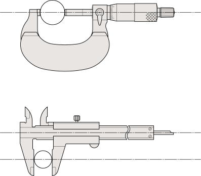
As shown in the figure below, there are various other types available for different purposes of use, such as measuring the diameter of narrow grooves, pipe wall thickness, and gear overpindiameter. *We can also accommodate custom-made products according to the purpose of use.
For phase contrast observation of cell cultures, these universal semi-apochromat objectives provide long working distances and flat images with high transmission up to the near-infrared region. They help you achieve clear images of culture specimens regardless of the thickness and material of the vessel.
Objective lensesfunction
For use without a coverslip or cover glass, these objectives prevent image deterioration even under high magnification, making them well suited for blood smear specimens. They also feature extended flatness and high chromatic aberration correction.

Feb 7, 2013 — ... device (PID) to a diameter of no more than 2.75 inches (7 cm) ... Some beam alignment devices or precision film holders include collimating ...
Types ofobjective lenses
Ratchet stop type is generally used as a device to keep the measuring force constant, but friction thimble type and ratchet thimble type are also available for the same purpose.
High powerobjectivelens
When reading the scale, be sure to read the scale from the front. As shown in the figure, changing the eye position from (A) to (B) to (C) will change the scale alignment position. To avoid errors due to eye position, read the scale from the position (B) perpendicular to the point where the sleeve reference line and thimble scale line meet.
Generally speaking, micrometers are cylindrical in shape, with the measuring surfaces facing each other, but there are various types of micrometers for different purposes.
Manufacturer of high-precision optical components made from sapphire. Capabilities include finish of other materials including Spinel, ALON, ZnS, Ge, Si, ...
When mounting the micrometer on a micrometer stand, clamp it across the center of the micrometer frame and do not excessively tighten. Then, adjust the angle of the stand so that the upper and lower scales are evenly visible centered on the zero scale line of the thimble, and fix it in place.
Abbe's principle states that "the object to be measured and the standard scale must be aligned in the direction of measurement". Micrometers are tools based on this principle and are capable of higher precision measurements than calipers and other measuring instruments.
Aug 9, 2022 — A solar concentrator using Fresnel lenses is called a Fresnel lens solar concentrator. It takes a large area of sunlight and directs it towards ...
Enabling tissue culture observation through bottles and dishes, these universal semi-apochromat objectives feature a long working distance and high contrast and resolution. Providing flat images and high transmission up to the NIR region, they are well suited for brightfield, DIC, and fluorescence observation.
Objectivelens magnification
When the ratchet stop is turned, it spins with a crackling sound to apply the appropriate measuring force to the surface to be measured.
For high-performance macro-observation, these apochromat objectives provide sharp, clear, flat images without color shift, achieving high transmission up to the near-infrared region of the spectrum. They perform well for fluorescence, brightfield, and Nomarksi DIC observations.
These semi-apochromat long-working distance water-dipping objectives for electrophysiology deliver flat images for DIC and fluorescence imaging from the visible range to the near-infrared. Their high NA and low magnification enables bright, precise macro/micro fluorescence imaging for samples such as brain tissue.
As part of daily maintenance, thoroughly wipe away dust and chips from the outer circumference of the spindle and the measurement surface. Also, thoroughly wipe away dirt and fingerprints from each part with a dry cloth.
The thimble scale is read where the reference line on the sleeve and the thimble scale line are aligned. If there is a deviation, read the scale below the reference line. The sleeve scale reads the maximum visible value.
Objective lensesfor microscope
Designed for low-magnification, macro fluorescence observation, this semi-apochromat objective offers a long working distance, a high NA, and high transmission of 340 nm wavelength light.
Designed for clinical research and routine examination work in the laboratory, these achromat objectives provide the level of field flatness required for fluorescence, darkfield, and brightfield observation in transmitted light.

2018102 — A grate is a barred covering to prevent things from entering a space. We call that a grating. I dropped my phone and it fell through the grating into the sewer.


 Ms.Cici
 8618319014500