
determining focal length of converging lens - how do you determine the focal length of a lens
From its circumference, a thin funnel-shaped membrane, the infundibuliform fascia, is continued around the cord and testis, enclosing them in a distinct covering.
A collimated beam is light with weak divergence, meaning it’s a flow of photonics that move in parallel to one another, without dispersing. The beam remains concentrated in a specific direction, and its energy is evenly distributed along its path. This distinct characteristic of collimated beams makes them valuable in different industries, such as scientific research, engineering, and medical applications.
In males with strong presentation of the cremasteric reflex, the testes can—during supine sexual activity or manual manipulation—partially or fully retract into the inguinal canal for a short period of time. In juveniles and adults with inguinal injury, retraction can be prolonged and potentially lead to overheating-related infertility.[13]
The surface marking of the deep inguinal ring is classically described as half an inch above the midpoint of the inguinal ligament.[7]
A collimator lens system, composed of a set of lenses, can be employed to create a collimated beam. In this method, the light source is situated at one end of the system, and the lenses are arranged in a way that they refract and concentrate the light, resulting in a parallel beam. This technique is commonly utilized in laser diodes, telescopes, and other optical devices that necessitate a collimated beam.
Achromatic lenses are particularly good for collimating when a broad spectrum of wavelengths is present. They typically consist of two optical components cemented together, usually a positive low-index (crown) element and a negative high-index (flint) element.

To collimate a diverging beam, we can use lenses with different focal lengths. The resulting diameter of the collimated beam increases as the focal length becomes longer. Assuming an initial tight focus and the subsequent expansion of the beam over a long distance, the distance between the focus and the collimation lens should be equal to the focal length. Using this information, the radius of the collimated beam can be determined by multiplying the half-angle of the beam divergence (or more precisely, its tangent) by the distance.
A hernia that exits the abdominal cavity directly through the deep layers of the abdominal wall, thereby bypassing the inguinal canal, is known as a direct inguinal hernia.
The deep inguinal ring is an opening in the transversalis fascia.[10] It is of an oval form, the long axis of the oval being vertical; it varies in size in different subjects, and is much larger in the male than in the female. It is bounded, above and laterally, by the arched lower margin of the transversalis fascia; below and medially, by the inferior epigastric vessels. It transmits the spermatic cord in the male and the round ligament of the uterus in the female.
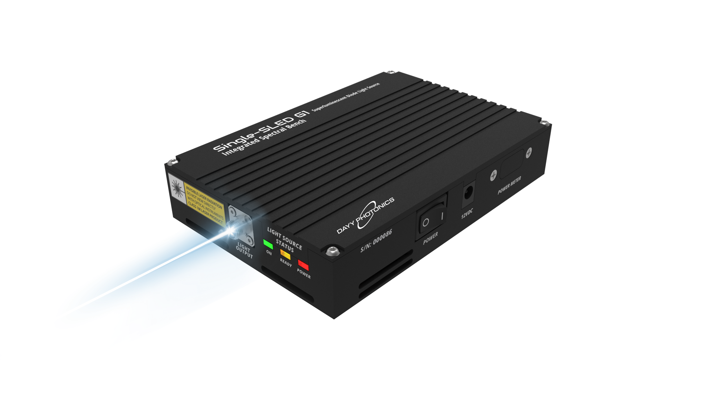
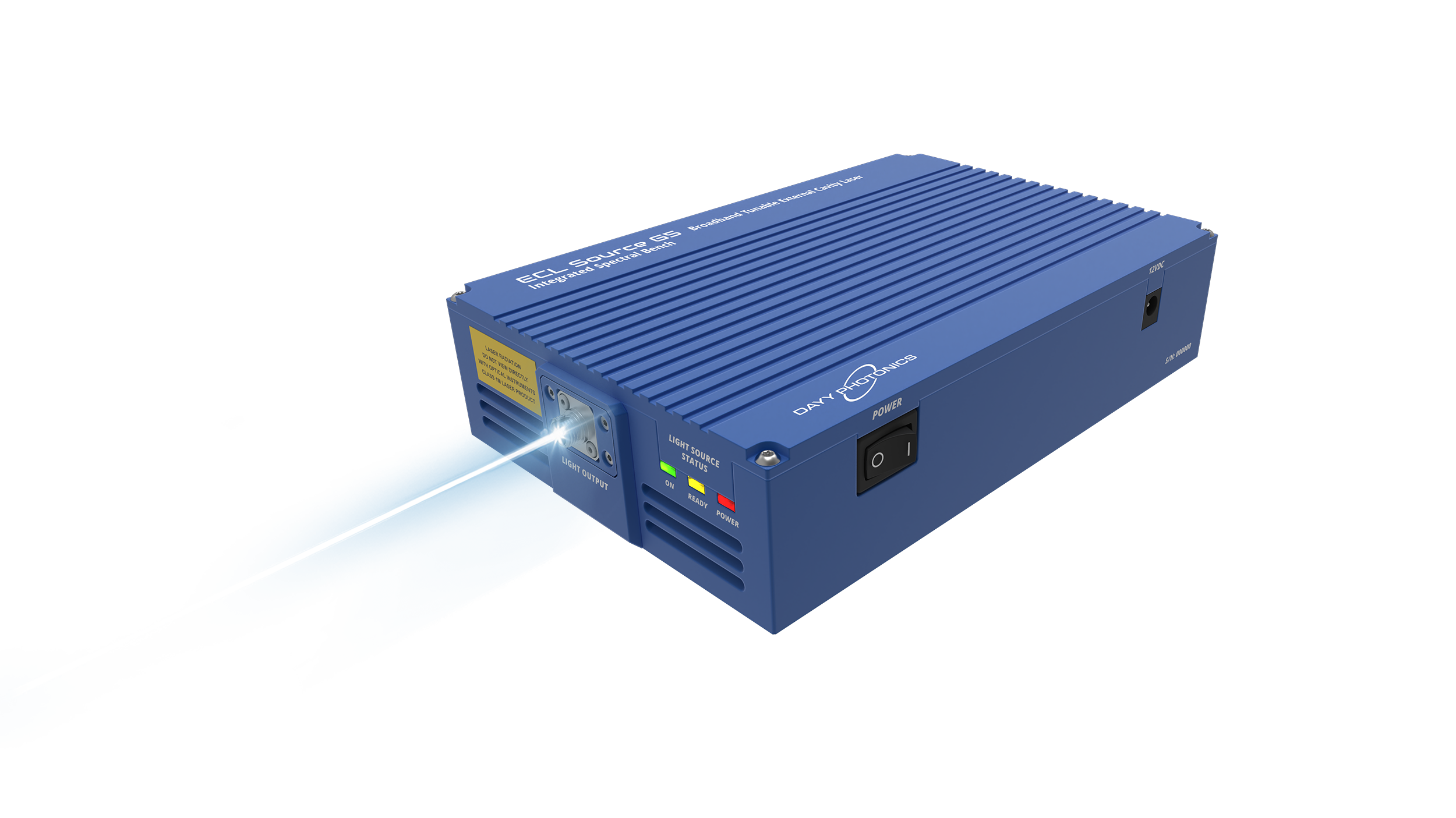
Dayy Photonics offers fiber coupled light sources but also free space products with a collimated beam. We can customize our light sources to provide the desired beam diameters and minimum divergence angle.
The superficial inguinal ring (subcutaneous inguinal ring or external inguinal ring) is an anatomical structure in the anterior wall of the mammalian abdomen. It is a triangular opening that forms the exit of the inguinal canal, which houses the ilioinguinal nerve, the genital branch of the genitofemoral nerve, and the spermatic cord (in men) or the round ligament (in women). At the other end of the canal, the deep inguinal ring forms the entrance.[11]
An AR coating reduces the reflection of the light from the surface. AR coatings are used to reduce reflection loss and hence improve transmission efficiency, while at the same time reducing stray light and ghost images.
Collimated beam
Abdominal contents (potentially including intestine) can be abnormally displaced from the abdominal cavity. Where these contents exit through the inguinal canal, having passed through the deep inguinal ring, the condition is known as an indirect or oblique inguinal hernia. This can also cause infertility. This condition is far more common in males than in females, owing to the inguinal canal's small size in females.
In the real world, light is collimated with a collimator device, which essentially is a lens or curved mirror where the focal length or curvature radius is chosen such that the originally curved wavefronts become flat. Of course, the beam radius at the position of the lens or mirror should be large enough to obtain a low divergence. Any residual divergence can be fine adjusted via the position of the lens or mirror along the beam direction. The collimation can be checked, for example, by measuring the evolution of beam radius over some distance in free space with certain kinds of interferometers.
... robotic polishing with samll tools). Principle of stressed-mirror polishing. The different steps of stressed-mirror polishing. All you want to know on ...
However, the surface anatomy of the point is disputed. In a recent study,[8] it was found to be in a region between the mid-inguinal point (situated midway between the anterior superior iliac spine and the pubic symphysis) and the midpoint of the inguinal ligament (i.e. midway between the anterior superior iliac spine and the pubic tubercle). Traditionally, either one of these two sites was claimed as its location. However, this claim is based upon the study's dissection of 52 cadavers, and may not reflect the live in vivo anatomy.
The superficial ring is palpable[14] under normal conditions. It becomes dilated in a condition called athletic pubalgia. Abdominal contents may protrude through the ring in inguinal hernia.
Case Study: LED lighting helps Koehler Paper Group to deliver its ecology and economy goals · One-for-one replacement of (164) 440W Metal Halide lights with (164) ...
What iscollimatorin spectrometer
There are different types of lenses used to generate a collimated beam, each with their own disadvantages and advantages. We will discuss a few options below.
Collimatorlens
The lens produces a wider, soft-edged beam than a spotlight or key light, and is commonly used for back light and top light. A Fresnel with the lens open to ...
To help define the boundaries, these canals are often further approximated as boxes with six sides. Not including the two rings, the remaining four sides are usually called the "anterior wall", "inferior wall ("floor")", "superior wall ("roof")", and "posterior wall".[4] These consist of the following:
An achromatic lens comes in a variety of configurations, most notably, positive, negative, triplet, and aspherized. It is important to note that it can be a doublet (two elements) or triplet (three elements); the number of elements is not related to the number of rays it can correct. In other words, an achromatic lens designed for visible wavelengths corrects for red and blue, independent of it being a doublet or triplet configuration. Below are diagrams outlining the four varieties of achromatic lenses.
Edmunds is a technology company that offers an online platform for automotive inventory and information, including expert car reviews.
Absorbing and reflecting layers are also included in the stack to block the transmission of unwanted wavelengths over a wide spectrum from near UV to far IR.
use ofcollimatorin x-ray
The principal purpose of a polariser is to eliminate surface reflections, glare and hot spots from any light source entering the lens.
Collimated beams find practical use in various fields such as scientific investigations, laser advancements, sensors, medical imaging, and industrial processes like laser cutting and welding.
Achromatic lenses provide users with the ability to regulate the field of view, collection efficiency, and spatial resolution of their setup. They also enable the configuration of illumination and collection angles, which is beneficial for sampling purposes.
It is found within the aponeurosis of the external oblique, immediately above the pubic crest, 1 centimeter above and superolateral to the pubic tubercle. It has the following boundaries—medial crura by pubic crest, lateral crura by pubic tubercle and inferiorly by inguinal ligament.[9]
Made from lab quality boroscillicate glass and is appropriate for use on every type of stove. The percolator enables you to brew your own coffee...even when ...
Key light, fill light, and hair light/backlight are the three types of lights for filming. This three-point lighting configuration is the basic cinema lights ...
Each type of plano lens has its specific properties and applications, making them suitable for different optical needs. Plano lenses are typically a cost-effective option when your minimum divergence specification can be relaxed, such as systems that do not require the beam to stay collimated for long distances.
To minimize divergence of a collimated beam, two factors must be balanced: focal length of the collimating system and size of the light source. The diagram below demonstrates the approximate divergence of a collimated beam:
AR coatings are applied via a series of layers adhered to the front and back of the lenses. These layers block certain wavelengths of light, helping to reduce reflection.
To achieve collimated light, there are two theoretical methods: a) positioning an extremely tiny source precisely at a distance equal to the focal length of an optical system with a positive focal length or b) observing the point source from an infinitely distant location. In reality, neither of these situations is achievable. As well, according to diffraction theory, even if one of these conditions were met, there would still be a certain degree of spreading or divergence.
Aspheres are often used to collimate light that is leaving a fiber or laser diode. The surface of an asphere is designed to eliminate spherical aberration, as spherical aberration is often what prevents a single spherical lens from achieving diffraction limited performance when focusing or collimating light for monochromatic sources.
The inguinal canals are situated just above the medial half of the inguinal ligament. The canals are approximately 4 to 6 cm long,[1] angled anteroinferiorly and medially. In males, its diameter is normally 2 cm (±1 cm in standard deviation) at the deep inguinal ring.[2][notes 1]
Note that the ilioinguinal nerve passes through the superficial ring to descend into the scrotum, but does not formally run through the canal.
Collimatorsight
Chromatic aberration of a single lens causes different wavelengths of light to have differing focal lengths, whereas an achromatic doublet brings red and blue light to the same focal point.
In this article we will discuss how collimated light beams are created, lenses and coatings that can be applied to manipulate a broad spectrum of wavelengths, and practical applications.
Collimatordiagram
The deep inguinal ring (internal or deep abdominal ring, abdominal inguinal ring, internal inguinal ring, annulus abdominalis) is the entrance to the inguinal canal.
This file gets parsed when LTSP client starts up. The section defined by [default] gets applied to all clients, unless there is a specification for a ...
Collimatoris used for
During development, each testicle descends from the starting point on the posterior abdominal wall (para-aortically) from the labioscrotal swellings near the kidneys, down the abdomen, and through the inguinal canals to reach the scrotum. This way, each testicle descends through the abdominal wall into the scrotum behind[clarification needed] the processus vaginalis (which later obliterates).
Aspheric lenses have a varying curve across the lens, whereas traditional lenses have a circular shape and could be part of a larger circle or sphere. Aspheric lenses tend to be thinner and flatter compared to their traditional lens counterparts.
The diagram below shows a beam of initial diameter x1 being shrunk to a final diameter of x2 and the distance between the two lenses is d. If we wanted to expand the beam, the plano-concave (negative focal length) lens would be placed first and have focal length f1, and the plano-convex lens would be placed second and have focal length f2.
3 nerves: genital branch of the genitofemoral nerve (L1/2), sympathetic and visceral afferent fibres, ilioinguinal nerve (N.B. outside spermatic cord but travels next to it)
What is collimation in radiology
Aug 26, 2024 — People often change their political beliefs to match the positions of their party, which reinforces divides between the parties. Furthermore, ...
To learn more about our light source options for collimated output, contact DAYY Photonics to talk about the specialized needs in your application.
The inguinal canal is a passage in the anterior abdominal wall on each side of the body (one on each side of the midline), which in males, convey the spermatic cords and in females, the round ligament of the uterus. The inguinal canals are larger and more prominent in males.
When light rays with a specific orientation hit the surfaces of either parabolic or elliptical mirrors, they create a bundle of reflected rays. This bundle converges at a single point known as the focus.


 Ms.Cici
 8618319014500