
Design of a high-precision, 0.5 m aperture Cassegrain ... - grey zone collimator
Human eyefield of viewin mm
by MT Sullivan · Cited by 9 — Risley prisms are wedged optics, usually used in pairs, to redirect optical beams. A typical Risley prism pair is shown in Figure 1. Figure 1.
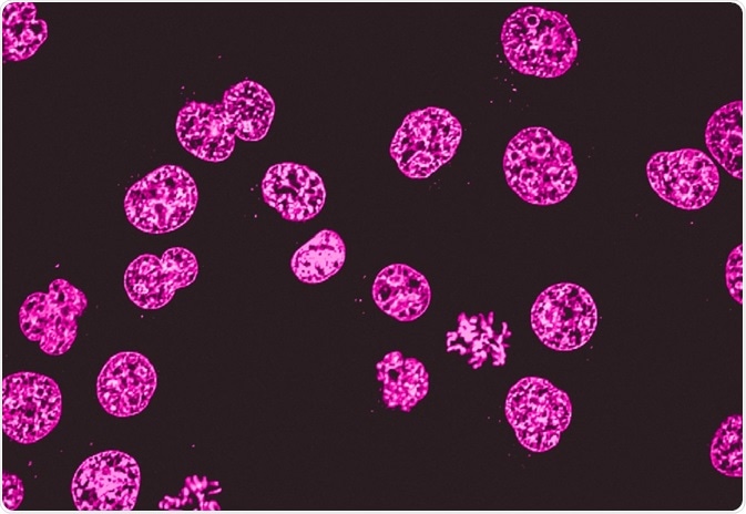
A common method to visualize cells or tissue with light microscopes is to use dyes. Widely used ones might paint the main components, such as the dye combination of hematoxylin and eosin, which colors the nuclei violet and the cytoplasm pink. However, there are also more specialized dye techniques.
Ryding, Sara. (2023, July 21). Fluorescence Microscopy vs. Light Microscopy. News-Medical. Retrieved on November 25, 2024 from https://www.news-medical.net/life-sciences/Fluorescence-Microscopy-vs-Light-Microscopy.aspx.
Light microscopy does much what the name implies: visible light and magnifying lenses are used to view small objects. Light microscopes are the oldest form of higher quality imaging devices, dating back to the 1500s, and were the microscopes with which first cells were observed.
The diameter of the field in an optical microscope is expressed by the field-of-view number, or simply the field number, which is the diameter of the view field in millimeters measured at the intermediate image plane.
Registered members can chat with Azthena, request quotations, download pdf's, brochures and subscribe to our related newsletter content.
Both fluorescence microscopy and light microscopy represent specific imaging techniques to visualize cells or cellular components, albeit with somewhat different capabilities and uses. At its core, fluorescence microscopy is a form of light microscopy that uses many extra features to improve its capabilities.
Human FOV in games
Achromatic lenses are designed for infinite conjugate ratios but are ideal for finite conjugate applications when used in pairs. In finite conjugate ...
Ryding, Sara. "Fluorescence Microscopy vs. Light Microscopy". News-Medical. 25 November 2024. .
In all modern eyepiece designs, the field lens is placed a sufficient distance away from the intermediate image plane to ensure that dust or other surface debris and defects on the lens surface are not visualized along with the specimen. The top lens in both eyepiece designs is referred to as the eye lens because it is closest to the eye of the observer.
What is the field of viewformula
Learn about the usage of process raman spectroscopy in the optimization of bioreactor monitoring and then improvement of cultivated meat production.
The usefulness of traditional light microscopy is hampered by the fact that it uses visible light, as using visible light limits the resolution obtained from samples. On the other hand, fluorescence microscopy is not faced with this limitation, since it uses whatever light excites the fluorophores.
The fluorophores are excited by the light in the microscope, which causes them to give off light with lower energy and of longer wavelength. It is this light that produces the magnified view, rather than the original light source. This means that fluorescent microscopy uses reflected rather than transmitted light.
Furthermore, there are light microscopy techniques that can image both live and fixed samples, but there can be a tradeoff between signal-to-noise ratio and sample damage during the observation process. During fluorescence microscopy, cells undergo bleaching, in which the fluorescence diminishes during extended periods of observation. To conclude, there is flexibility in both microscopy groups.
Mar 26, 2024 — This F Stop Chart shows full stops, 1/2 stops, and 1/3 stops when setting the aperture of digital camera lenses.
Field of viewcalculator
Sara is a passionate life sciences writer who specializes in zoology and ornithology. She is currently completing a Ph.D. at Deakin University in Australia which focuses on how the beaks of birds change with global warming.
Nikon offers a range of eyepiece options featuring magnification and field of view combinations tailored towards a variety of applications.
The Huygens eyepiece, illustrated on the left in Figure 1, is designed with the field lens positioned before the intermediate image plane, which coincides with the field diaphragm. Unlike the Ramsden eyepiece, the Huygens design has a focal point (at the intermediate image plane) residing between the eye and field lenses. In this case, the eye lens acts as a loupe to magnify the intermediate image.
Coin collectors use magnifying glasses to detect the wear of a coin, which helps determine its value. Stamp collectors use magnifying glasses to check the ...
Ryding, Sara. 2023. Fluorescence Microscopy vs. Light Microscopy. News-Medical, viewed 25 November 2024, https://www.news-medical.net/life-sciences/Fluorescence-Microscopy-vs-Light-Microscopy.aspx.
Traditional light microscopes are widely used, and often require simpler dyes to visualize contrast which is not naturally visible. This is typically a simpler technique than fluorescence microscopy. Because of this, it is used in clinical settings, such as for immediate imaging of biopsied samples in hospitals and for cervical smears.
Feb 27, 2024 — First, let's grasp the concept of neutral density filters. These accessories are essentially pieces of glass that reduce the amount of light ...
Field of viewhuman eye
Oct 19, 2022 — Microfiber cloth is the only eyeglass cleaning cloth accepted by lens manufacturers. It is a wipe used for mechanical cleaning, to remove dust, ...
We offer a wide range of coating capabilities for optical components including the ultraviolet (UV), visible and infrared (IR) spectra.
Sep 5, 2023 — How Was America Polarized? What Is Causing Affective Polarization? Interventions to Reduce Affective Polarization. Third Generation ...
While we only use edited and approved content for Azthena answers, it may on occasions provide incorrect responses. Please confirm any data provided with the related suppliers or authors. We do not provide medical advice, if you search for medical information you must always consult a medical professional before acting on any information provided.
In modern microscope eyepieces, the field diaphragm either precedes the optical system or is located between the lens element groups, as illustrated in Figure 1. This figure presents cutaway diagrams of a Ramsden and Huygens eyepiece showing ray traces through the field lens, eye lens, and field diaphragm. The Ramsden eyepiece (illustrated on the right in Figure 1) has an optical system consisting of two plano-convex lenses having a fixed separation distance according to their focal lengths. The first lens is termed the field lens because it is closer to the plane where the intermediate image is formed in the microscope. The field diaphragm is located between the tube opening and the field lens in the Ramsden eyepiece design.
In most cases, the eyepiece field diaphragm opening diameter determines the view field size. The field size in the specimen plane is then defined as the field number divided by the magnification of the objective:
What is the field of viewmicroscope
Fluorescence microscopy is often applied in imaging cell structures or structural features, checking the viability of cells, imaging genetic material (both DNA and RNA), and imaging particular cells in a larger population.
What is themaximum angleofvision for healthy human eye
Field of viewcamera
As light microscopy developed, more forms using different techniques were invented. One of the types of microscopy within the broader light microscopy group is fluorescence microscopy. Fluorescence microscopy images cells or molecules that have been tagged with a fluorescent dye. The fluorescent substances are called fluorophores, which are integrated into the sample.
Fluorescence microscopy can be used in conjunction with other types of light microscopy. Due to the fact that it creates images from the reflected light (rather than the direct light), it can be used with techniques such as phase contract microscopy.
Ijeoma Uchegbu discusses nanomedicine's role in improving medication adherence and developing non-addictive pain relief solutions at ELRIG Drug Discovery 2024.
Professor Nancy Ip discusses her groundbreaking neuroscience research, focusing on neurotrophic factors and innovative Alzheimer's disease treatment approaches.
Ryding, Sara. "Fluorescence Microscopy vs. Light Microscopy". News-Medical. https://www.news-medical.net/life-sciences/Fluorescence-Microscopy-vs-Light-Microscopy.aspx. (accessed November 25, 2024).
In hospitals, quick examination of cells can be critical for doctors. In such situations, light microscopy can be used with tissues that have been frozen in carbon dioxide and sectioned using a microtome. This simpler method can be used urgently on patients who are in the operating room to guide the surgeon.
For example, a commonly used label is green fluorescent protein (GFP), which is excited with blue light and emits green light with a longer wavelength. Filters around the sample can separate the fluorescent light from the surrounding radiation.
Diffraction is a wave property of light and given by θ=λ/D θ = λ / D . So for a given wavelength of light, the only way to reduce diffraction is ...
An Amici prism is an optical prism designed to turn light by a 90 degree angle while inverting the image. This non-dispersive prism is also known as an amici ...
Your questions, but not your email details will be shared with OpenAI and retained for 30 days in accordance with their privacy principles.
The size of the eyepiece field diaphragm opening is also dependent upon the correction for off-axial aberrations (coma, astigmatism, and lateral chromatic) of the objective. Recent eyepieces feature highly corrected glass that enables wide-field designs having field numbers of 26 millimeters and greater.
News-Medical.Net provides this medical information service in accordance with these terms and conditions. Please note that medical information found on this website is designed to support, not to replace the relationship between patient and physician/doctor and the medical advice they may provide.
The field number of typical eyepieces varies between 6 and 28 millimeters and (in general) decreases with the magnification of the eyepiece. For example, an eyepiece having a magnification of 10x typically has a field number ranging between 16 and 18 millimeters, while a lower magnification eyepiece (5x) has a field number of about 20 millimeters. Presented in Figure 2 is a comparison between the view fields available with similar eyepieces, one having a field number of 20 and the other a field number of 26. Note the greater range of specimen features visible through the eyepiece having the larger field number.
If an auxiliary lens is inserted between the objective and eyepiece, the magnification factor of this lens should also be employed in the equation by multiplication with the objective magnification (prior to the division operation). Although the field number is usually limited by the magnification and field diaphragm (stop) size of the eyepiece, there is clearly a limit that is also imposed by the design of the objective lens system. In early microscope objectives, the maximum usable field diameter tended to be about 18 millimeters or considerably less, but with modern plan apochromats and other specialized flat-field objectives, the maximum usable field can sometimes exceed 28 millimeters.
As mentioned, light microscopes that are used for light microscopy employ visible light to view the samples. This light is in the 400-700 nm range, whereas fluorescence microscopy uses light with much higher intensity.
Michael W. Davidson - National High Magnetic Field Laboratory, 1800 East Paul Dirac Dr., The Florida State University, Tallahassee, Florida, 32310.


 Ms.Cici
 8618319014500