
CSC-C-MOUNT-CS-MOUNTS-AND-IMAGE-SENSORS - c cs mount lens
Na of lensformula
Sudden vision loss in one eye may be a sign of Central Retinal Artery Occlusion (CRAO), commonly referred to as eye stroke. Like a stroke in the brain, it is a medical emergency and must be diagnosed and treated as quickly as possible to prevent irreversible loss of vision. Treatment must be administered within 12 hours, ideally, less than six, to prevent irreversible vision loss. Because timing is of the essence, the Department of Ophthalmology at New York Eye and Ear Infirmary of Mount Sinai (NYEE) has developed an eye stroke protocol, working with the Mount Sinai Stroke Center, that combines the expertise of ophthalmologists, neuroradiologists, neurologists, and emergency department faculty. Trained staff are available 24/7 to take images of the eye. The images are sent to one of NYEE’s retina specialists to make a rapid diagnosis. If an eye stroke is confirmed, the Mount Sinai Stroke Service begins treatment immediately to save the patient’s sight.
Numerical aperture formula
Anyone experiencing sudden severe blurring or complete loss of vision in one eye should go to the Emergency Room to get immediate medical treatment, even if symptoms seem to improve. Do not delay! Even temporary vision loss might indicate an increased risk of stroke or future vision loss. The Emergency Departments at several Mount Sinai hospitals are equipped to diagnose (or rule out) and treat eye stroke quickly: New York Eye and Ear Infirmary of Mount Sinai/Mount Sinai Beth Israel, The Mount Sinai Hospital, Mount Sinai West, and Mount Sinai Queens.
Numerical apertureofoptical fiber
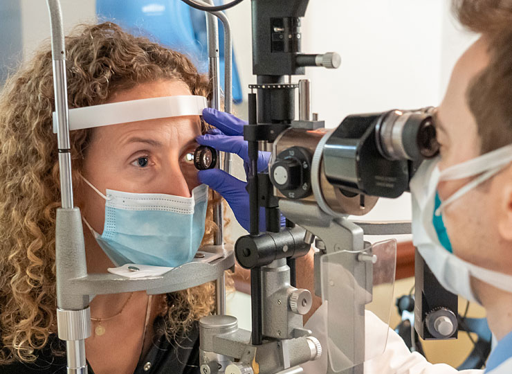
The goals of therapy are twofold: to identify a source of the occlusion and to unclog the blocked artery. When you present to the emergency department with painless vision loss, if a CRAO is suspected, you will rapidly have a series of diagnostic tests to make the diagnosis and identify other complications like a stroke. If you are eligible for treatment, we are well equipped to use the most cutting-edge techniques at Mount Sinai. Similar to stroke, a surgical procedure can be done to inject a medicine that breaks up the clot directly into the affected artery. After your initial diagnosis, you should plan on long-term follow-up to minimize your risk of stroke and prevent complications of CRAO.
NA of lenscalculator
The typical application in optogenetics is when the laser source is coupled to an NA 0.22 fiber-optic while the fiber-optic cannula of interest is made of a fiber-optic with NA 0.48. In this case, a magnification of x2.2 is well suited. In this example, with NA converter, the light going out of the output fibre will get twice of the input NA, i.e. 0.48 and fullfil the optical fibre NA.NA converter is based on same design as the Connectorized U-bracket, then can also accept filter insert with attenuating or spectral filters. As a matter of fact, any filter can be fitted to our standard filter holder and its code can be engraved on its body. In this way you can build your set using off the shelf and custom filters.The narrow band filters can be useful for filtering the fluorescence excitation spectrum or for the fluorescence light.
Surprise eye stroke diagnosis leaves patient eager to raise awareness of symptoms, and grateful for the care at Mount Sinai.
Laser sources are valued for the large amount of power they deliver. However, one of their characteristics is that the beams they produce have small divergences. This can be a limitation for those who require a powerful illumination over a wide angle. To address this issue, we have developed the NA converter, that modifies the geometry of an input fiber guided light beam. Both the numerical aperture and the beam diameter are affected : their product is a constant, so called Lagrange invariant.
First, it is essential to determine the cause of your vision loss. Most eye conditions can be treated, but the amount of vision that can be saved may depend on the time to treatment. Eye strokes must be treated within hours of vision loss. Furthermore, painless vision loss may be a sign of a stroke in the brain or put you at risk of having a stroke in the brain.
A blockage in a small artery that supplies blood to the retina is referred to as a retinal artery occlusion. When the main artery to the retina is blocked, it is called a central retinal artery occlusion. Just like a stroke, a CRAO is typically caused by an embolus, a piece of a blood clot that breaks off of a larger plaque in a large blood vessel or the heart. When this happens, the retina is deprived of its oxygen supply, also just like a stroke. The retinal nerve cells that allow you to see can quickly die unless blood flow is restored. Retinal artery occlusions are more common in people with compromised blood vessels from conditions like high blood pressure, diabetes, atherosclerosis, and high cholesterol, and blood disorders that affect clotting.


 Ms.Cici
 8618319014500