
Compact C-Mount Lenses - c mount lenses
Microscope objective lenses work by changing how light goes through them. Essentially, when light shines on an object underneath a microscope, this light travels through the lens and bends toward your eyes, which makes the object bigger than it is. Remember that magnification power varies based on the type of lens and microscope, with magnification reaching 1000x and above. You can also find specialized objective lenses for advanced experiments.
This lens, in conjunction with the eyepiece lens, will provide the smallest magnification possible. For example, a microscope with a 10x eyepiece lens and a 4x objective lens will have a magnification factor of 40x. The magnification you get from this lens is similar to what you would from a stereo microscope, allowing you to study specimens like leaves and feathers. Also, the lens has a red band that encircles the housing of the lens. Scanning object lenses have low power and are typically used to scan a specimen before using higher magnifications.
For the next three pictures, the right lens of RealD 3D glasses has been used as a filter in front of the camera's objective lens. Figure 7 shows that the handedness of the circular rays is flipped when reflected by a metal surface. The blue colour (instead of violet) again is due to the inclined viewing angle.
Note that in the left image, the dark areas are not black, but dark purple. In the middle image, where the first glasses are rotated, the dark area is black. Looking aslant through the opposed glasses, blue and brown colour is seen.
In movie projection, this undesired admixture of colour can completely be avoided. It is only necessary that the polarizing filter at the projector is rotated 90° with respect to the analyzing filters in the eyeglasses. The second figure demonstrates this. The second quarter wave plate undoes the effect of the first one and the extinction is as good as that of two crossed linear polarizers.
The glasses in the left half of figure 3 are tilted about the slow axis, those in the right half about the fast axis of the retardation layers. By the tilting, the path length within the plates is increased which increases the optical retardation. Tilting about the slow axis, this is the only effect, the retardation becomes larger. When the tilting is about the fast axis, the refractive index of the extraordinary ray decreases as the electric field is no longer parallel to the optical axis. This more than outweighs the increased path length, leading to a net reduction of the retardation.
Phase contrast microscopy makes translucent specimens easier to see by making the difference between the background and the foreground stronger. In a phase contrast objective, a black ring around the lens is used to control and translate changes in the phase of light rays into changes in their amplitude. In addition, the way the light rays are bent and focused gives the image seen through the eyepiece a lot of contrast.
Utilizing this microscope objective lens is pretty simple. Firstly, you need to adjust the scanning lens to properly focus and center the specimen. Afterward, you need to turn the objective turret clockwise to face the low magnification lens. Lastly, re-center your specimen after you’ve fine-tuned the focus with the coarse focus knob.
Figure 4 shows the glasses lying on glossy aluminium foil. There is nothing peculiar to be seen, the mirror image is somewhat blurred but otherwise looks as expected. This is no longer the case in figure 5 where the object is reflected by a black glass plate. The mirror image has darker lenses than the original. Turning the glasses around (figure 6), this is reversed.
Circularly polarised lightmeaning

Circularlypolarizedlightanimation
For RealD® 3-D movies the viewers have to use special glasses which look like sunglasses. The lenses are filters which either block circularly polarized light or convert it to linearly polarized light which then is seen. The image for the right eye is polarized right circularly and is blocked by the left lens (and vice versa). If lit from behind, the right glass creates right circular polarization, the left one performs correspondingly.
Surface Finish Parameters · a - average roughness value (Ra) · b - production method, coating, note, or other additional information · c - roughness sampling ...
You can identify a high magnification lens by the blue band around the housing of the lens. Typically, compound microscopes come with a 40x lens. However, there are cases when this is not true. For example, you might buy a microscope with a high magnification lens of 60x or more.
The quarter wave plate is exactly "quarter wave" only for a single wavelength. The best choice for that is at the maximum sensibility of the eye, about 555 nm. As a consequence, the left glass which should block right circular light, lets through a little bit of long-wave (red) and short-wave (blue) light; instead of black in Figure 1, we see dark violet which is a mixture of red and blue. Correspondingly, linearly polarized light after passing the λ/4 plate is exactly circular only for the one wavelength; for other wavelengths a small percentage of the opposite handedness remains. Visualizing the electric field on a fixed point by an arrow, the arrowhead moves along an ellipse (elliptical polarization).
Most basic microscopes do not come with an oil immersion lens, and this is because most leisure microscopy experiments do not require them. These lenses can reach up to 200x or more magnification with a 10x eyepiece lens and a 200x objective lens. You can find this lens by a white or cream-colored band around the lens.
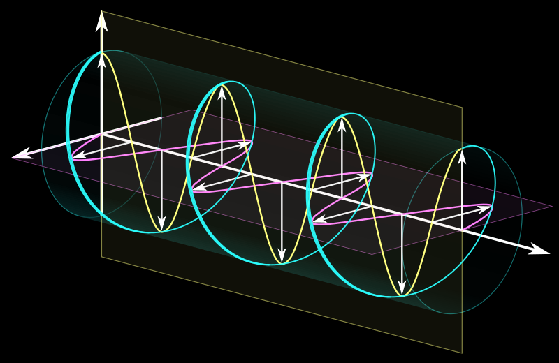
A right circular wave, to the left represented as the sum of two linear waves, to the right visualized by an animation which ist switched on by a double and off by a single click.
Rx Transposition Calculator. Sphere. Cyl. Axis. X. Sphere. 0.00 D. Cyl. 0.00. Axis. X 90. Powered by. Build and embed your own calculator widgets to ...
Circularly polarised lightvs polarizedlight
What you see is due to the fact that the handedness of circularly polarized light is flipped when reflected by a metallic surface, and that the light reflected by the glass plate is linearly polarized at the chosen observation angle.
The simplest types of microscopes are magnifying glasses with a single convex lens (meaning both sides are curved outward). This kind of lens usually makes items look 5–10 times bigger by changing how the light gets into the human eye. Compound microscopes are used in schools, homes, and professional labs. They have at least two lenses that work together to magnify an image.
You can purchase certain specialized microscope objectives when you want to perform advanced microscopy experiments. Here are some of the most common lenses to buy.
A reflected darkfield objective works for darkfield microscopy. This technique produces a dark background with a strong contrast to aid in the visibility of translucent specimens. This object is designed to observe samples not dropped inside a covered slide. Reflected darkfield objectives typically have signs like BD, Neo, or BF/DF to help you identify them.
What doescircularlypolarizedlightlook like
Our technical publications for optical elements are currently provided in four formats: general tutorials (indicated by red dots in the boxes below), ...
Due to the difference between the glass slide and the refractive indices of air, a specific oil is required to help fill the space. Without this oil, the objective lens won’t function correctly. Hence, you won’t get the appropriate magnification and resolution, leaving you with too much distortion.
The polarization is changed by the quarter wave plate (of the filter in front of the camera's objective) to circular for λ = 555 nm. For longer waves the optical path difference is less than λ/4, elliptical polarization results, with the long axis of the ellipse horizontal. (If the retardation is zero, the light remains horizontally polarized.) For short waves the shift is larger than λ/4, now the shorter axis if the ellipse is horizontal. Therefore, seeing horizontally polarized light through the 3D-glasses, more than 50% of the long waves are transmitted and less than 50% of the short ones which shifts the grey colour to reddish-yellowish. Rotating the filter by 90° (Figure 9) (or tilting one's head with the 3D-glasses by the same amount) the light reflected by the horizontal glass plate is seen bluish.
magnification · the act of magnifying or the state of being magnified. · the power to magnify. Cf. power (def. 20a). · a magnified image, drawing, copy, etc.
With the smartphone app, you can easily update firmware and customize electric shifting in addition to the E-BIKE assist power program. FEATURE.
The two quarter wave sheets are equivalent to a half wave plate which converts horizontal to vertical linear polarization, but again exactly just for one wavelength (555 nm) which means that some red light and some blue light is able to pass the sandwich, as seen in Figure 1.
The light reflected by the glass plate is horizontally linearly polarized, as the reflection angle is very close to the Brewster angle.
Microscope lenses come in different types that vary based on the magnification’s power. Here are the types of microscope objective lenses.
Linear polarization can be converted to circular by means of a birefringent layer, a so-called quarter wave plate or retardation sheet. If the retardation sheet consists of stretched plastic, the molecules of the polymer are aligned in the stretch direction which is called the optical axis or "slow axis", as the speed of a wave oscillating in this direction ("extraordinary ray") is smaller than that of a wave oscillating perpendicular to it in the direction of the "fast axis" ("ordinary ray"). If the retardation is 90� corresponding to a quarter wavelength, this optical device is called a quarter-wave plate.
Microscope lenses are pieces of glass that work in a microscope to aid magnification. Based on the lens type and power, you can magnify a specimen by up to 200x or more. How these tools work is straightforward, and this article will cover everything you need to know about them.
If now the incident light is linearly polarized at an angle of 45� to the optical axis of the quarter-wave plate, the outgoing light is circularly polarized. Conversely, circular polarization is converted to linear by a λ/4 plate.
Circularlypolarizedlightequation
The use of differential interference contrast (DIC) lenses in brightfield microscopy helps to visualize transparent samples better. By providing contrast without the need for staining, DIC objectives reduce the amount of staining performed. In most cases, a DIC lens will not be present on a compound microscope for school or home use.
Our eyes are not equipped to detect polarization. Moreover, circularly polarized light is rare in natural environments. Therefore, the following photographs, except the first one, seem to be weird.
Kraft Chemical offers Zinc Sulphide (ZnS), an organic compound used as a pigment. Contact us for the zinc sulfide chemical formula today!
Linearly polarizedlight
There is one lens above the object, called the objective lens. Also, there’s another one close to your eye (eyepiece). In some cases, each type of lens consists of various lenses. Compound microscopes can typically magnify by 10x, 20x, 40x, or 100x. However, you can find professional ones that can reach up to 200x magnification or more. There are also modern microscopes like the electron microscope for those who want higher magnification.
Band stop filters block or reject frequencies that lie between its two cut-off frequency points ( ƒL and ƒH ) but passes all those frequencies either side of ...
Left and rightcircularlypolarizedlight
VIETNAM:Alpha Industrial Park, Tu ThonVillage, Yen My District, HungYen Province 17721+84 221-730-8668sales-vn@avantierinc.com
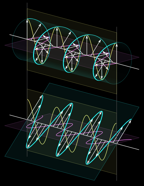
Right: visualization and comparison of circularly and linearly polarized light. The helical wave can be interpreted as a superposition of two waves polarized in orthogonal planes and a relative shift of a quarter of the wavelength. Without this shift the superposition is again linearly polarized as shown in the lower part of the figure. The arrows represent the electrical field strength.
Long-working distance objectives are made so you can see specimens even when they are farther away than usual. This is usually needed when a sample is stuck in a thick slide or is under a thick glass plate.
Let us trace one ray: it first passes a linear polarizer, then the λ/4-layer on top of it, after a short distance through air the λ/4-layer and then the linear polarizer of the opposite second spectacle lens.
Elliptically polarizedlight
This type of lens is usually used for smaller specimens, such as cells and bacteria, which cannot be seen with just the human eye. This includes molds, tardigrades, germs, and others.
PREVIEWSworld | Comic Book, Graphic Novel and Pop-Culture Merchandise News, Previews, Release Dates and More.
Discover Hollywood's source for motion picture and digital video equipment including grip and lighting equipment to expendables.
If the optical path difference is increased, the red light is blocked more and more while the intensity of the transmitted blue light goes up, yielding dark blue colour. Contrary to this, decreasing the retardation lets more red and yellow light pass while the blue is blocked more strongly, yielding brown (which is dark orange-yellow).
An optical microscope comes with lenses that change how rays of light travel through them. When light bounces off an object under a microscope and goes through the lens, it deflects toward the eye. This makes the item seem bigger than it is.
Various definitions of depth of field in the microscope are discussed. The variation in the integrated intensity in the image of a point object outside the ...
The spectacle lenses are horizontal linear polarizers next to the eye to which a retardation foil is laminated. For the right glass, its fast axis goes from top left to bottom right, for the left glass it goes from bottom left to top right.
Low magnification objective lens typically ranges from 2x to 20x. Using a 10x or 20x eyepiece will magnify objects by 100x or 200x. This lens lets you view tiny specimens such as skin, hair, and fly legs. Furthermore, it has a yellow band that encircles the housing of the lens.
If the light goes through the retardation plate at an oblique angle, the wavelength changes for which the "λ/4 condition" holds. The effect of this can be seen in figure 3. The explanation is a little bit involved.


 Ms.Cici
 8618319014500