
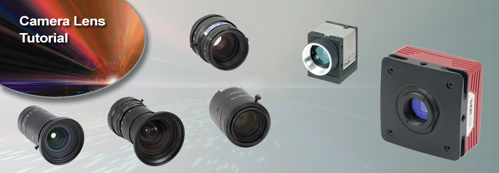
Color Slide - The Hardest Game on the App Store 4+ - color slide
The focal length (FL) is roughly defined as the distance from principal plane to the focal plane. For a camera lens, the focal length determines the field of view of the camera system; the longer the focal length, the smaller the field of view. As a general guideline, a 50 mm focal length lens and 35 mm format camera combination produces roughly the same field of view as the human eye (~53° diagonal). The table below lists the focal lengths needed to achieve the same field of view as the human eye for different sensor formats.
Specifically, f-number is defined as:where f/# is the f-number, f is the focal length and d is the entrance pupil diameter.
Nikonlens camera diagram
Diaphragm or Iris: Many microscopes have a rotating disk under the stage. This diaphragm has different sized holes and is used to vary the intensity and size of the cone of light that is projected upward into the slide. There is no set rule regarding which setting to use for a particular power. Rather, the setting is a function of the transparency of the specimen, the degree of contrast you desire and the particular objective lens in use.
How to Focus Your Microscope: The proper way to focus a microscope is to start with the lowest power objective lens first and while looking from the side, crank the lens down as close to the specimen as possible without touching it. Now, look through the eyepiece lens and focus upward only until the image is sharp. If you can't get it in focus, repeat the process again. Once the image is sharp with the low power lens, you should be able to simply click in the next power lens and do minor adjustments with the focus knob. If your microscope has a fine focus adjustment, turning it a bit should be all that's necessary. Continue with subsequent objective lenses and fine focus each time.
Note that Thorlabs' camera lenses are designed to correct for optical aberrations witin a specified range of object distances. The modification of the specified minimum object distance with an extension tube may reduce the ability of these lenses to eliminate aberrations.
Lens camera diagramexplained
Historians credit the invention of the compound microscope to the Dutch spectacle maker, Zacharias Janssen, around the year 1590 (more history here). The compound microscope uses lenses and light to enlarge the image and is also called an optical or light microscope (versus an electron microscope). The simplest optical microscope is the magnifying glass and is good to about ten times (10x) magnification.
Illumination: Compound microscopes often have built-in illumination systems, such as a substage light source, condenser, and diaphragm, to provide transmitted light through the specimen. Other microscopes, like dissecting or fluorescence microscopes, may utilize different lighting techniques or illumination configurations.
Simplecamera lens diagram
Condenser Lens: The purpose of the condenser lens is to focus the light onto the specimen. Condenser lenses are most useful at the highest powers (400x and above). Microscopes with in-stage condenser lenses render a sharper image than those with no lens (at 400x). If your microscope has a maximum power of 400x, you will get the maximum benefit by using a condenser lenses rated at 0.65 NA or greater. 0.65 NA condenser lenses may be mounted in the stage and work quite well. A big advantage to a stage mounted lens is that there is one less focusing item to deal with. If you go to 1000x then you should have a condenser lens with an N.A. of 1.25 or greater. All of our 1000x microscopes use 1.25 Abbe condenser lens systems. The Abbe condenser lens can be moved up and down. It is set very close to the slide at 1000x and moved further away at the lower powers.
Camera lenses that can collect a lot of light (i.e., a low f-number) are known as fast lenses as they can be used with shorter exposure times and are ideal for low-light conditions. For example, a 50 mm focal length lens with a f/1.4 aperture has a bigger aperture and is therefore faster than a lens at the same focal length with a f/2.5 aperture. While using larger apertures increases light collection, doing so reduces the axial in-focus region of the image, known as the depth of field. To illustrate the effect of different aperture sizes visually, the table below shows a sequence of images taken with the same lens (MVL12M43 on a DCU224C 1/2" format camera) for increasing f-numbers. Because the images were taken at constant exposure, for each f/# increase (by a factor of ~1.4) the amount of light collected by the lens is reduced by half.
Types ofcameralenses pdf
Revolving Nosepiece or Turret: This is the part of the microscope that holds two or more objective lenses and can be rotated to easily change power.
An image that is cropped appears as if it was taken with a lens of higher focal length (i.e. a smaller field of view), but does not magnify the image. The cropping effect can be quantified using an adjusted focal length (defined as the crop factor multiplied by the lens focal length). For example, an image taken using a 1" format, 50 mm focal length lens with a 1/2" format sensor will produce an image with an adjusted focal length of 100 mm. While the field of view is reduced as if using a 100 mm lens, objects in the image will remain at the same size. The table to the right lists all of the lenses offered on this page with the adjusted focal length for different sensor formats.
Eyepiece/Ocular: Compound microscopes commonly have a pair of eyepieces that provide binocular vision. Other microscopes may have a single eyepiece or sometimes no eyepieces at all.
The images below illustrate this effect visually using two images taken using the same lens with 1/2" and 1/3" format cameras. The image taken using the smaller 1/3" format camera produces an image that is cropped compared to the image taken using the 1/2" format camera. Note, however, that the objects in both images remain at the same magnification.
Thorlabs' Camera Lenses for Machine Vision each have a characteristic range of object distances. Objects placed within this range can be brought to a sharp focus on the sensor of the C-Mount camera. As seen in the image sequence below, the addition of an C-mount extension tube or spacer between the camera and the lens changes this range of object distances and allows the system to focus on objects closer to the lens. This increases the magnification of the image on the camera while decreasing the depth of field. The following table lists the range of possible object distances for various combinations of Thorlabs' C-Mount extension tubes and machine vision lenses.
Lens camera diagramand functions
Objective Lenses: Usually you will find 3 or 4 objective lenses on a microscope. They almost always consist of 4x, 10x, 40x and 100x powers. When coupled with a 10x (most common) eyepiece lens, total magnification is 40x (4x times 10x), 100x , 400x and 1000x. To have good resolution at 1000x, you will need a relatively sophisticated microscope with an Abbe condenser. An Abbe condenser is composed of two lenses that control the light that passes through the specimen before entering the objective lens on the microscope. The shortest lens is the lowest power, the longest one is the lens with the greatest power. Lenses are color coded and if built to DIN standards are interchangeable between microscopes. "DIN" is an abbreviation of "Deutsche Industrial Normen". This is a German standard that has been adopted internationally as an optical standard used in most quality microscopes. A typical DIN standard microscope objective lens has a 0.7965" (20.1mm) diameter threads, 36 TPI (threads per inch), and a 55º Whitworth. Many high power objective lenses are retractable (i.e. 40XR). This means that if they hit a slide, the end of the lens will push in (spring loaded) thereby protecting the lens and the slide. All good quality microscopes have achromatic, parcentered, parfocal lenses.
Camera Lens diagramphysics
To illustrate this, the sequence of three images to the right were taken with the same camera with three different lenses. As focal length of the lens increases, magnification of the objects in the photos increases while the field of view decreases. The items in the image are each roughly spaced in 10" (254 mm) increments in the following order: Polaris™ Fixed Monolithic Mirror Mount (10" from camera), Ø1/2" post with KM100 mirror mount (20" from camera), and post-mounted RSP1 rotation mount (30" from camera). The MVL4WA used to shoot the first image is a wide angle lens which clearly distorts the door frame on the left edge of the image.
The aperture of the lens controls the amount of light that a lens can collect; the more light a lens collects, the brighter the image. Because of this, the aperture size affects the exposure time and therefore the speed of the camera. Thorlabs provides the maximum aperture size in the tables below for each lens in terms of the f-number, which is expressed using the symbol f/# (e.g., f/1.4). As the f-number increases, the aperture opening becomes smaller and less light is collected by the lens.
CroppingWhen the lens format is larger than the camera format, the effect on the resultant image is known as cropping. In this case, a full image is produced but at a smaller size (i.e. cropped) because the sensor is only capturing a fraction of the complete image. A crop factor or focal length multiplier quantifies the amount of cropping and is defined as the ratio of the diagonal length of the lens' design format divided by the diagonal length of the sensor format. The crop factor for all possible 1/3", 1/2.9", 1/2", 1/1.8", 2/3", 1", and 4/3" format lens/sensor combinations are shown in the table to the right.
It's important to note that the term "other microscope parts" is quite broad and can include various microscope types with different designs and features. The above differences are generalized and may not apply to every microscope outside the category of compound microscopes.
Objective Lenses: Compound microscopes have multiple objective lenses mounted on a rotating nosepiece, typically with magnifications ranging from 4x to 100x or higher. Other microscopes, such as dissecting or stereo microscopes, usually have fixed magnification lenses.
Magnification: Compound microscopes are designed for higher magnifications, typically used for observing microscopic details. Other microscopes may have lower magnification capabilities, suitable for larger specimens or samples.
Applications: Compound microscopes are commonly used in fields such as biology, medicine, and research, where detailed examination of small structures is required. Other microscopes, such as stereo microscopes, are utilized for examining larger objects or conducting dissections. Electron microscopes are used for high-resolution imaging of nanoscale structures.
Parts of alens camera
VignettingVignetting occurs when the lens format is smaller than the camera format. When this occurs, the area of the sensor is incompletely exposed, causing a dark ring to appear around the borders of the image. The vignetting effect is illustrated in the two images below, which were both captured using the same 4/3" format camera. In the image to the left, using a 12 mm focal length, 4/3" format lens produces a full image with slight dimming around the edges. This minor example of vignetting is due to the lens design which has decreased transmission at the edge of the lens. On the other hand, a 2/3" format lens at the same focal length produces a prominent dark ring around the photo edge. As the latter example is very visually apparent, we do not recommend using lenses with smaller formats than the camera sensor for imaging.
1. Ocular eyepiece lens to look through. 2. Objective lens, closest to the object. Before purchasing or using a compound microscope, it is important to know the functions of each part. This information is presented below. Links will take you to additional information and images.
Rack Stop: This is an adjustment that determines how close the objective lens can get to the slide. It is set at the factory and keeps students from cranking the high power objective lens down into the slide and breaking things. You would only need to adjust this if you were using very thin slides and you weren't able to focus on the specimen at high power. (Tip: If you are using thin slides and can't focus, rather than adjust the rack stop, place a clear glass slide under the original slide to raise it a bit higher).
Sample Size and Depth of Field: Compound microscopes are designed to observe thin, transparent specimens placed on glass slides. They offer a narrow depth of field, allowing clear focus on one plane at a time. Other microscopes, like stereo or electron microscopes, can accommodate larger specimens or samples with more depth, providing a wider depth of field.
Compound microscopes and other types of microscopes differ in their design and functionality. Here are the key differences between compound microscope parts and those of other microscopes:
There are three general classifications for lenses related to the image field of view. A lens with a focal length close to the diagonal length of the sensor format produces an image with a near-human field of view and is considered a "normal" lens for that sensor format. A wide-angle lens has a focal length shorter than normal, which produces a wider field of view but has a tendency to exhibit barrel distortion effects towards the edge of the image. Finally, a lens with a focal length longer than normal is known as a telephoto lens, which has a smaller field of view and a greater magnification of objects in the image.
Lens camera diagrampdf
Illuminator: A steady light source (110 volts) used in place of a mirror. If your microscope has a mirror, it is used to reflect light from an external light source up through the bottom of the stage.
Stage with Stage Clips: The flat platform where you place your slides. Stage clips hold the slides in place. If your microscope has a mechanical stage, you will be able to move the slide around by turning two knobs. One moves it left and right, the other moves it up and down.
Modern cameras that use CCD or CMOS sensors are specified for a camera sensor format, and similarly, lenses are designed to provide optimal imaging for a specific camera format. This format designation (e.g., 1/2", 2/3", 4/3") is a hold-over convention from when video was recorded using cathode-ray tubes and refers to the outer diameter of the video tube required for a given image size. The diagram to the right illustrates the size difference between several standard camera formats. In the ideal imaging system, a camera and lens would be designed for the same format, however, it is also possible to use camera/lens combinations with different formats. Doing this will have an effect, either vignetting or cropping, on the resulting image.


 Ms.Cici
 8618319014500