
Collimated Light's Non-Zero Beam Divergence - laser beam divergence
Our products are to be used for Research Use Only. They may not be used for any other purpose, including, but not limited to, use in humans, therapeutic or diagnostic use, or commercial use of any kind. Our products may not be transferred to third parties, resold, modified for resale, or used to manufacture commercial products or to provide a service to third parties without our prior written approval.
Designed for low-magnification, macro fluorescence observation, this semi-apochromat objective offers a long working distance, a high NA, and high transmission of 340 nm wavelength light.
Our products are to be used for Research Use Only. They may not be used for any other purpose, including, but not limited to, use in humans, therapeutic or diagnostic use, or commercial use of any kind. Our products may not be transferred to third parties, resold, modified for resale, or used to manufacture commercial products or to provide a service to third parties without our prior written approval.
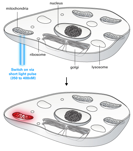
Living Colors PAmCherry is a photoactivatable fluorescent protein (PAFP) derived from the red fluorescent protein mCherry. PAmCherry is non-fluorescent until it is exposed to 350- to 400-nm light. By selecting which cells or cellular regions to activate, you can track cells, organelles, or proteins of interest against a dark background using this photoactivatable fluorescent protein.
The mCherry monoclonal antibody detects PAmCherry-N1 in the lysate of mammalian cells. HEK 293 cells were transiently transfected with pPAmCherry-N1. Cell lysates (corresponding to 30,000 cells) were prepared from HEK 293 cells transiently expressing PAmCherry-N1 (Lane 1) or a negative control (untransfected cells; Lane 2). Both lysates and a positive control (5 ng recombinant mCherry; Lane 3) were separated by SDS-PAGE and analyzed by Western blot using the mCherry monoclonal antibody at the recommended dilution of 1:1000.
These extended apochromat objectives offer high NA, wide homogenous image flatness, 400 nm to 1000 nm chromatic aberration compensation, and the ability to observe phase contrast. Use them to observe transparent and colorless specimens such as live cells, biological tissues, and microorganisms.
pPAmCherry-C1 Vector is a mammalian expression vector encoding PAmCherry, a photoactivatable mutant of the fluorescent protein mCherry. PAmCherry is non-fluorescent until photoactivated by a short exposure to light at a wavelength between 350 nm and 400 nm. The excitation/emission wavelengths of photoactivated PAmCherry are 564 nm and 595 nm. Genes cloned into the MCS will be expressed as fusions to the C-terminus of PAmCherry if they are in the same reading frame as PAmCherry and there are no intervening stop codons.
To clean a microscope objective lens, first remove the objective lens and place it on a flat surface with the front lens facing up. Use a blower to remove any particles without touching the lens. Then fold a piece of lens paper into a narrow triangular shape. Moisten the pointed end of the paper with small amount of lens cleaner and place it on the lens. Wipe the lens in a spiral cleaning motion starting from the lens’ center to the edge. Check your work for any remaining residue with an eyepiece or loupe. If needed, repeat this wiping process with a new lens paper until the lens is clean. Important: never wipe a dry lens, and avoid using abrasive or lint cloths and facial or lab tissues. Doing so can scratch the lens surface. Find more tips on objective lens cleaning in our blog post, 6 Tips to Properly Clean Immersion Oil off Your Objectives.
The ocular lens is located at the top of the eyepiece tube where you position your eye during observation, while the objective lens is located closer to the sample. The ocular lens generally has a low magnification but works in combination with the objective lens to achieve greater magnification power. It magnifies the magnified image already captured by the objective lens. While the ocular lens focuses purely on magnification, the objective lens performs other functions, such as controlling the overall quality and clarity of the microscope image.
The mCherry monoclonal antibody detects PAmCherry-N1 in the lysate of mammalian cells. HEK 293 cells were transiently transfected with pPAmCherry-N1. Cell lysates (corresponding to 30,000 cells) were prepared from HEK 293 cells transiently expressing PAmCherry-N1 (Lane 1) or a negative control (untransfected cells; Lane 2). Both lysates and a positive control (5 ng recombinant mCherry; Lane 3) were separated by SDS-PAGE and analyzed by Western blot using the mCherry monoclonal antibody at the recommended dilution of 1:1000.
Please see the product's Certificate of Analysis for information about storage conditions, product components, and technical specifications. Please see the Kit Components List to determine kit components. Certificates of Analysis and Kit Components Lists are located under the Documents tab.
Optimized for polarized light microscopy, these semi-apochromat objectives provide flat images with high transmission up to the near-infrared region of the spectrum. They are designed to minimize internal strain to meet the requirements of polarization, Nomarski DIC, brightfield, and fluorescence applications.
Our products are to be used for Research Use Only. They may not be used for any other purpose, including, but not limited to, use in humans, therapeutic or diagnostic use, or commercial use of any kind. Our products may not be transferred to third parties, resold, modified for resale, or used to manufacture commercial products or to provide a service to third parties without our prior written approval.
Takara Bio USA, Inc. provides kits, reagents, instruments, and services that help researchers explore questions about gene discovery, regulation, and function. As a member of the Takara Bio Group, Takara Bio USA is part of a company that holds a leadership position in the global market and is committed to improving the human condition through biotechnology. Our mission is to develop high-quality innovative tools and services to accelerate discovery.
pPAmCherry-Mito Vector is a mammalian expression vector encoding PAmCherry, a photoactivatable mutant of the fluorescent protein mCherry, fused to the mitochondrial targeting sequence derived from the precursor subunit VIII of human cytochrome C oxidase. PAmCherry is non-fluorescence until photoactivated by a short exposure to light at a wavelength between 350 nm and 400 nm. The excitation/emission wavelengths of photoactivated PAmCherry are 564 nm and 595 nm.
Our products are to be used for Research Use Only. They may not be used for any other purpose, including, but not limited to, use in humans, therapeutic or diagnostic use, or commercial use of any kind. Our products may not be transferred to third parties, resold, modified for resale, or used to manufacture commercial products or to provide a service to third parties without our prior written approval.
Designed for clinical research and routine examination work in the laboratory, these achromat objectives provide the level of field flatness required for fluorescence, darkfield, and brightfield observation in transmitted light.
PAmCherry subcellular localization vectors are available that target PAmCherry to the cell membrane, mitochondria, actin, or tubulin, respectively. Plasmid and lentiviral N- and C-terminal vectors are also available to create your own PAmCherry-tagged protein of interest.
This super-corrected apochromat objective corrects a broad range of color aberrations to provide images that capture fluorescence in the proper location. Delivering a high degree of correction for lateral and axial chromatic aberration in 2D and 3D images, it offers reliability and accuracy for colocalization analysis.
This semi-apochromat objective series provides flat images and high transmission up to the near-infrared region of the spectrum. Acquiring sharp, clear images without color shift, they offer the desired quality and performance for fluorescence, brightfield, and Nomarksi DIC observations.
Our products are to be used for Research Use Only. They may not be used for any other purpose, including, but not limited to, use in humans, therapeutic or diagnostic use, or commercial use of any kind. Our products may not be transferred to third parties, resold, modified for resale, or used to manufacture commercial products or to provide a service to third parties without our prior written approval.
Our products are to be used for Research Use Only. They may not be used for any other purpose, including, but not limited to, use in humans, therapeutic or diagnostic use, or commercial use of any kind. Our products may not be transferred to third parties, resold, modified for resale, or used to manufacture commercial products or to provide a service to third parties without our prior written approval.
pLVX-PAmCherry-C1 Vector is an HIV-1-based, lentiviral expression vector. Lentiviral particles derived from the vector allow you to infect cells and express your gene of interest fused to PAmCherry, a photoactivatable mutant of the fluorescent protein mCherry. PAmCherry is non-fluorescent until photoactivated by a short exposure to light at a wavelength between 350 nm and 400 nm. The excitation/emission wavelengths of photoactivated PAmCherry are 564 nm and 595 nm. Genes cloned into the MCS will be expressed as fusions to the C-terminus of PAmCherry if they are in the same reading frame as PAmCherry and there are no intervening stop codons.
For use without a coverslip or cover glass, these objectives prevent image deterioration even under high magnification, making them well suited for blood smear specimens. They also feature extended flatness and high chromatic aberration correction.
These super apochromat objectives provide spherical and chromatic aberration compensation and high transmission from the visible to the near infrared. Using silicone oil or water immersion media, which have refractive indexes closely matching that of live cells, they achieve high-resolution imaging deep in living tissue.
Our products are to be used for Research Use Only. They may not be used for any other purpose, including, but not limited to, use in humans, therapeutic or diagnostic use, or commercial use of any kind. Our products may not be transferred to third parties, resold, modified for resale, or used to manufacture commercial products or to provide a service to third parties without our prior written approval.
Microscope objectives come in a range of designs, including apochromat, semi-apochromat, and achromat, among others. Our expansive collection of microscope objectives suits a wide variety of life science applications and observation methods. Explore our selection below to find a microscope objective that meets your needs. You can also use our Objective Finder tool to compare options and locate the ideal microscope objective for your application.
These semi-apochromat objectives enable phase contrast observation while providing a high level of resolution, contrast, and flatness for unstained specimens.
• Creating and saving shopping carts • Keeping a list of your products of interest • Saving all of your favorite pages on the site* • Accessing restricted content
Offering our highest numerical aperture values, these apochromat objectives are optimized for high-contrast TIRF and super resolution imaging. Achieve wide flatness with the UPLAPO-HR objectives’ high NA, enabling real-time super resolution imaging of live cells and micro-organelles.
PAmCherry-Mito makes it easy to follow the behavior of a subset of mitochondria. Activating the mitochondria in just one region of the cell makes it possible to follow their movements into dark (nonactivated) areas of the cell. U2OS cells were transiently transfected with pPAmCherry-Mito. PAmCherry-Mito was activated in a small region of a cell, and the cells were imaged every 10 sec for 15 min using a HeNe 543 nm laser for excitation and a standard red fluorescence emission filter set.
pLVX-PAmCherry-N1 Vector is an HIV-1-based, lentiviral expression vector. Lentiviral particles derived from the vector allow you to infect cells and express your gene of interest fused to PAmCherry, a photoactivatable mutant of the fluorescent protein mCherry. PAmCherry is non-fluorescence until photoactivated by a short exposure to light at a wavelength between 350 nm and 400 nm. The excitation/emission wavelengths of photoactivated PAmCherry are 564 nm and 595 nm. Genes cloned into the MCS will be expressed as fusions to the N-terminus of PAmCherry if they are in the same reading frame as PAmCherry and there are no intervening stop codons.

What does it take to generate good science? Careful planning, dedicated researchers, and the right tools. At Takara Bio, we thoughtfully develop exceptional products to tackle your most challenging research problems, and have an expert team of technical support professionals to help you along the way, all at superior value.
These apochromat objectives are dedicated to Fura-2 imaging that features high transmission of 340 nm wavelength light, which works well for calcium imaging with Fura-2 fluorescent dye. They perform well for fluorescence imaging through UV excitation.
The mCherry monoclonal antibody detects PAmCherry-N1 in the lysate of mammalian cells. HEK 293 cells were transiently transfected with pPAmCherry-N1. Cell lysates (corresponding to 30,000 cells) were prepared from HEK 293 cells transiently expressing PAmCherry-N1 (Lane 1) or a negative control (untransfected cells; Lane 2). Both lysates and a positive control (5 ng recombinant mCherry; Lane 3) were separated by SDS-PAGE and analyzed by Western blot using the mCherry monoclonal antibody at the recommended dilution of 1:1000.
pPAmCherry-Actin Vector is a mammalian expression vector encoding PAmCherry, a photoactivatable mutant of the fluorescent protein mCherry, fused to human cytoplasmic beta-actin. PAmCherry is non-fluorescence until photoactivated by a short exposure to light at a wavelength between 350 nm and 400 nm. The excitation/emission wavelengths of photoactivated PAmCherry are 564 nm and 595 nm.
Our products are to be used for Research Use Only. They may not be used for any other purpose, including, but not limited to, use in humans, therapeutic or diagnostic use, or commercial use of any kind. Our products may not be transferred to third parties, resold, modified for resale, or used to manufacture commercial products or to provide a service to third parties without our prior written approval.
pPAmCherry-Mem Vector is mammalian expression vector encoding a fusion protein of PAmCherry and the N-terminal 20 amino acids of neuromodulin (GAP-43). PAmCherry is a photoactivatable mutant of the fluorescent protein mCherry. PAmCherry is non-fluorescence until photoactivated by a short exposure to light at a wavelength between 350 nm and 400 nm. The excitation/emission wavelengths of photoactivated PAmCherry are 564 nm and 595 nm. The GAP-43 fragment contains a signal for posttranslational palmitoylation of cysteins 3 and 4 that targets the fusion protein to the plasma membrane.
Objective lenses are responsible for primary image formation, determining the quality of the image produced and controlling the total magnification and resolution. They can vary greatly in design and quality.
For relief contrast observation of living cells, including oocytes, in plastic vessels, our universal semi-apochromat objectives feature a long working distance. These also provide high image flatness and high transmission up to the near-infrared region.
Photoactivated PAmCherry-Mito shows strong red fluorescence and localizes correctly to the mitochondria. U2OS cells were transiently transfected with pPAmCherry-Mito. PAmCherry-Mito was activated using an Argon 458 nm laser (scan speed: 400 Hz), and the cells were imaged using a HeNe 543 nm laser for excitation and a standard red fluorescence emission filter set.
Optimized for multiphoton excitation imaging, these objectives achieve high-resolution 3D imaging through fluorescence detection at a focal point of a large field of view. They enable high-precision imaging of biological specimens to a depth of up to 8 mm for in vivo and transparent samples.
Unsure of what microscope objective is right for you? Use our guide on selecting the right microscope objective to weigh your options.
For high-performance macro-observation, these apochromat objectives provide sharp, clear, flat images without color shift, achieving high transmission up to the near-infrared region of the spectrum. They perform well for fluorescence, brightfield, and Nomarksi DIC observations.
The mCherry monoclonal antibody detects PAmCherry-N1 in the lysate of mammalian cells. HEK 293 cells were transiently transfected with pPAmCherry-N1. Cell lysates (corresponding to 30,000 cells) were prepared from HEK 293 cells transiently expressing PAmCherry-N1 (Lane 1) or a negative control (untransfected cells; Lane 2). Both lysates and a positive control (5 ng recombinant mCherry; Lane 3) were separated by SDS-PAGE and analyzed by Western blot using the mCherry monoclonal antibody at the recommended dilution of 1:1000.
Designed for clinical research and routine examination in labs using phase contrast illumination, these achromat objectives offer excellent field flatness.
Takara Bio USA, Inc. provides kits, reagents, instruments, and services that help researchers explore questions about gene discovery, regulation, and function. As a member of the Takara Bio Group, Takara Bio USA is part of a company that holds a leadership position in the global market and is committed to improving the human condition through biotechnology. Our mission is to develop high-quality innovative tools and services to accelerate discovery. FOR RESEARCH USE ONLY. NOT FOR USE IN DIAGNOSTIC PROCEDURES (EXCEPT AS SPECIFICALLY NOTED).
Many microscopes have several objective lenses that you can rotate the nosepiece to view the specimen at varying magnification powers. Usually, you will find multiple objective lenses on a microscope, consisting of 1.25X to 150X.
Takara Bio is proud to offer GMP-grade manufacturing capabilities at our award-winning facility in Kusatsu, Shiga, Japan.
These semi-apochromat and achromat objectives are designed for integrated phase contrast observation of cell cultures. They are used in combination with a pre-centered phase contrast slider (CKX3-SLP), eliminating centering adjustments when changing the objective magnification.
pPAmCherry-N1 Vector is a mammalian expression vector encoding PAmCherry, a photoactivatable mutant of the fluorescent protein mCherry. PAmCherry is non-fluorescent until photoactivated by a short exposure to light at a wavelength between 350 nm and 400 nm. The excitation/emission wavelengths of photoactivated PAmCherry are 564 nm and 595 nm. Genes cloned into the MCS will be expressed as fusions to the N-terminus of PAmCherry if they are in the same reading frame as PAmCherry and there are no intervening stop codons.
Designed for phase contrast observation of cell cultures in transmitted light, these achromat objectives combine field flatness and easy focusing with cost efficiency. They are well suited for routine microscopy demands.
Enabling tissue culture observation through bottles and dishes, these universal semi-apochromat objectives feature a long working distance and high contrast and resolution. Providing flat images and high transmission up to the NIR region, they are well suited for brightfield, DIC, and fluorescence observation.
For clinical research requiring polarized light microscopy and pathology training, these achromat objectives enable transmitted polarized light observation at an affordable cost.
For relief contrast observation of living cells, including oocytes, in plastic vessels using transmitted light, these achromat objectives provide excellent field flatness.
The mCherry monoclonal antibody detects PAmCherry-N1 in the lysate of mammalian cells. HEK 293 cells were transiently transfected with pPAmCherry-N1. Cell lysates (corresponding to 30,000 cells) were prepared from HEK 293 cells transiently expressing PAmCherry-N1 (Lane 1) or a negative control (untransfected cells; Lane 2). Both lysates and a positive control (5 ng recombinant mCherry; Lane 3) were separated by SDS-PAGE and analyzed by Western blot using the mCherry monoclonal antibody at the recommended dilution of 1:1000.
For phase contrast observation of cell cultures, these universal semi-apochromat objectives provide long working distances and flat images with high transmission up to the near-infrared region. They help you achieve clear images of culture specimens regardless of the thickness and material of the vessel.
Photoactivatable fluorescent proteins such as PAmCherry are particularly useful for determining protein half-life and protein transport pathways because molecules synthesized after activation are not able to fluoresce. Therefore, what you observe is a snapshot of the protein molecules that were present at the time of activation.
The mCherry monoclonal antibody detects PAmCherry-N1 in the lysate of mammalian cells. HEK 293 cells were transiently transfected with pPAmCherry-N1. Cell lysates (corresponding to 30,000 cells) were prepared from HEK 293 cells transiently expressing PAmCherry-N1 (Lane 1) or a negative control (untransfected cells; Lane 2). Both lysates and a positive control (5 ng recombinant mCherry; Lane 3) were separated by SDS-PAGE and analyzed by Western blot using the mCherry monoclonal antibody at the recommended dilution of 1:1000.
pPAmCherry-Tubulin Vector is a mammalian expression vector encoding PAmCherry, a photoactivatable mutant of the fluorescent protein mCherry, fused to human alpha-tubulin. PAmCherry is non-fluorescence until photoactivated by a short exposure to light at a wavelength between 350 nm and 400 nm. The excitation/emission wavelengths of photoactivated PAmCherry are 564 nm and 595 nm.
Takara Bio USA, Inc.United States/Canada: +1.800.662.2566 • Asia Pacific: +1.650.919.7300 • Europe: +33.(0)1.3904.6880 • Japan: +81.(0)77.565.6999FOR RESEARCH USE ONLY. NOT FOR USE IN DIAGNOSTIC PROCEDURES. © 2023 Takara Bio Inc. All Rights Reserved. All trademarks are the property of Takara Bio Inc. or its affiliate(s) in the U.S. and/or other countries or their respective owners. Certain trademarks may not be registered in all jurisdictions. Additional product, intellectual property, and restricted use information is available at takarabio.com.
These semi-apochromat long-working distance water-dipping objectives for electrophysiology deliver flat images for DIC and fluorescence imaging from the visible range to the near-infrared. Their high NA and low magnification enables bright, precise macro/micro fluorescence imaging for samples such as brain tissue.
Takara Bio USA, Inc. provides kits, reagents, instruments, and services that help researchers explore questions about gene discovery, regulation, and function. As a member of the Takara Bio Group, Takara Bio USA is part of a company that holds a leadership position in the global market and is committed to improving the human condition through biotechnology. Our mission is to develop high-quality innovative tools and services to accelerate discovery.
The mCherry monoclonal antibody detects PAmCherry-N1 in the lysate of mammalian cells. HEK 293 cells were transiently transfected with pPAmCherry-N1. Cell lysates (corresponding to 30,000 cells) were prepared from HEK 293 cells transiently expressing PAmCherry-N1 (Lane 1) or a negative control (untransfected cells; Lane 2). Both lysates and a positive control (5 ng recombinant mCherry; Lane 3) were separated by SDS-PAGE and analyzed by Western blot using the mCherry monoclonal antibody at the recommended dilution of 1:1000.
These extended apochromat objectives offers a high numerical aperture (NA), wide homogenous image flatness, and 400 nm to 1000 nm chromatic aberration compensation. They enable high-resolution, bright image capture for a range of applications, including brightfield, fluorescence, and confocal super resolution microscopy.
Living Colors PAmCherry is a photoactivatable fluorescent protein (PAFP) derived from the red fluorescent protein mCherry. PAmCherry is non-fluorescent until it is exposed to 350- to 400-nm light. By selecting which cells or cellular regions to activate, you can track cells, organelles, or proteins of interest against a dark background using this photoactivatable fluorescent protein. The activated PAmCherry excitation maximum is 564 nm, and the emission maximum is 595 nm, which allows you to visualize and monitor photoactivated PAmCherry with the same filter sets used for other red fluorescent proteins, such as DsRed variants and mCherry.
The mCherry monoclonal antibody detects PAmCherry-N1 in the lysate of mammalian cells. HEK 293 cells were transiently transfected with pPAmCherry-N1. Cell lysates (corresponding to 30,000 cells) were prepared from HEK 293 cells transiently expressing PAmCherry-N1 (Lane 1) or a negative control (untransfected cells; Lane 2). Both lysates and a positive control (5 ng recombinant mCherry; Lane 3) were separated by SDS-PAGE and analyzed by Western blot using the mCherry monoclonal antibody at the recommended dilution of 1:1000.


 Ms.Cici
 8618319014500