Collecting Light: The Importance of Numerical Aperture in ... - light microscope objectives
What isfocal length of lens
Brightfield Microscope is used in several fields, from basic biology to understanding cell structures in cell Biology, Microbiology, Bacteriology to visualizing parasitic organisms in Parasitology. Most of the specimens to be viewed are stained using special staining to enable visualization. Some of the staining techniques used include Negative staining and Gram staining.
1st Vision has made calculating your lens focal length a bit easier! As in engineering, its good to know the background formulas, but in practicality, like to simplify life with tools
Cameralensdistance calculator
The basic formula to calculate the lens focal length is as follows: FL = (Sensor size * WD) / FOVUsing the values from our application,
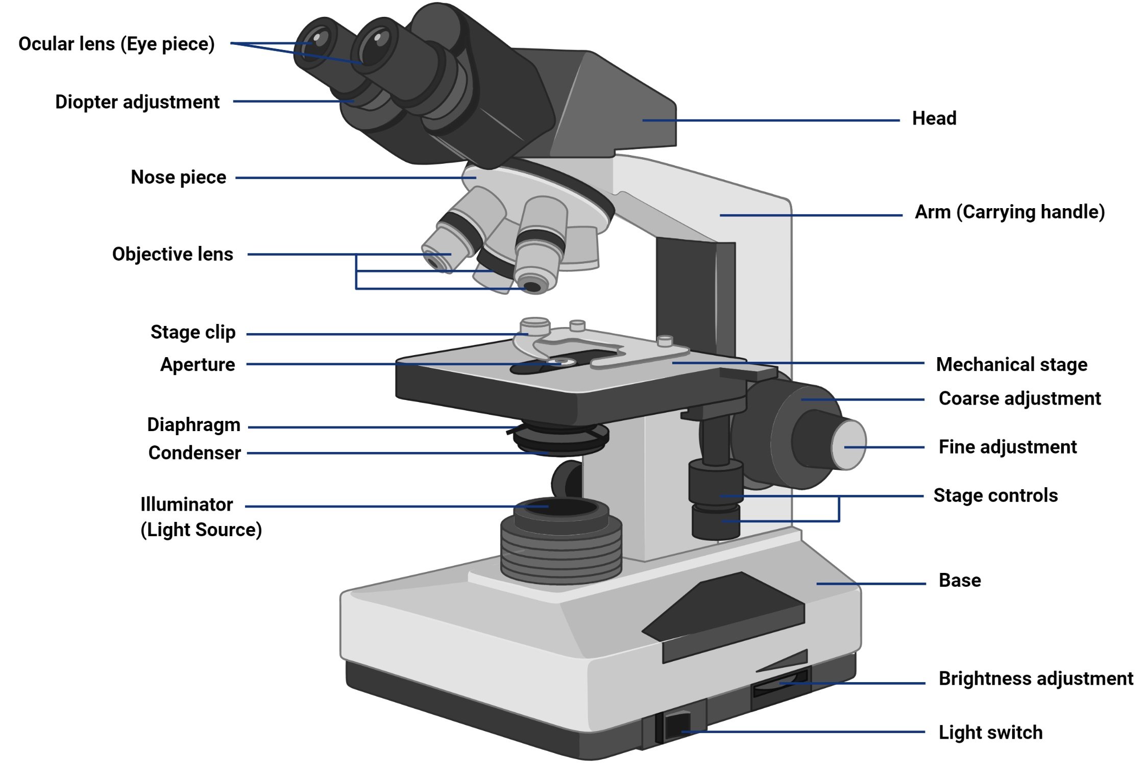
Brightfield Microscope is an optical microscope that uses light rays to produce a dark image against a bright background. It is the standard microscope that is used in Biology, Cellular Biology, and Microbiological Laboratory studies.
Calculatefocal lengthfrom image
The specimens used are prepared initially by staining to introduce color for easy contracting characterization. The colored specimens will have a refractive index that will differentiate it from the surrounding, presenting a combination of absorption and refractive contrast.
How to calculatefocal lengthPhysics
For this exercise, we want to image an object that is 400mm from the front of the lens to the object and desire a field of view of 90mm. We have selected a camera with the Sony Pregius CMOS IMX174 sensor. This uses a 1/1.2" format which measures 10.67mm x 8mm.
You will find our lens calculator HERE. Alternatively as select a camera, you will find an icon to the right which will automatically populate the calculator. Below is a short video showing how to use this resource from the camera pages.
Select Accept to consent or Reject to decline non-essential cookies for this use. You can update your choices at any time in your settings.
LinkedIn and 3rd parties use essential and non-essential cookies to provide, secure, analyze and improve our Services, and to show you relevant ads (including professional and job ads) on and off LinkedIn. Learn more in our Cookie Policy.
How to calculatefocal length ofparabola
Contact us to discuss your application and help make a recommendation! 1st Vision can provide a complete solution including lenses, cables and lighting.
For a specimen to be the focus and produce an image under the Brightfield Microscope, the specimen must pass through a uniform beam of the illuminating light. Through differential absorption and differential refraction, the microscope will produce a contrasting image.
How to calculatefocal length ofconvexlens

In order to select the correct focal length lens which is denoted in millimeters (i.e 25mm focal length), we need additional information on the camera sensor. Camera sensors come in various "Image formats". The chart below indicates some common formats which relate to the sensor size. The sensor size can be found on the actual sensor datasheets if not available in a given chart.
In any industrial imaging application, we have the task of selecting several main components to solve the problem at hand. The first being an industrial camera and second, a lens to acquire the given image. In many cases, our working distance of our lens is constrained and may have to mount the camera closer or further from the object plane. Once set, this defines our working distance (WD) for the lens. In addition, we have a given field of view (basically the dimension across the image) of the desired object.
Lenses are only available off the shelf in various focal lengths (i.e 25mm, 35mm, 50mm), so this calculate is theoretical and may need an iteration to adjust working distance. Alternatively, if your application can have a slightly smaller or larger FOV, the closest focal length lens to your calculation may be suitable.
This microscope is used to view fixed and live specimens, that have been stained with basic stains which gives a contrast between the image and the image background. It is specially designed with magnifying glasses known as lenses that modify the specimen to produce an image seen through the eyepiece.
The functioning of the microscope is based on its ability to produce a high-resolution image from an adequately provided light source, focused on the image, producing a high-quality image.


 Ms.Cici
 8618319014500