
Choosing the Right Microscope - what are microscopes
10 importance ofmicroscope
Cells vary in size. With few exceptions, individual cells are too small to be seen with the naked eye, so scientists use microscopes to study them. A microscope is an instrument that magnifies an object. Most images of cells are taken with a microscope and are called micrographs.
The optics of the lenses of a light microscope changes the orientation of the image. A specimen that is right-side up and facing right on the microscope slide will appear upside-down and facing left when viewed through a microscope, and vice versa. Similarly, if the slide is moved left while looking through the microscope, it will appear to move right, and if moved down, it will seem to move up. This occurs because microscopes use two sets of lenses to magnify the image. Due to the manner in which light travels through the lenses, this system of lenses produces an inverted image (binoculars and a dissecting microscope work in a similar manner, but include an additional magnification system that makes the final image appear to be upright).
Cytotechnologists play vital roles in saving people’s lives. When abnormalities are discovered early, a patient’s treatment can begin sooner, which usually increases the chances of successful treatment.
Why is microscope importantessay
A cell is the smallest unit of life. Most cells are so small that they cannot be viewed with the naked eye. Therefore, scientists must use microscopes to study cells. Electron microscopes provide higher magnification, higher resolution, and more detail than light microscopes. The unified cell theory states that all organisms are composed of one or more cells, the cell is the basic unit of life, and new cells arise from existing cells.

Why is microscope importantpdf
In multicellular organisms, several cells of one particular kind interconnect with each other and perform shared functions to form tissues (for example, muscle tissue, connective tissue, and nervous tissue), several tissues combine to form an organ (for example, stomach, heart, or brain), and several organs make up an organ system (such as the digestive system, circulatory system, or nervous system). Several systems functioning together form an organism (such as an elephant, for example).
The microscopes we use today are far more complex than those used in the 1600s by Antony van Leeuwenhoek, a Dutch shopkeeper who had great skill in crafting lenses. Despite the limitations of his now-ancient lenses, van Leeuwenhoek observed the movements of protists (a type of single-celled organism) and sperm, which he collectively termed “animalcules.”
5 importance ofmicroscope
unified cell theory: the biological concept that states that all organisms are composed of one or more cells, the cell is the basic unit of life, and new cells arise from existing cells
In this article from our Professional Photographers on Light Metering series, photojournalist C.S. Muncy explains how he uses a handheld meter in the field to get precise exposures with black-and-white film.
A cell is the smallest unit of a living thing. A living thing, like you, is called an organism. Thus, cells are the basic building blocks of all organisms.
Most student microscopes are classified as light microscopes (Figure 3.2 a). Visible light both passes through and is bent by the lens system to enable the user to see the specimen. Light microscopes are advantageous for viewing living organisms, but since individual cells are generally transparent, their components are not distinguishable unless they are colored with special stains. Staining, however, usually kills the cells.
Why is microscope importantto society
I love shooting in black and white with an overcast sky; the light tends to hit subjects more naturally and the shadows are slightly subdued. When shooting on bright days, you have to use a scrim (or at least a reflector) to achieve similar results.
Every environment offers both great opportunities and challenges for photographers trying to get just the right exposure. Finding the right way to measure the light hitting your subject can mean the difference between a mediocre image and a great one. In this article, I'm going to address one of the more basic ways to find a proper exposure using your handheld ambient light meter.
To meter for the first two photos, I held the meter as close to the subject as possible and took a number of readings. You can use the "memory" function and take an average reading from multiple measurements. The white bulb at the top of the meter was pointed towards me, measuring the light falling on the subject rather than being reflected from it. Your in-camera meter is great when you're running and gunning, but if you have the time, a handheld meter can provide far more accurate readings. When you have the time to take multiple readings, you will come away with a far more precise exposure.
Why is microscope importantin biology
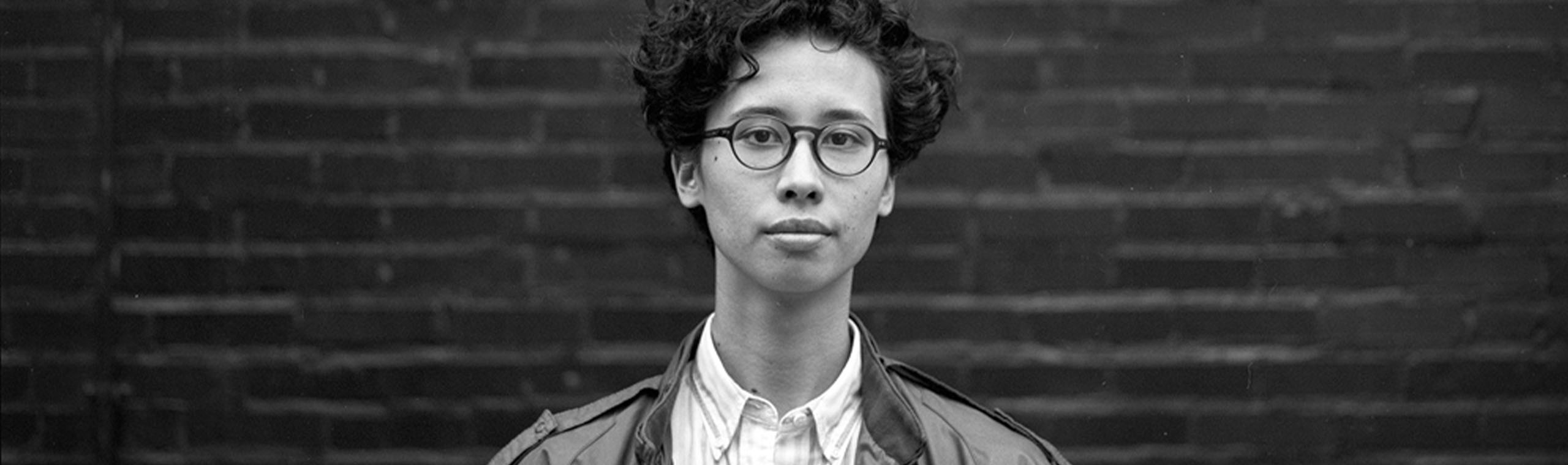
The third image would have been a bit more challenging if I had been using the in-camera meter, which only measures the light reflected off the subject. Because she was relatively pale, but standing against a wall that was painted black, I likely would have ended up with an overexposed image. By measuring the light falling on her, rather than reflecting from her, I was able to get a more accurate idea of what the proper exposure time should be. On brighter days, you have to be careful not to hold the meter in your own shadow, as this can affect your measurements and result in an overexposed image.
Light microscopes commonly used in the undergraduate college laboratory magnify up to approximately 400 times. Two parameters that are important in microscopy are magnification and resolving power. Magnification is the degree of enlargement of an object. Resolving power is the ability of a microscope to allow the eye to distinguish two adjacent structures as separate; the higher the resolution, the closer those two objects can be, and the better the clarity and detail of the image. When oil immersion lenses are used, magnification is usually increased to 1,000 times for the study of smaller cells, like most prokaryotic cells. Because light entering a specimen from below is focused onto the eye of an observer, the specimen can be viewed using light microscopy. For this reason, for light to pass through a specimen, the sample must be thin or translucent.
By the late 1830s, botanist Matthias Schleiden and zoologist Theodor Schwann were studying tissues and proposed the unified cell theory, which states that all living things are composed of one or more cells, that the cell is the basic unit of life, and that all new cells arise from existing cells. These principles still stand today.
Cytotechnologist: Have you ever heard of a medical test called a Pap smear? In this test, a doctor takes a small sample of cells from the uterine cervix of a patient and sends it to a medical lab where a cytotechnologist stains the cells and examines them for any changes that could indicate cervical cancer or a microbial infection.
In contrast to light microscopes, electron microscopes use a beam of electrons instead of a beam of light. Not only does this allow for higher magnification and, thus, more detail (Figure 3.4), it also provides higher resolving power. Preparation of a specimen for viewing under an electron microscope will kill it; therefore, live cells cannot be viewed using this type of microscopy. In addition, the electron beam moves best in a vacuum, making it impossible to view living materials.
Why is microscope importantin science
A second type of microscope used in laboratories is the dissecting microscope (Figure 3.2 b). These microscopes have a lower magnification (20 to 80 times the object size) than light microscopes and can provide a three-dimensional view of the specimen. Thick objects can be examined with many components in focus at the same time. These microscopes are designed to give a magnified and clear view of tissue structure as well as the anatomy of the whole organism. Like light microscopes, most modern dissecting microscopes are also binocular, meaning that they have two separate lens systems, one for each eye. The lens systems are separated by a certain distance, and therefore provide a sense of depth in the view of their subject to make manipulations by hand easier. Dissecting microscopes also have optics that correct the image so that it appears as if being seen by the naked eye and not as an inverted image. The light illuminating a sample under a dissecting microscope typically comes from above the sample, but may also be directed from below.
Most meters offer several options. Ambient light is "non-directional," and I tend to measure for it when I'm in an environment offering diffused light. In the images below, I was shooting on a mildly overcast day using Kodak 125PX film with a Mamiya 645 Pro and an 80mm lens opened up to f/2.8. For the purpose of these instructions, all photos were taken manually and the exposure measurements were taken with a Sekonic L-478DR LiteMaster Pro meter.
10 importance ofmicroscopePDF
In a 1665 publication called Micrographia, experimental scientist Robert Hooke coined the term “cell” (from the Latin cella, meaning “small room”) for the box-like structures he observed when viewing cork tissue through a lens. In the 1670s, van Leeuwenhoek discovered bacteria and protozoa. Later advances in lenses and microscope construction enabled other scientists to see different components inside cells.
C.S. Muncy is a freelance photojournalist based out of New York City with a client list that includes The New York Times, The Wall Street Journal, Newsday, The New York Daily News, and The Village Voice. As a freelancer, he’s covered such subjects as the BP oil spill, the fight for same-sex civil rights, the takeover of the state capitol in Madison, Wisconsin, and Occupy Wall Street. A graduate of the Defense Information School, he enlisted in the United States Air Force in 2002 and is currently a photographer with the New York Air National Guard.
Concepts of Biology - 1st Canadian Edition Copyright © 2015 by Charles Molnar and Jane Gair is licensed under a Creative Commons Attribution 4.0 International License, except where otherwise noted.
Read more from C.S. Muncy and other professional photographers in our Professional Photographers on Light Metering series:
There are many types of cells, and all are grouped into one of two broad categories: prokaryotic and eukaryotic. Animal cells, plant cells, fungal cells, and protist cells are classified as eukaryotic, whereas bacteria and archaea cells are classified as prokaryotic. Before discussing the criteria for determining whether a cell is prokaryotic or eukaryotic, let us first examine how biologists study cells.
In a scanning electron microscope, a beam of electrons moves back and forth across a cell’s surface, rendering the details of cell surface characteristics by reflection. Cells and other structures are usually coated with a metal like gold. In a transmission electron microscope, the electron beam is transmitted through the cell and provides details of a cell’s internal structures. As you might imagine, electron microscopes are significantly more bulky and expensive than are light microscopes.

To give you a sense of the size of a cell, a typical human red blood cell is about eight millionths of a meter or eight micrometers (abbreviated as µm) in diameter; the head of a pin is about two thousandths of a meter (millimeters, or mm) in diameter. That means that approximately 250 red blood cells could fit on the head of a pin.
When shooting with digital you need to be meticulously accurate with your light readings. Unlike film, which has a wider latitude when it comes to your exposure, digital (especially JPEGs) requires you to be spot on. What looks good on the finder may be, in reality, wildly overexposed on your computer screen. A handheld meter isn't always practical, especially in high-impact scenes, but for photos where you need your exposure right on target, nothing beats having one on hand.
Cytotechnologists (cyto– = cell) are professionals who study cells through microscopic examinations and other laboratory tests. They are trained to determine which cellular changes are within normal limits or are abnormal. Their focus is not limited to cervical cells; they study cellular specimens that come from all organs. When they notice abnormalities, they consult a pathologist, who is a medical doctor who can make a clinical diagnosis.


 Ms.Cici
 8618319014500