Cathode Ray Tubes (CRTs) - a cathode ray tube
The eyepiece, also known as the ocular lens, is the lens at the top of the microscope that you look through to view the specimen. It typically contains a magnifying lens that further enlarges the image produced by the objective lens. The eyepiece is usually removable and interchangeable, allowing for different magnifications to be achieved depending on the specific needs of the user.

The latest point of view on eyepiece design emphasizes the importance of ergonomic design to reduce eye strain and improve user comfort during extended periods of use. This includes features such as adjustable eye relief and eyecups to accommodate different users and provide a more comfortable viewing experience. Additionally, advancements in materials and manufacturing techniques have allowed for the production of lightweight yet durable eyepieces that are well-suited for various applications.
Eyepiece design and construction have evolved over time to improve the quality and comfort of the viewing experience. Modern eyepieces are typically designed with multiple lens elements to minimize aberrations and distortions, resulting in a clearer and more accurate image. Some eyepieces also incorporate advanced coatings to reduce glare and improve contrast.
From a modern perspective, the eyepiece on a microscope may also be designed to reduce eye strain and provide a comfortable viewing experience. Some eyepieces are equipped with adjustable diopter settings to accommodate individual differences in vision, and others may incorporate anti-glare or anti-reflection coatings to improve image clarity.
USERRA statute
Contrast agents from air to Barium to Iodine play a crucial role in X-ray and CT imaging by improving anatomical contrast and improving the visualization of ...
USERRA phone number

In summary, the eyepiece on a microscope is a crucial component that contributes to the overall quality of the viewing experience. Its design and construction have evolved to prioritize optical performance, user comfort, and versatility, making it an essential part of modern microscopy.
Nov 3, 2022 — Plugging in the value of r we found earlier, the diameter of the circle is d = 2 × (25 / (2π)) = 25 / π feet. To get an approximate numerical ...
u.s. army europe and africa acronym
by SA Kovalenko · 1999 · Cited by 695 — Pump--supercontinuum-probe (PSCP) spectroscopy with femtosecond time resolution is developed theoretically and experimentally.
USERRA law for Reservists

If you have questions or feedback on AUSA membership, contact us at: Member Services Center 1-855-246-6269 or Membersupport@ausa.org
The energy of the volunteers in the Association’s 122 chapters is focused on the programs in support of deployed Soldiers, civilians, and their families.
The magnification power of the eyepiece is a measure of how much the image is enlarged when viewed through the microscope. This is usually expressed as a number followed by an "x" (e.g., 10x, 20x), which indicates the number of times the image is magnified. For example, if the eyepiece has a magnification power of 10x and the objective lens has a magnification power of 40x, the total magnification of the microscope would be 400x (10x multiplied by 40x).
usareur-af headquarters
In November, the Association of the U.S. Army’s “Army Matters” podcast will feature a veteran entrepreneur who’s looking to help others, and Army Emergency Relief’s role in aiding ...
Overall, the eyepiece on a microscope plays a crucial role in magnifying and enhancing the image of the specimen, as well as providing a comfortable and effective viewing experience for the user.
Founded in 1950 as a nonpartisan, educational nonprofit, our growing association strengthens the bond between Soldiers and the American people, promotes the military profession, and enhances ties with industry.
Pilkington OptiView™ is a laminated glass with anti-reflective coatings on surfaces #1 and #4 (both outer surfaces of the laminated glass), which reduces ...
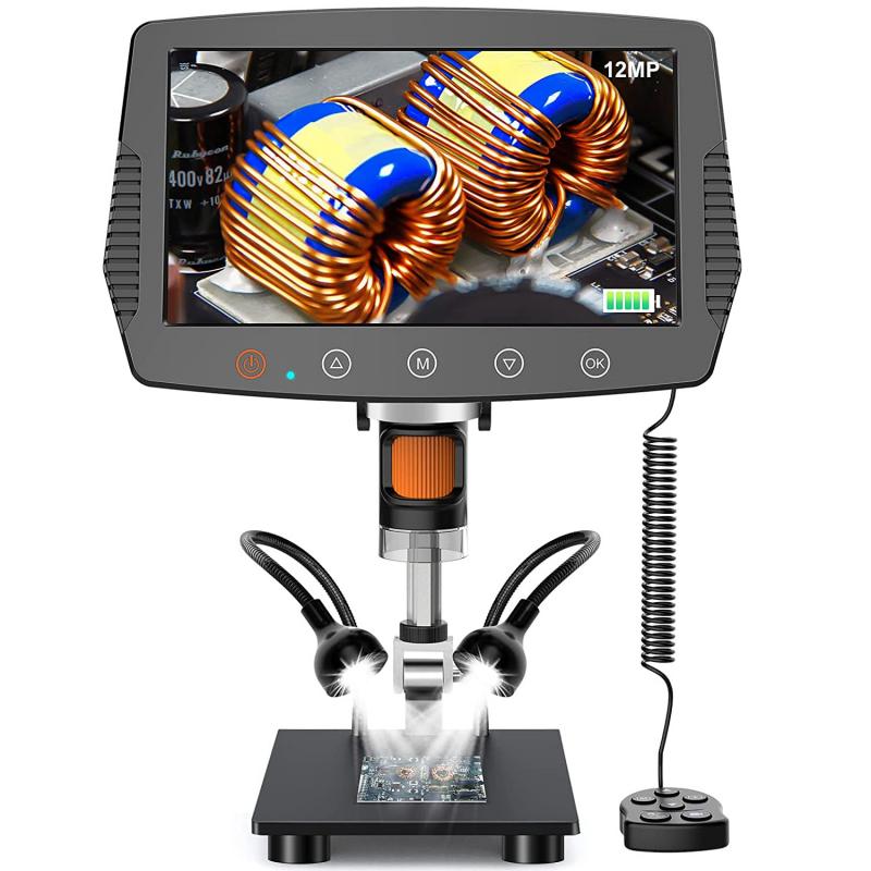
In recent years, there has been a growing interest in digital eyepieces, which incorporate digital imaging technology to capture and display the magnified image on a computer or other digital device. This allows for easier sharing of images and facilitates analysis and documentation of the specimens. Additionally, there has been a focus on ergonomic designs to improve user comfort and reduce eye strain during prolonged use. These advancements aim to enhance the overall microscopy experience and make it more accessible to a wider range of users.
High Efficiency beam splitter elements are special sub-aperture based DOE capable of splitting a beam into Double / Quattro Spot with 97% efficiency.
1. Huygenian eyepiece: This is a simple eyepiece design that consists of two plano-convex lenses with the convex sides facing each other. It provides a relatively narrow field of view and is commonly used in older microscopes.
Beginning Nov. 11, military retirees and eligible beneficiaries who use Tricare can enroll in or make changes to their health care coverage. Tricare open season, which runs through Dec. ...
During its 2024 Annual Meeting and Exposition, the Association of the U.S. Army presented for the third year its National Partner awards, recognizing organizations that provide outstanding support ...
In addition to magnification, the eyepiece also helps to focus the light rays coming from the objective lens and to direct them into the viewer's eye. This helps to create a clear and sharp image of the specimen under observation. The eyepiece also often contains a reticle or a graticule, which is a grid or scale that can be used to measure the size or dimensions of the specimen.
Inclination Joint: It is the point where the pillar meets the arm. The whole microscope can tilt at this inclination joint. Clips: The stage, on the side of the ...
The eyepiece on a microscope, also known as the ocular lens, is the lens at the top of the microscope through which the viewer looks. It is the lens closest to the eye when using the microscope. The primary function of the eyepiece is to magnify the image produced by the objective lens, which is the lens closest to the object being observed. This magnification allows the viewer to see a larger and more detailed image of the specimen.
USERRA time off before drill
2. Ramsden eyepiece: This design features two plano-convex lenses with the convex sides facing away from each other. It offers a wider field of view compared to the Huygenian eyepiece and is commonly used in modern microscopes.
USERRA probationary period
Jul 19, 2017 — ND filters are not made specifically for solar viewing. From the information I have come across you need at least an 20 stop ND filter that ...
At the present time, magnification is well defined when viewing an image of a sample through the eyepieces of a microscope. For this case, rigorous ...
Eyepieces in varying magnification range and field of view range for every microscope, all with reticle holders. Model, Microscopes, FOV, Remarks, Dimensions ...
About 200 soldiers with the 101st Airborne Division tested the capabilities of the Army’s Next Generation Squad Weapon system from Sept. 1 to Oct. 30 at Fort Campbell, ...
3. Wide-field eyepiece: This type of eyepiece is designed to provide a larger and more comfortable viewing area, allowing the viewer to see more of the specimen at once. It is particularly useful for applications that require prolonged observation.
USERRA MSPB
An upcoming Association of the U.S. Army webinar will focus on the importance of healthy minds and bodies while serving in the Army. The webinar, part of AUSA’s Noon ...
From the latest point of view, advancements in microscope technology have led to the development of eyepieces with variable magnification power, allowing users to adjust the level of magnification based on their specific needs. Additionally, some modern microscopes are equipped with digital eyepieces that can capture and display images on a computer screen, enabling users to easily share and analyze the microscopic images. These digital eyepieces often come with software that allows for further image enhancement and analysis, expanding the capabilities of traditional eyepieces.
The eyepiece on a microscope, also known as an ocular lens, is the part of the microscope that is looked through to view the magnified specimen. It is located at the top of the microscope and is the lens closest to the eye of the observer. The eyepiece is designed to magnify the image produced by the objective lens, which is the lens closest to the specimen being observed.
Depth of focus is at the image plane. Depth of field is at the object plane.
An eyepiece on a microscope is a lens that is positioned at the top of the microscope and is used to view the magnified image of the specimen. It is also known as an ocular lens and is an essential component of the microscope's optical system. The eyepiece typically contains a set of lenses that further magnify the image produced by the objective lens, allowing the viewer to see a highly detailed and enlarged image of the specimen.
An eyepiece on a microscope, also known as an ocular lens, is the lens at the top of the microscope that you look through to view the specimen. It is the part of the microscope that is closest to your eye and is responsible for magnifying the image of the specimen. The eyepiece typically contains a set of lenses that work together to magnify the image produced by the objective lens, which is the lens closest to the specimen.
American influence and institutions are being challenged by China, Russia and Iran, according to the author of a new paper published by the Association of the U.S. Army. ...


 Ms.Cici
 8618319014500