
Buyers Guide | What is OD (Optical Density) Laser Safety? - optical density
Ramanspectroscopy principle
TweezersSHOP ALL TWEEZERSCeramic TweezersHigh Precision TweezersOther TweezersPlastic TweezersVacuum TweezersWafer Tweezers
Ramanspectroscopy principle and instrumentation PDF
When the NA of the sub-stage condenser and the objective are different, then we can use the following formula to calculate resolution;
Calibration StandardsSHOP ALL CALIBRATION STANDARDSGeller Reference StandardsCoverglasses / CoverslipsDiamond Knives - Histo DissectionEyepiece GraticulesFinder Grids
Slide & Block StorageSlide StainingSHOP ALL SLIDE STAININGStains for Light MicroscopyStage MicrometersTissue EmbeddingTissue Processing Consumables
Dry IceFibre Optic IlluminatorsFlowmetersFume CabinetsGrinders, Polishers & PressesSHOP ALL GRINDERS, POLISHERS & PRESSESGrinding, Polishing & Press MachinesAccessoriesIncubators & OvensKnifemakersLam Plan - Sample PreparationLiquid Nitrogen DewarsMagnetic Field Cancelling
Oils & GreasesSafety GlovesSafety ProductsSpecimen PreparationStorage BoxesSHOP ALL STORAGE BOXESGel-Pak BoxesMembrane Boxes
JavaScript seems to be disabled in your browser. For the best experience on our site, be sure to turn on Javascript in your browser.
What is a raman spectrometerand how does it work
Grids - Athene by Agar ScientificSHOP ALL GRIDS - ATHENE BY AGAR SCIENTIFICStandard Square PatternThin BarThick Bar/Thin BarSlot and Multiple SlotThick SlotHexagonalRound Hole PatternSingle HoleFoldingOtherK-kits for Liquid TEMLight Element Support GridsMaterial ProcessingPhotographic Films & Papers
Replication MaterialsSpecimen Stub Storage BoxesSpecimen Stubs & MountsSpecimen Stubs - ModularScintillatorsSputter Targets
Photons interacting with molecules most commonly scatter elastically. This is called Rayleigh scattering. Rayleigh scattered photons have the same wavelength as the incident light. However, approximately 1 out of a million photons are inelastically scattered...an effect first described by Sir Chandrasekhara Raman in 1922. This is the principle from which Raman spectroscopy derives.
AdhesivesBags & LabelsBeakers, Tubes & ContainersCleaning ProductsSHOP ALL CLEANING PRODUCTSAir DustersCleaners, Solvents & CreamsCloths & WipesPolishesTools
AperturesCalibration & Test SpecimensSHOP ALL CALIBRATION & TEST SPECIMENSGeller Reference StandardsCertified Particle Size StandardsCritical Dimension StandardsMagnification CalibrationResolution & Grey Level Test SpecimensConsumables KitsFilaments
MicroscopesSHOP ALL MICROSCOPESAsbestos MicroscopesBiological MicroscopesIndustrial MicroscopesMounting MediaSectioningSlides & Accessories
Adam Equipment Balances & ScalesCell Manipulation InstrumentationSHOP ALL CELL MANIPULATION INSTRUMENTATIONElectroporatorMicromanipulatorsMicroinjectorsMicrocapillariesVibration ProtectionAccessoriesDiamond Saws & Cutting
What is a raman spectrometerused for
AdhesivesBags & LabelsBeakers, Tubes & ContainersCleaning ProductsSHOP ALL CLEANING PRODUCTSAir DustersCleaners, Solvents & CreamsCloths & WipesPolishesTools
ChemicalsSHOP ALL CHEMICALSBuffersElectrophoresisHiFliQ® FPLC ColumnsProtein Ark ResinsStains for Electron MicroscopyStains for Light MicroscopyCryogenicCutting Wheels & Blades
AperturesCalibration & Test SpecimensSHOP ALL CALIBRATION & TEST SPECIMENSGeller Reference StandardsCertified Particle Size StandardsCritical Dimension StandardsMagnification CalibrationResolution & Grey Level Test SpecimensConsumables KitsFilaments
When referring to optics and microscopy, resolution is simply defined as the minimum distance at which two separate points in a field of view can be distinguished as distinct.
Raman SpectrometerPrice
Support Films - Lacey Carbon Support Films - QuantifoilSupport Films - SiliconTissue Processing ChemicalsTissue Processing ConsumablesVacuum Coating MaterialsVacuum Oils & GreasesX-ray Microanalysis Standards
The factors which determine resolution include the wavelength of light used to image a specimen- the shorter the wavelength, the more detail will be resolved. The next factor to consider is Numerical Aperture (NA). If you read my previous article on NA, you will remember that this is dependent on not only the refractive index of the medium between the specimen and the objective, but also the angular aperture of the objective.
We have looked at numerical aperture (NA) in a previous article and indeed, NA is related to resolution. However, resolution isn’t just dependant on the NA of an objective, but on the NA of the microscope system as a whole. This takes into account the NA of the sub-stage condenser for example. The higher the NA of the whole optical components of the microscope, then the greater the detail you will be able to resolve.
Cell Manipulation by Calibre ScientificSHOP ALL CELL MANIPULATION BY CALIBRE SCIENTIFICElectroporatorMicromanipulatorsMicroinjectorsMicrocapillariesVibration ProtectionAccessories for Cell Manipulation
TweezersSHOP ALL TWEEZERSCeramic TweezersHigh Precision TweezersOther TweezersPlastic TweezersVacuum TweezersWafer Tweezers
Of course, not every molecule or functional group exhibits Raman scattering. The following factors influence Raman scattering:
Dry IceFibre Optic IlluminatorsFlowmetersFume CabinetsGrinders, Polishers & PressesSHOP ALL GRINDERS, POLISHERS & PRESSESGrinding, Polishing & Press MachinesAccessoriesIncubators & OvensKnifemakersLam Plan - Sample PreparationLiquid Nitrogen DewarsMagnetic Field Cancelling
Sample HoldersSectioningStainingSupport Films - Carbon Support Films - Forming MaterialsSupport Films - Formvar / PioloformSupport Films - Formvar CarbonSupport Films - GrapheneSupport Films - Holey Carbon
Cell Manipulation by Calibre ScientificSHOP ALL CELL MANIPULATION BY CALIBRE SCIENTIFICElectroporatorMicromanipulatorsMicroinjectorsMicrocapillariesVibration ProtectionAccessories for Cell Manipulation
John William Strutt, 3rd Baron Rayleigh (1842-1919) was an English physicist who co-discovered the noble gas argon (and was subsequently awarded the Nobel Prize in 1904 for this achievement). Much of Strutt’s work was based on acoustics, but he also invented the ‘Rayleigh Criterion’ which explains that when two Airy Discs are far enough apart to be resolved as separate entities within an image, then they are said to meet the Rayleigh Criterion. When two Airy Discs are closer than the Rayleigh criteria, then they merge and cannot be resolved as separate details.
MicroscopesSHOP ALL MICROSCOPESAsbestos MicroscopesBiological MicroscopesIndustrial MicroscopesMounting MediaSectioningSlides & Accessories
With Raman scattering, the incident light interacts with matter and its wavelength is either shifted lower or higher (red or blue shifted, respectively). Red shifted photons are the most common, having been subject to a "Stokes shift". What has happened is that the photon has interacted with the electron cloud of the functional groups bonds, exciting an electron into a virtual state. The electron then relaxes into an excited vibrational or rotational state (see the diagram). This causes the photon to lose some of its energy and it is detected as Stokes Raman scattering. This loss of energy is directly related to the functional group, the structure of the molecule to which it is attached, the types of atoms in that molecule and the molecule's environment.
The above limit of resolution in optical microscopy meant that objects smaller than 200 nm could not be resolved. However, techniques have been developed which have pushed this limit back even further. These methods are known as ‘Super-Resolution Microscopy’ and have achieved resolution down to tens of nanometres. There are many different methods which come under the umbrella-term of super-resolution microscopy and I will cover the main ones in a forthcoming article.
Support Films - Lacey Carbon Support Films - QuantifoilSupport Films - SiliconTissue Processing ChemicalsTissue Processing ConsumablesVacuum Coating MaterialsVacuum Oils & GreasesX-ray Microanalysis Standards
Calibration StandardsSHOP ALL CALIBRATION STANDARDSGeller Reference StandardsCoverglasses / CoverslipsDiamond Knives - Histo DissectionEyepiece GraticulesFinder Grids
It should be noted that Raman scattering is a very weak effect as most photons are Rayleigh scattered. However, the intensity of the effect can be dramatically increased using resonance Raman spectroscopy. In resonance Raman spectroscopy, the wavelength of the exciting laser light coincides with the absorbance maximum of the molecule or functional group. Therefore, the photon can excite an electron to near an electronic excited state rather than a virtual excited state. This results in an increase in the Raman scattering intensity by a factor up to a million. This transition is therefore dominant in the resultant Raman spectrum: the Raman spectrum is of the molecule whose absorbance corresponds to the wavelength of the laser. This is why Raman microspectrometers are often offered with multiple lasers with differing laser wavelengths: different samples may be excited at different wavelengths in order to obtain the strongest Raman signal.
Resolution in microscopy is dependent on a number of factors. To achieve the maximum resolution using a microscope, the whole system has to be correctly aligned and the properties of each of the optical components of a microscope should be complimentary to each other. For correct alignment, check back soon for my next article entitled ‘Koehler Illumination: A History and Practical Set Up’.
There are two linked mathematical concepts which need to be taken into consideration when dealing with resolution; ‘Airy Discs’ and the ‘Rayleigh Criterion’.
Surface enhanced Raman spectroscopy (SERS) is another method of signal enhancement for Raman spectroscopy. The most important factor of this technique is sample preparation. Sample molecules are adsorbed onto specially prepared metal surfaces. These surfaces are "rough" on the nanoscale and are usually either gold or silver. Raman spectra can then be taken using standard methodologies but with signal enhancement of approximately a trillion. However, the correct laser wavelength must be selected that best interacts with the metal surface in order to see the SERS effect.
George Biddell Airy (1801-1892) was an English mathematician and astronomer (he was the ‘Astronomer Royal’ from 1835 to 1881). In 1835, his paper entitled ‘On the Diffraction of an Object-Glass with Circular Aperture’ was published in the Transactions of the Cambridge Philosophical Society. There are some 19 equations in Airy’s 1835 paper which, you will be pleased to read, I won’t go into here! Although Airy was writing from the point of view of an astronomer and describing rings of light surrounding the image of a star, these observations are relevant to many other optical systems, including the microscope. The Airy Disc appears (from above) as a bright central point of light with rings or ripples around (think of the ripples of a stone as it drops into still water). The central point of light is the optimally focussed light which can be determined by a circular aperture in a perfectly aligned system. The ‘Airy Pattern’ is a more appropriate terminology when it comes to describing the optics and resolution of a microscope. The Airy Disc assumes a single point or source of light, whereas, in reality, we are dealing with numerous points of light in a single specimen view.
During an experiment using Raman spectroscopy, light of a single wavelength is focused onto a sample. Most commonly a laser is used as it is a powerful monochromatic source. The photons from the laser interact with the molecules of the sample and are scattered inelastically. The scattered photons are collected and a spectrum is generated from the scattered photons.
This means that some vibrational or rotational transitions, which exhibit low polarizability, and will not be Raman active. They will not appear in a Raman spectra.
Replication MaterialsSpecimen Stub Storage BoxesSpecimen Stubs & MountsSpecimen Stubs - ModularScintillatorsSputter Targets
Raman microspectroscopy is where a Raman microspectrometer is used in place of a standard Raman spectrometer. A Raman microspectrometer consists of a specially designed Raman spectrometer integrated with an optical microscope. This allows the experimenter to acquire Raman spectra of microscopic samples or microscopic areas of larger samples. The advantages are that much less samples is required and certain effects may also be enhanced over very localized regions.
Ramanspectroscopy instrumentation
Grids - Athene by Agar ScientificSHOP ALL GRIDS - ATHENE BY AGAR SCIENTIFICStandard Square PatternThin BarThick Bar/Thin BarSlot and Multiple SlotThick SlotHexagonalRound Hole PatternSingle HoleFoldingOtherK-kits for Liquid TEMLight Element Support GridsMaterial ProcessingPhotographic Films & Papers
Grids - SEM FinderGrinding & PolishingMaterials EmbeddingSHOP ALL MATERIALS EMBEDDINGCold Mounting ResinsHot Mounting ResinsMounting Tabs & AdhesivesPreparation
Raman spectroscopy is the study of the interaction between light and matter where light is inelastically scattered: a process upon which Raman spectroscopy is based.
Oils & GreasesSafety GlovesSafety ProductsSpecimen PreparationStorage BoxesSHOP ALL STORAGE BOXESGel-Pak BoxesMembrane Boxes
The limit of resolution in light microscopy is approximately 200 nm. Remember that NA= n • sin(θ), where n is the refractive index and θ is half of the angular aperture. The resolution limit is calculated using the following information;
AperturesCalibration StandardsCalibration Standard - Lattice PlaneCirclip Injector and CirclipsCoated GridsCryo PreparationDiamond Knives - DiATOMEFilamentsGrid Boxes & StorageGrids - FinderGrids - Omniprobe
When electromagnetic energy interacts with a material, it can either be reflected, absorbed, transmitted, or scattered. One type of scattering is Raman scattering and is the basis for Raman spectroscopy. Raman spectroscopy is useful as the Raman scattered light yields data of the vibrational modes of the sample molecules. A Raman spectrum thus enables the identification of molecules and their functional groups, similar to IR spectroscopy, but with visible range light.
Polishing & Grinding MaterialsSHOP ALL POLISHING & GRINDING MATERIALSAbrasive DiscsDiamond DiscsDiamond Polishing CompoundsDiamond Suspensions & SpraysPolishing Cloths & PadsPolishing CompoundsAccessories
ChemicalsSHOP ALL CHEMICALSBuffersElectrophoresisHiFliQ® FPLC ColumnsProtein Ark ResinsStains for Electron MicroscopyStains for Light MicroscopyCryogenicCutting Wheels & Blades
Ramanspectroscopy PDF
Slide & Block StorageSlide StainingSHOP ALL SLIDE STAININGStains for Light MicroscopyStage MicrometersTissue EmbeddingTissue Processing Consumables
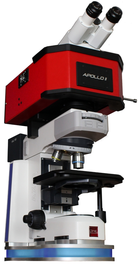
Raman Microspectrometer DesignUses of Micro Raman Spectrometers Units of Micro Raman SpectroscopyCRAIC Apollo™ MicroRaman Spectrometer
Grids - AgarSHOP ALL GRIDS - AGARSquare MeshRectangular Mesh Parallel BarFoldingHexagonal MeshThin BarVery Fine MeshSingle & Triple SlotSingle HoleTabbedResin Embedding - AcrylicResin Embedding - EpoxyResin Embedding - London ResinResin Embedding Consumables
MicroscopesSHOP ALL MICROSCOPESMic-Fi Digital MicroscopesMicrowave ProcessorsNanoparticle DepositionOhaus Analytical & Precision BalancesPelco EquipmentpH MeasurementPlatform RockersServicing & Repair
What is a raman spectrometerin chemistry
Adam Equipment Balances & ScalesCell Manipulation InstrumentationSHOP ALL CELL MANIPULATION INSTRUMENTATIONElectroporatorMicromanipulatorsMicroinjectorsMicrocapillariesVibration ProtectionAccessoriesDiamond Saws & Cutting
Polishing & Grinding MaterialsSHOP ALL POLISHING & GRINDING MATERIALSAbrasive DiscsDiamond DiscsDiamond Polishing CompoundsDiamond Suspensions & SpraysPolishing Cloths & PadsPolishing CompoundsAccessories
AperturesCalibration StandardsCalibration Standard - Lattice PlaneCirclip Injector and CirclipsCoated GridsCryo PreparationDiamond Knives - DiATOMEFilamentsGrid Boxes & StorageGrids - FinderGrids - Omniprobe
Grids - AgarSHOP ALL GRIDS - AGARSquare MeshRectangular Mesh Parallel BarFoldingHexagonal MeshThin BarVery Fine MeshSingle & Triple SlotSingle HoleTabbedResin Embedding - AcrylicResin Embedding - EpoxyResin Embedding - London ResinResin Embedding Consumables
MicroscopesSHOP ALL MICROSCOPESMic-Fi Digital MicroscopesMicrowave ProcessorsNanoparticle DepositionOhaus Analytical & Precision BalancesPelco EquipmentpH MeasurementPlatform RockersServicing & Repair
Sample HoldersSectioningStainingSupport Films - Carbon Support Films - Forming MaterialsSupport Films - Formvar / PioloformSupport Films - Formvar CarbonSupport Films - GrapheneSupport Films - Holey Carbon
Grids - SEM FinderGrinding & PolishingMaterials EmbeddingSHOP ALL MATERIALS EMBEDDINGCold Mounting ResinsHot Mounting ResinsMounting Tabs & AdhesivesPreparation
So, where does the figure of ‘1.22’ come from? If you really want to know, this figure is the approximate first zero of the Bessel function of the first kind of order one divided by Pi. Bessel functions are used when solving problems in spherical or cylindrical coordinate systems. For the purpose of calculating resolution in microscopy, it is simpler to assume 1.22 as a constant!


 Ms.Cici
 8618319014500