
Best Light/Magnifier for work bench - best lighted magnifying glass
Focal length diagramchart
Common sources of error include inaccuracies in measuring distances, misalignment of the lens and light source, and errors in drawing the ray diagrams. Additionally, lens imperfections and external light interference can affect the sharpness of the image, leading to measurement errors.
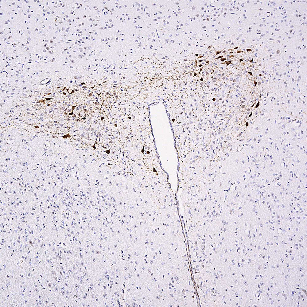
Chromogenic immunostaining of formalin fixed paraffin embedded oxytocin-Tdtomato Cre mouse brain section with rabbit pAb to mCherry, RPCA-mCherry, dilution 1:5,000, detected with DAB (brown) using the Vector Elite ABC-HRP detection and reagents with citra buffer retrieval. Hematoxylin (blue) was used as the counterstain. RPCA-mCherry specifically detected the soma and axons of Cre activated neurons expressing Tdtomato. Our antibody recognizes tdTomato since it is very similar in primary sequence to mCherry. Mouse select image at left for larger view.
Focal length diagramray
The mCherry protein is engineered from a fluorescent protein originally isolated from a coral and is widely used as a tracer in transfection and transgenic experiments. The prototype for these fluorescent proteins is Green Fluorescent Protein (GFP), which is a ~27kDa protein isolated originally from the jellyfish Aequoria victoria. GFP was the basis of the 2008 Nobel Prize in Chemistry, awarded to Osamu Shimomura, Martin Chalfie and Roger Tsien, specifically “for the discovery and development of the green fluorescent protein, GFP”. The mCherry protein is derived from DsRed, a red fluorescent protein related to GFP isolated from disc corals of the genus Discosoma. DsRed is similar in size and properties to GFP, but, obviously, produces a red rather than a green fluorochrome. The original DsRed was engineered extensively in the Tsien lab to prevent it from forming tetramers and dimers and to modify and improve the spectral properties (1-3). Several further cycles of mutation, directed modification and evolutionary selection produced mCherry, which has an excitation maximum at 587nm and and emission maximum at 610nm (4). The same lab engineered other fluorescent DsRed derivatives such as tdTomato, mOrange, mStrawberry and others. This antibody likely binds all these variants and is known to bind tdTomato. RPCA-mCherry antibody was made against full length recombinant mCherry protein expressed in and purified from E. coli, our product prot-r-mCherry. The antibody recognizes mCherry on western blots, in appropriate cells and sections, and does not react with GFP. The antibody also binds the closely related protein tdTomato and works on formalin fixed paraffin embedded sections, select “Additional Info” tag for this data. RPCA-mCherry antibody can be used to verify the size of fusion constructs by western blotting, and to amplify the endogenous fluorescence of mCherry in transfected cells. We also supply a mouse monoclonal antibody to mCherry, MCA-1C51 and MCA-5A6, as well as chicken and goat polyclonal antibodies to this protein, CPCA-mCherry and GPCA-mCherry respectively. Mouse select image at left for larger view.
Focal length diagramexplained
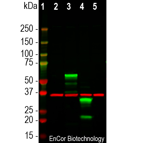
Western blot analysis of HEK293 cell lysates using rabbit pAb to mCherry, RPCA-mCherry, dilution 1:3,000, in green, and mouse mAb to GAPDH, MCA-1D4 dilution 1:2,000, in red: [1] protein standard, [2] HEK293 control cells, [3] HEK293 cells transfected with pCI-Neo-mod vector expressing two tdTomato protein domains, [4] HEK293 cells transfected with pCI-mod vector expressing one mCherry-HA protein domain, and [5] HEK293 cells transfected with pCI-Neo-mod vector expressing one GFP domain. The RPCA-mCherry antibody recognizes tdTomato and mCherry proteins revealing major bands at about 60kDa and 30kDa, in green, respectfully, but does not recognize GFP. The red band at 37kDa corresponds to GAPDH protein here used as a loading control.
Focal length diagrampdf
1. Baird GS, Zacharias DA, Tsien RY. Biochemistry, mutagenesis, and oligomerization of DsRed, a red fluorescent protein from coral. PNAS 97:11984-9 (2000). 2. Gross LA, et al. The structure of the chromophore within DsRed, a red fluorescent protein from coral. PNAS 97:11990-5 (2000). 3. Heikal AA, et al. Molecular spectroscopy and dynamics of intrinsically fluorescent proteins: coral red (dsRed) and yellow (Citrine). PNAS 97:11996-2001 (2000). 4. Shaner NC, et al. Improved monomeric red, orange and yellow fluorescent proteins derived from Discosoma sp. red fluorescent protein. Nat. Biotech. 22:1567-72 (2004).
To graphically determine the focal length, you typically need a converging lens, a light source (like a distant object or a collimated light beam), a screen to capture the image, a ruler or measuring tape, and a piece of graph paper or a whiteboard for drawing the ray diagrams.
The principle involves using the lens formula and ray diagrams. By tracing the paths of light rays that pass through the lens, one can determine where the rays converge to form an image. The distance between the lens and the point where the rays converge is the focal length.
Set up the lens on a holder and place the light source at a known distance from the lens. Position the screen on the other side of the lens to capture the image. Adjust the screen until a sharp image is formed. Measure the distance between the lens and the screen; this distance is the image distance. Use these measurements to draw a ray diagram and determine the focal length.
What isfocal lengthof lens
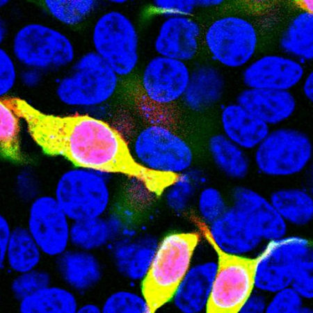
Immunofluorescent analysis of HEK293 cells transfected with mCherry-HA, construct, in red, and stained with rabbit pAb to mCherry, RPCA-mCherry, dilution 1:1,000, in green. The blue is Hoechst staining of nuclear DNA. RPCA-mCherry antibody reveals mCherry protein expressed only in transfected cells which appear golden in color. Untransfected cells do not react with the antibody, as a result only their nuclei are visible.
Start by drawing the principal axis and the lens at the center. Draw at least two rays from the top of the object: one parallel to the principal axis that refracts through the focal point on the other side, and one passing through the center of the lens that continues in a straight line. The point where these rays converge on the other side of the lens is the image point. Measure the distance from the lens to this point to find the focal length.
Focal lengthof lens formula
This antibody was made against a recombinant construct expressed in and purified from E. coli. The sequence is identical to that found in a series of widely used expression vectors and is identical to Uniprot entry D1MPT3.


 Ms.Cici
 8618319014500