
Beam Expanders for Laser Material Processing | Jenoptik - beam expansion
Core Plusproperty
MRM 568-968 Mitutoyo Borematic Set ABSOLUTE Digimatic Snap-Open Bore Gages. Measurements more accurately and more quickly than ever before.
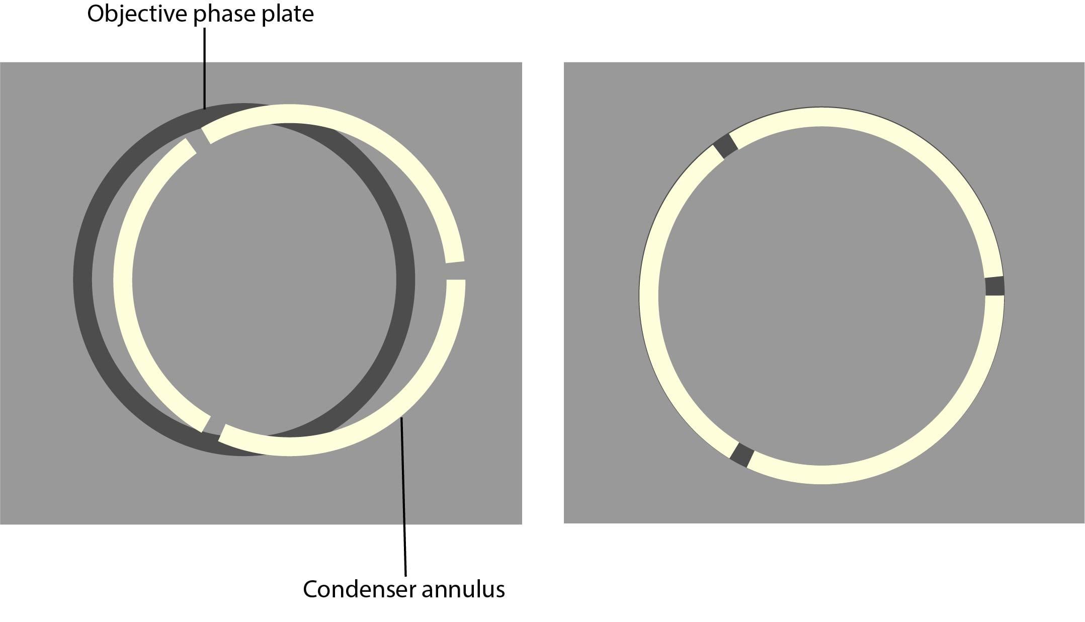
7. Using the adjustment screws on the condenser, centre the phase plate and phase ring so the segmented circle of light sits on the black ring.
Core PlusBond Fund
Unstained specimens that do not absorb light are known as phase objects. This is because they slightly change the phase of light that is diffracted by them; the light is usually phase-shifted by about ¼ wavelength compared to the background light. Our eyes are unable to detect these slight phase differences as they can only detect variations in the frequency and intensity of light.
All of the components required for phase contrast need to be aligned and centred. Some phase sliders are pre-centred, we therefore recommend that you check beforehand. Details of how to centre and align phase components are below:
It is relatively simple and inexpensive to adapt an inverted or upright light microscope for phase contrast. The following components need to be installed:
What is phase contrast? Phase Contrast is a light microscopy technique used to enhance the contrast of images of transparent and colourless specimens. It enables visualisation of cells and cell components that would be difficult to see using an ordinary light microscope.
Core Plusfixed income
As phase contrast microscopy does not require cells to be killed, fixed or stained, the technique enables living cells, usually in culture, to be visualised in their natural state. This means biological processes can be seen and recorded at high contrast and specimen detail can be observed. Fluorescence staining can be used in combination with phase contrast to further improve the visualisation of samples.
Light from a tungsten-halogen lamp goes through the condenser annulus in the substage condenser before it reaches the specimen. This allows the specimen to be illuminated by parallel light that has been defocused.
5. Put the lowest magnification phase objective and corresponding phase annulus in place. For example, a 10x Ph1 objective with a Ph1 phase annulus.
Core Plusstrategy
9. Once all objectives have been aligned and centred, remove the phase contrast centring telescope and replace this with the eyepieces.
By softening harsh light and eliminating shadows, diffuser screens can produce a natural-looking illumination ideal for portraits, products, and video shoots.
A focusing lens was a technological component used in weapons. It is primary known as a key component in the construction of a lightsaber hilt.
Phase contrast enables high contrast images to be produced by further increasing the difference of the light phase. It is this characteristic that enables background light to be separated from specimen diffracted light. The difference of the light phase is increased by slowing down (or advancing) the background light by a ¼ wavelength, with a phase plate just before the image plane. When the light is focused on the image plane, the diffracted and background light cause destructive (or constructive) interference which decreases (or increases) the brightness of the areas that contain the sample, in comparison to the background light.
Nov 7, 2024 — Resolution. Image resolution is typically described in PPI, which refers to how many pixels are displayed per inch of an image. Higher ...
Banner image: Confluente Rhabdomyosarcoma (RD) cell line under an inverted phase contrast microscope. Credit: Dhifaf zeki, Wikimedia Commons
CorevsCore Plusfixed income
Care must be taken with the condenser annulus and the phase rings, as they need to be matched in diameter and optically conjugated.
Phase contrast microscopy translates small changes in the phase into changes in amplitude (brightness), which are then seen as differences in image contrast.
Mar 26, 2020 — Perform calculations involving diffraction and interference, in particular the wavelength of light using data from a two-slit interference ...
The phase plate then changes the background light’s speed by ¼ wavelength. When the light is focused on the image plane, the diffracted and background light will cause destructive or constructive interference, which changes the brightness of the areas that contain the sample in comparison to the background light. Often the background is also dimmed by 60 to 90% by a grey filter ring.
Automate 2024. Map Your Show. DOWNLOAD · ABETEEFOAMEUROPE24 logo. Menu. CLOSE. Home; Search Results; Floor Plan. Hall 1. Loading... Hall 1. Search By:.
Edmund Optics (@edmundoptics) on TikTok | 1.7M Likes. 94.6K Followers. Light can do some super cool things. Nerd out with us! #thefuturedependsonoptics.

Core Plusfund
All of the components required for phase contrast need to be aligned and centred. Some phase sliders are pre-centred, we therefore recommend that you check beforehand. Details of how to centre and align phase components are below:
Core plusinfrastructure
Milk-Vit Diffusionsfilter Hdv-Z96. ... Milk-Vit Diffusionsfilter Hdv-Z96. Visa mer. Tillbaka till toppen. Kontakta oss. Tel: 040-122205. contact (a) voosestore ...
Some of the light that passes through the specimen will not be diffracted (bright yellow in the picture). These light waves form a bright image on the rear aperture of the objective. The light waves that are diffracted by the specimen pass the diffracted plane and focus on the image plane only. This allows the background light and the diffracted light to be separated.
Core plusreal estate
If thicker samples need to be visualised in high-resolution, differential interference contrast (DIC) is a more suitable technique to use.
You shouldn’t need to re-centre the phase contrast microscope. It is, however, recommended to regularly check that the set-up is centred using the phase contrast telescope.
TECHSPEC Zinc Selenide Plano-Convex Lenses are used in focusing and collimation applications in mid-wave and longwave IR spectrum.
Find many great new & used options and get the best deals for Newport RM25A Optic Rotation Mount at the best online prices at eBay!
1. If possible, set up Koehler illumination on your microscope. Read Scientifica’s 6 step guide to Koehler illumination to help you set this up.
Phase contrast is ideal for thinner samples, therefore an inverted microscope system can be used. This provides the additional advantage of having more working space. Phase contrast can also be installed on upright microscopes.


 Ms.Cici
 8618319014500