
CMOS/CCD Sensors and Camera Systems, Second Edition - camera cmos
CameraFOV calculator
A camera with 25 mm diagonal FOV can only image an area this large if the magnification is 1x. With a typical life science magnification of 40x, the camera FOV decreases by a factor of 40, resulting in a 625 µm diagonal FOV.
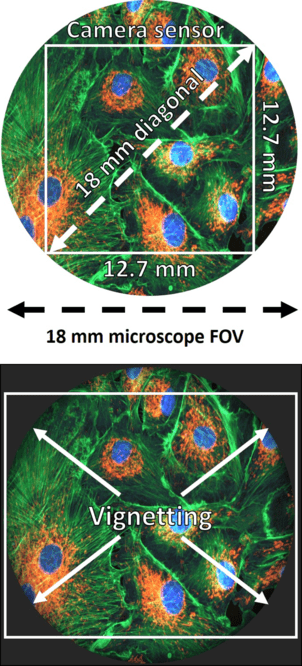
Some rectangular camera sensors can often occupy the microscope FOV more effectively depending on the size, but if the camera FOV is larger than the microscope FOV there will be vignetting, an effect seen at the corners of the image due to the lack of light, as seen in Fig.3 Bot. While this may capture more of the microscope FOV, the substantial image artifacts at the corners of each image can lead to decreased image quality.
Camera field of view anglecalculator
Waldmann Lighting™ Tevisio and Televisiomax magnifier lights combine the latest in LED technology with magnification. Ideal for the lab, research, or any work surface where glare-free close-up work is necessary.
CCDs and EMCCDs typically had smaller sensors measuring around 11 mm, this limits what you can image and is well below the maximum of most microscopes.
Angle of viewvsfield of view
Magnifier Lamp is the ideal tool to view objects at three times magnification in bright light, with high-contrast and a sharply defined focus
CMOS cameras offer much larger sensors, typically around 18.8 mm diagonal which matches up well to certain microscope models. Some microscopes can go above 20 mm diagonal, so CMOS cameras are also available with sensor sizes of 22 mm, 25 mm, and even 29 mm.
Similarly to resolution, FOV is dependent on both the microscope and the camera, both of which have upper limits on their max FOV. By pairing a large FOV camera with a large FOV microscope much of your sample can be captured at once, while using a smaller FOV camera with a large FOV microscope will limit the amount of data you can receive even if the microscope can deliver much more.
A microscope C-mount or F-mount adaptor is needed to connect a scientific camera to the microscope camera port. The mount threading is standardized which means that a C-mount adaptor will connect to all scientific cameras that connect via C-mount. However, the adaptors are microscope specific which means that although any C-mount camera will connect to a C-mount adaptor, the adaptor will only fit microscopes of the matching brand.
We keep science moving forward by offering over 2.5 million products and extensive support services to the research, production, healthcare, and science education markets. Count on us for an unrivaled selection of lab, life sciences, safety, and facility management supplies—including chemicals, equipment, instruments, diagnostics, and much more—along with exceptional customer care from an industry-leading team that’s proud to be part of Thermo Fisher Scientific.
Figure 1: Different camera FOVs displayed on the same sample. While CCD/EMCCDs have small FOVs, CMOS cameras range from 18.8-29 mm in FOV, and can be square or rectangular.
Camera field of view angleformula
Figure 3: Matching camera FOV to microscope FOV. Top . An 18 mm FOV microscope is matchedby an 18 mm FOV camera, which fits within the microscope circular FOV. Bot . Having a camerawith a larger FOV than the microscope leads to more image capture but introduces vignetting artifacts.
Camera field of viewsimulator
The area across which your camera can image is known as the field of view or FOV, the larger the FOV the more of your sample you can see. Having a large FOV allows you to take more efficient images containing more data, and take fewer images in order to capture the entire sample. But as with all camera specifications, changes to FOV will affect other vital factors such as resolution and imaging speed.
Lensangle of viewchart
Waldmann Lighting™ RING LED magnifier light provides 6 diopters (2.5X) of magnification surrounded by 63 glare-free LEDs.
By recognizing that FOV requirements can be highly variable, we are able to better serve the needs of our customers and offer a broad range of camera FOV options.
Camera field of view anglenikon
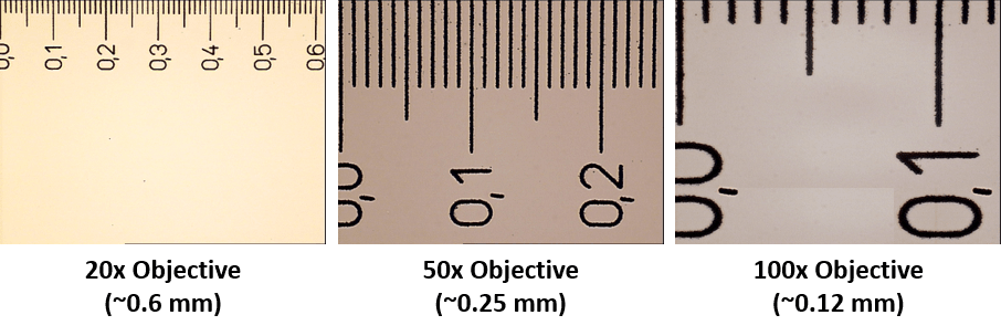
The greater the magnification, the smaller the FOV, as shown in Figure 2. However, high resolutions depend on high magnifications (see our resolution article for more), and high camera speeds can also be obtained with smaller FOVs. So in order to image at a large FOV, it will affect other factors in your imaging. Luckily, most biological samples are small (from cells to molecules) and often don 't need the entire FOV the camera can offer.
Adapters can have lenses in them to magnify or demagnify the image before it reaches the camera. This can be used to better match the camera FOV to the microscope FOV. For example, if the camera has an 11 mm diagonal FOV but the microscope is capable of an 18 mm FOV, a 0.67x adaptor would demagnify the image and allow it to be displayed on the 11 mm camera. However, this increase in FOV comes at the cost of reduced resolution.
Field of viewhuman eye
The camera FOV depends on two factors: the size of the camera sensor and the total magnification. Sensor size can be measured in a number of ways but a commonly used measure is the size of the diagonal across a sensor, as some sensors are square and others rectangular. This is typically displayed in millimeters, and a range of camera sensor sizes can be seen in Figure 1.
One thing to keep in mind is that a microscope FOV is circular, and a camera FOV is square/rectangular, as seen in Fig.3 Top. Pairing an 18 mm FOV camera with a 18 mm FOV microscope does result in some areas that are not imaged, but this is largely a common factor with all imaging systems unless circular camera sensors emerge in the future. So for now, the camera FOV should aim to fit within the microscope FOV, meaning that it is best to match the FOV between camera and microscope.
The development of larger FOV microscopes and scientific cameras that can take advantage of the F-mount is relatively recent - at the time of writing only one commercially available 25 mm microscope exists. Most modern microscopes have a 19 mm or 22 mm FOV and are therefore still able to use the C-mount. The largest format spinning disk confocal systems are also limited to a 22 mm FOV.
Ideal for use on workbenches, lab tables, desks or any work surface. The Waldmann Lighting™ Omnivue and OmnivueMax LED Magnifier Lights offers precise positioning and ergonomic easy viewing.
The field of view of the camera determines how much of your sample you can see. With larger FOVs, cameras can image more effectively and capture a sample in fewer images. However, FOV is decreased at higher magnifications and in order to improve speed, and it should be matched to the model of microscope. By keeping this in mind, you can maximize your FOV and perform more efficient imaging.
If the goal is simply to attach the camera to the microscope, a 1x adaptor contains no additional lenses and provides no additional magnification or demagnification. This is often the preferred method as it introduces no additional lenses into the system. Every extra lens reduces the number of photons reaching the camera by 3-4% so many researchers will try to avoid this.
These larger sensors typically have more pixels, so while an EMCCD would have 0.25 megapixels (MP), CMOS cameras contain anywhere from 1-15 MP depending on the pixel size.
Adaptors can also affect the microscope and camera FOV depending on the type of adaptor used. A C-mount adaptor is the most popular microscope camera adaptor and is restricted to a maximum 22 mm FOV. The F-mount adaptor is a larger format adaptor capable of reaching >30 mm FOV.
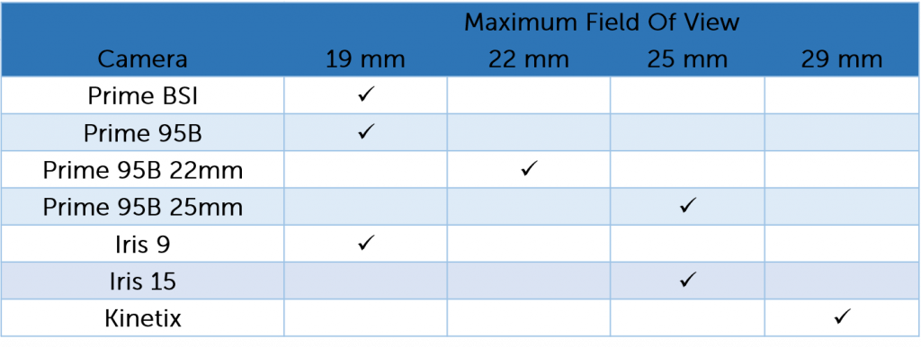
At Teledyne Photometrics, we aim to create cameras that can optimally match the FOV of all modern microscopes (Table 1). For this reason, the Prime 95B Series comprises a 19 mm camera, a 22 mm camera and a 25 mm camera. Additionally, the Prime BSI and Iris 9 both fit a 19 mm microscope FOV and the Iris 15 fits a 25 mm microscope FOV. The Kinetix is our largest format sensor which is able to be used to get the maximum FOV out of any system up to 29 mm.


 Ms.Cici
 8618319014500